Drugs for hemophilia

CHAPTER 54


Drugs for hemophilia


Hemophilia is a rare genetic bleeding disorder seen almost exclusively in males. About 70% of cases result from inheriting a defective gene from the mother. The other 30% result from a spontaneous gene mutation.


Hemophilia has two forms: hemophilia A and hemophilia B. In hemophilia A, there is a deficiency of clotting factor VIII (aka antihemophilic factor). In hemophilia B, there is a deficiency of clotting factor IX (aka Christmas factor, named for Stephen Christmas, the first boy diagnosed with the disease). Hemophilia A is about 6 times more prevalent than hemophilia B, occurring in 1 of every 5000 males, compared with 1 of every 30,000 males for hemophilia B.


When hemophilia is managed well, the prognosis is good. Patients starting treatment today can live healthy, near-normal lives. The foundation of treatment is clotting factor replacement, which may be given on a regular schedule (to prevent bleeds from occurring) or “on demand” (to stop an ongoing bleed). Unfortunately, although treatment is highly effective, it is also very expensive: For patients undergoing prophylactic treatment, the cost for clotting factors alone ranges between $140,000 and $300,000 a year.


Basic considerations


Pathophysiology


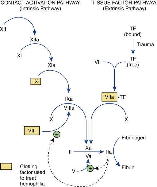
In people with hemophilia, there is a failure of hemostasis, the process by which bleeding is stopped. As discussed in Chapter 52, hemostasis occurs in two stages: (1) formation of a platelet plug followed by (2) production of fibrin, a protein that reinforces the platelet plug. In patients with hemophilia, platelet aggregation proceeds normally, but fibrin production does not. The underlying problem is a deficiency of clotting factors—specifically, factor VIII (in hemophilia A) and factor IX (in hemophilia B). As indicated in Figure 54–1, both factors are part of the contact activation (intrinsic) coagulation pathway, and both—in their activated forms—are needed to catalyze the conversion of factor X to its active form (factor Xa), which in turn catalyzes the conversion of prothrombin to thrombin, which catalyzes the formation of fibrin. If either factor VIII or factor IX is deficient, the contact activation pathway will not work properly, causing clot formation to be delayed. As a result, bleeding will continue longer than in the population at large.


It should be noted that the degree of factor deficiency—and hence the likelihood of serious bleeding—depends on the nature of the underlying gene mutation. In some patients, the mutation produces a severe deficiency, resulting in a high probability of prolonged bleeding. In others, the mutation causes mild deficiency, and hence the tendency to bleed is low.


Inheritance pattern


The genes for factors VIII and IX are recessive, and both are carried on the X chromosome. Because males have only one X chromosome, a male with a defective gene will have hemophilia. In contrast, a female with a defective gene on one X chromosome will usually be an asymptomatic carrier, since she still has a functioning gene on her other X chromosome. Be aware, however, that although females are usually asymptomatic carriers, there are two situations in which females can have hemophilia: (1) a female could be born with defective genes on both X chromosomes, which is rare; and (2) a female who was born with one defective gene could experience inactivation of the good gene. Boys whose mothers are carriers have a 1 in 2 chance of inheriting the disease. Girls whose mothers are carriers have a 1 in 2 chance of being carriers themselves. Males with hemophilia cannot pass the disease on to their sons, but all of their daughters will be carriers. The risk of acquiring hemophilia is shared by all races and ethnic groups.


Clinical features


Hemophilia may be severe, moderate, or mild, depending on the degree of clotting factor deficiency. Patients with severe hemophilia may experience life-threatening hemorrhage in response to minor trauma, whereas those with mild hemophilia may experience little or no excessive bleeding. The defining characteristics of severe, moderate, and mild hemophilia are summarized in Table 54–1.


Severe hemophilia.

In patients with severe hemophilia, the concentration of clotting factor VIII or IX is very low—less than 1% of normal. As a result, these patients experience frequent bleeds within joints and soft tissues, especially muscle. Trauma or surgery can cause profuse hemorrhage. Joint bleeding occurs most often in the knee, followed in turn by the elbow, ankle, shoulder, and hip. Bleeding in these joints causes swelling and intense pain. With recurrent episodes, permanent injury to the joint develops. In addition to occurring in joints, bleeding may occur in muscles, mucous membranes (eg, nosebleeds), the GI and urinary tracts, near the pharynx (which can cause life-threatening restriction of airflow), and within the skull (which carries a 30% risk of death). Among patients with hemophilia A, about 60% have severe disease. In contrast, among patients with hemophilia B, only 20% to 45% have severe disease. Although severe hemophilia can be devastating, most patients can live normal and productive lives, thanks to the availability of safe factor concentrates for replacement therapy.


Overview of therapy


Whenever possible, treatment should be guided by a team of specialists at a hemophilia treatment center. Typically, the team consists of a hematologist, orthopedist, dietitian, psychologist, physical therapist, occupational therapist, genetics counselor, infectious disease specialist, social worker, and nurse coordinator.


The cornerstone of treatment is replacement therapy with factor VIII (hemophilia A) or factor IX (hemophilia B). In the past, factor replacement was performed only to terminate a bleeding episode. Today, however, there is increasing emphasis on primary prophylaxis, especially for young children. Why? Because, by minimizing bleeding episodes, prophylaxis can minimize long-term damage to joints.


For some patients with mild hemophilia A, bleeding can be stopped with desmopressin, a drug that promotes release of factor VIII from the vascular endothelium. Desmopressin has the advantage of being much cheaper than factor VIII, and can be administered by nasal spray as well as by IV infusion. Keep in mind, however, that repeated use of desmopressin can deplete stored factor VIII, making the drug ineffective until more factor VIII is made.


Antifibrinolytic drugs (ie, drugs that prevent the breakdown of fibrin) can be used as adjuncts to factors VIII and IX in special situations, such as tooth extractions. Two antifibrinolytic drugs are currently available: aminocaproic acid and tranexamic acid.


In some patients receiving factor VIII or factor IX, antibodies against the factor develop. These antibodies, referred to as inhibitors, prevent the factor from working. When inhibitors are present, bleeding can be stopped by infusing activated factor VII. Other treatments are also available, as discussed below under Managing Patients Who Develop Inhibitors.


Pain management


How should we manage bleeding-related pain? For mild pain, acetaminophen [Tylenol, others] is the drug of choice. For severe pain, an opioid analgesic may be needed. Regardless of pain severity, aspirin should be avoided! Why? First, aspirin causes irreversible inhibition of platelet aggregation, and can thereby increase bleeding risk. Second, aspirin can induce GI ulceration and bleeding, an obvious problem.


Can we use nonsteroidal anti-inflammatory drugs (NSAIDs) other than aspirin? As a rule, these agents should also be avoided. Why? First, like aspirin, most NSAIDs inhibit platelet aggregation (although the inhibition is reversible rather than irreversible). Second, like aspirin, most NSAIDs can promote GI ulceration and bleeding (although the risk is somewhat lower than with aspirin).


What about the second-generation NSAIDs, known as cyclooxygenase-2 (COX-2) inhibitors? As discussed in Chapter 71, the COX-2 inhibitors (eg, celecoxib) do not suppress platelet aggregation, and they cause less GI ulceration and bleeding than traditional NSAIDs. Accordingly, these agents are clearly preferred to traditional NSAIDs, although their safety in hemophilia has not been proved.


Immunization


Children with hemophilia should undergo the normal immunization schedule (see Chapter 68). Some clinicians inject vaccines subQ, rather than IM, to avoid muscle hemorrhage. However, since the efficacy of subQ vaccination is not certain, and since most patients tolerate IM injections without bleeding, IM vaccination is generally preferred. The risk of bleeding after IM injection can be reduced by prolonged application of pressure.


To minimize the risk of hepatitis (see below), all patients with newly diagnosed hemophilia should be vaccinated for hepatitis A and hepatitis B, as should all other patients with hemophilia who are not seropositive for hepatitis A or B. Family members who administer clotting factors at home should also be immunized, provided they test negative for hepatitis.


Preparations used to treat hemophilia


Factor VIII concentrates


Factor VIII concentrates are the mainstay of hemophilia A treatment. What’s a concentrate? It’s simply a powdered formulation in which the amount of factor VIII is very high. When treatment is needed, the powder is dissolved in a sterile solution and administered IV.


All factor VIII concentrates available today are very safe. They carry essentially no risk of HIV/AIDS, and little or no risk of hepatitis.


Production methods and product safety

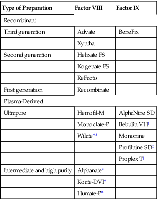
Factor VIII concentrates are made in two basic ways: (1) purification from human plasma and (2) production in cell culture using recombinant DNA technology. Recombinant factor VIII is somewhat safer than plasma-derived factor VIII, but is also more expensive. All factor VIII products, whether recombinant or plasma derived, are equally effective. Available products are listed in Table 54–2.


Plasma-derived factor VIII.

Prior to 1985, factor VIII produced from donor plasma often contained viral contaminants. As a result, nearly all people with hemophilia developed hepatitis and/or HIV/AIDS. Today, however, the risk of viral contamination is exceedingly low. Why? First, donated plasma is now screened for viral pathogens—specifically, human immunodeficiency virus (HIV), hepatitis A virus (HAV), hepatitis B virus (HBV), hepatitis C virus (HCV), and parvovirus B19. Second, techniques for inactivating lipid-coated viruses (HIV, HBV, and HCV) are now employed. Unfortunately, viruses that lack a lipid coat, such as HAV and parvovirus B19, are not eliminated. Nonetheless, no case of virus transmission has been reported with any of the products now used in the United States.


There is one additional concern: prions. These strange proteins, which are responsible for Creutzfeldt-Jakob disease (CJD, the human form of “mad cow disease”), are not susceptible to any known inactivation technique. Hence, the possibility of transmitting CJD remains.


As indicated in Table 54–2, plasma-derived factor VIII is available in varying degrees of purity. The ultrapure products (eg, Hemofil-M) are prepared using monoclonal antibodies.

< div class='tao-gold-member'>

Only gold members can continue reading. Log In or Register to continue

Stay updated, free articles. Join our Telegram channel

Jul 24, 2016 | Posted by in NURSING | Comments Off on Drugs for hemophilia

Full access? Get Clinical Tree

Get Clinical Tree app for offline access