Drugs for epilepsy

CHAPTER 24


Drugs for epilepsy


The term epilepsy refers to a group of chronic neurologic disorders characterized by recurrent seizures, brought on by excessive excitability of neurons in the brain. Symptoms can range from brief periods of unconsciousness to violent convulsions. Patients may also experience problems with learning, memory, and mood, which can be just as troubling as their seizures.


In the United States, about 2.3 million people have epilepsy, according to the Centers for Disease Control and Prevention (CDC). Every year, 140,000 new cases are diagnosed. The incidence is highest in the very young and the elderly. Between 60% and 70% of patients can be rendered seizure free with drugs. Unfortunately, this means that 30% to 40% cannot. The total direct and indirect costs of epilepsy are estimated at $15.5 billion a year.


The terms seizure and convulsion are not synonymous. Seizure is a general term that applies to all types of epileptic events. In contrast, convulsion has a more limited meaning, applying only to abnormal motor phenomena, for example, the jerking movements that occur during a tonic-clonic (grand mal) attack. Accordingly, although all convulsions may be called seizures, it is not correct to call all seizures convulsions. Absence seizures, for example, manifest as brief periods of unconsciousness, which may or may not be accompanied by involuntary movements. Since not all epileptic seizures involve convulsions, we will refer to the agents used to treat epilepsy as antiepileptic drugs (AEDs), rather than anticonvulsants.


Seizure generation


Seizures are initiated by synchronous, high-frequency discharge from a group of hyperexcitable neurons, called a focus. A focus may result from several causes, including congenital defects, hypoxia at birth, head trauma, brain infection, stroke, cancer, and genetic disorders. Seizures are seen when discharge from a focus spreads to other brain areas, thereby recruiting normal neurons to discharge abnormally along with the focus.


The overt manifestations of any particular seizure disorder depend on the location of the seizure focus and the neuronal connections to that focus. (The connections to the focus determine the brain areas to which seizure activity can spread.) If seizure activity invades a very limited part of the brain, a partial or local seizure occurs. In contrast, if seizure activity spreads to a large portion of the brain, a generalized seizure develops.


An experimental procedure referred to as kindling may explain how a focal discharge is eventually able to generate a seizure. Experimental kindling is performed by implanting a small electrode into the brain of an animal. The electrode is used to deliver localized stimuli for a brief interval once a day. When stimuli are first administered, no seizures result. However, after repeated once-a-day delivery, these stimuli eventually elicit a seizure. If brief, daily stimulations are continued long enough, spontaneous seizures will begin to occur.


The process of kindling may tell us something about seizure development in humans. For example, kindling may account for the delay that can take place between injury to the head and eventual development of seizures. Furthermore, kindling may explain why the seizures associated with some forms of epilepsy become more frequent as time passes. Also, the progressive nature of kindling suggests that early treatment might prevent seizure disorders from becoming more severe over time.


Types of seizures


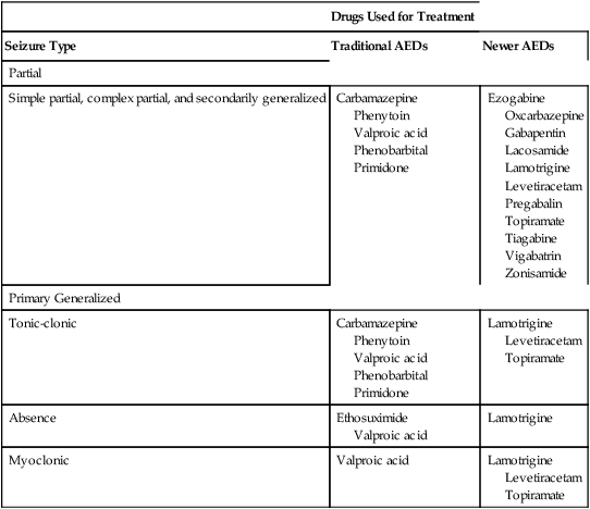
Seizure can be divided into two broad categories: partial (focal) seizures and generalized seizures. In partial seizures, seizure activity begins focally in the cerebral cortex and usually undergoes limited spread to adjacent cortical areas. In generalized seizures, focal seizure activity is conducted widely throughout both hemispheres. As a rule, partial seizures and generalized seizures are treated with different drugs (Table 24–1).


Partial seizures


Partial seizures fall into three groups: simple partial seizures, complex partial seizures, and partial seizures that evolve into secondarily generalized seizures.


Generalized seizures


Generalized seizures may be convulsive or nonconvulsive. As a rule, they produce immediate loss of consciousness. The major generalized seizures are discussed briefly below.


How antiepileptic drugs work


We have long known that AEDs can (1) suppress discharge of neurons within a seizure focus and (2) suppress propagation of seizure activity from the focus to other areas of the brain. However, until recently we didn’t know how these effects were achieved. It now appears that nearly all AEDs act through five basic mechanisms: suppression of sodium influx, suppression of calcium influx, promotion of potassium efflux, blockade of receptors for glutamate, and potentiation of gamma-aminobutyric acid (GABA).


Suppression of sodium influx.

Before discussing AED actions, we need to review sodium channel physiology. Neuronal action potentials are propagated by influx of sodium through sodium channels, which are gated pores in the cell membrane that control sodium entry. For sodium influx to occur, the channel must be in an activated state. Immediately following sodium entry, the channel goes into an inactivated state, during which further sodium entry is prevented. Under normal circumstances, the inactive channel very quickly returns to the activated state, thereby permitting more sodium entry and propagation of another action potential.


Several AEDs, including phenytoin, carbamazepine, valproic acid, and lamotrigine, reversibly bind to sodium channels while they are in the inactivated state, and thereby prolong channel inactivation. By delaying return to the active state, these drugs decrease the ability of neurons to fire at high frequency. As a result, seizures that depend on high-frequency discharge are suppressed.


Basic therapeutic considerations


Therapeutic goal and treatment options


The goal in treating epilepsy is to reduce seizures to an extent that enables the patient to live a normal or near-normal life. Ideally, treatment should eliminate seizures entirely. However, this may not be possible without causing intolerable side effects. Therefore, we must balance the desire for complete seizure control against the acceptability of side effects.


Epilepsy may be treated with drugs or with nondrug therapies. As noted, drugs can benefit 60% to 70% of patients. This means that, of the 2.3 million Americans with epilepsy, between 690,000 and 920,000 cannot be treated successfully with drugs. For these people, nondrug therapy may well help. Three options exist: neurosurgery, vagus nerve stimulation, and the ketogenic diet. Of the three, neurosurgery has the best success rate, but vagus nerve stimulation is used most widely. All three nondrug therapies are discussed in Box 24–1.


imageBOX 24–1    SPECIAL INTEREST TOPIC


NONDRUG THERAPIES FOR EPILEPSY: NEUROSURGERY, VAGUS NERVE STIMULATION, AND THE KETOGENIC DIET


Neurosurgery: the cure that’s rarely used


For patients with temporal lobe epilepsy, surgery is highly effective, but used infrequently. At this time, surgical intervention is the only cure for epilepsy. (Drugs may control symptoms, but they don’t cure.) The safety and efficacy of surgery have been documented in literally hundreds of studies. Among patients with forms of epilepsy that can be treated surgically, the procedure can render between 60% and 90% seizure free—and, even when seizures do continue, their frequency is often decreased. This degree of success is all the more remarkable when we consider that, in order to qualify for surgery, candidates must first be proved refractory to drugs. Put another way, surgery is only performed on patients who have epilepsy that is especially hard to treat. Yet, despite its proven efficacy, surgery remains grossly underutilized: Each year, only 500 or so surgeries are performed in the United States, although more than 100,000 patients are eligible. This is especially unfortunate because, among people who are refractory to drugs, surgery can greatly improve seizure control, thereby improving quality of life, along with attendance at work and at school.


Temporal lobe surgery is not without risk. Between 5% and 10% of patients experience adverse effects, including infection, visual field defects, memory loss, and paralysis. The death rate is very low—about 0.2%. Prior to surgery, patients undergo a battery of tests designed to locate the seizure focus as well as nearby areas associated with language and other critical functions. This information allows the surgeon to remove as little tissue as possible, thereby minimizing disruption of normal brain function.


Vagus nerve stimulation: fighting impulses with impulses


The vagus nerve stimulator (VNS) is the first medical device for reducing seizures. The only commercial VNS available—the VNS Therapy System (formerly known as the Neuro-Cybernetic Prosthesis System)—received FDA approval in 1997. The system is intended for use in conjunction with drugs by patients with severe, uncontrolled seizures. Responses to vagal stimulation develop slowly: Initial responses usually occur in 3 months, and full responses take even longer to develop. Vagal nerve stimulation is now the most widely used nondrug therapy for drug-resistant seizures.


The heart of the VNS is a small, programmable pulse generator that is implanted under the collarbone, much like a cardiac pacemaker. Subcutaneous leads connect the generator to the left branch of the vagus nerve in the neck. Stimulation is typically applied for 30 seconds every 5 minutes around the clock. When needed, stimulation parameters (voltage, frequency, duration) can be adjusted externally by the physician. By holding a small magnet over the generator, patients can activate the device manually if they feel a seizure coming on. In addition, patients can use the magnet to turn the generator off. VNS batteries last 3 to 5 years. Replacement is done in an outpatient procedure that takes 30 to 60 minutes.


In clinical trials, some patients responded dramatically and most showed at least some improvement. However, with a few patients, seizures increased. Specific results were as follows:


Vagal stimulation does not eliminate the need for drugs—but it can permit a simpler regimen. Up to 50% of patients can decrease the number of drugs they are taking (eg, two instead of three; one instead of two). Please note, however, that stimulation does not permit a reduction in dosage of the drugs that remain.


Vagal stimulation is well tolerated by most patients, although side effects occur often. During stimulation, patients experience hoarseness (100%), coughing (50%), voice alteration (73%), and shortness of breath (25%). In addition, there is a 2% to 3% risk of infection at the implant site. Stimulation does not cause cognitive effects and, perhaps surprisingly, does not cause autonomic effects (eg, bradycardia, GI disturbances, hypotension).


How does vagal stimulation decrease seizure frequency? No one knows. What we do know is that vagal fibers project to the brainstem, and from there to areas of the brain involved in seizure generation. When we stimulate the vagus, the resultant impulses in some way interrupt or prevent abnormal neuronal firing.


In 2005, the VNS Therapy System was approved for treating depression. This application is discussed in Chapter 32.


The ketogenic diet: it’s tough but it works


The ketogenic diet for epilepsy can decrease seizure frequency, but it’s hard to implement and potentially dangerous. The diet was introduced in the 1920s, but fell out of use when AEDs became available. Today, the diet is under renewed study as a way to control seizures when drug therapy fails—and even as first-line therapy for some patients. Because the diet is both difficult and hazardous, close medical supervision is essential.


The ketogenic diet has two cornerstones: high intake of fat and very low intake of carbohydrates. Fats—usually butter or heavy cream—comprise 80% of daily calories. The remaining 20% come from carbohydrates and proteins. With strict adherence to the diet, ketosis develops in a few days. However, with just a minor deviation from the diet (eg, ingestion of two cookies), ketosis will be lost in hours.


How does a high-fat, low-carbohydrate diet reduce seizures? The answer is unclear. For years, we believed that benefits derived from causing ketoacidosis. (Because carbohydrate availability is low, the body burns fat to meet energy needs. Burning fat produces large amounts of ketone bodies—beta-hydroxybutyric acid, acetoacetic acid, and acetone—whose presence creates a state of ketoacidosis.) However, although the diet does indeed cause ketoacidosis, there are no data showing that ketoacidosis is the reason for seizure control. In fact, another high-fat diet (a modified form of the Atkins diet), which does not cause ketoacidosis, can nonetheless reduce seizure occurrence. The bottom line? High intake of fat and low intake of carbohydrates are probably both playing a role in seizure control.


The principal candidates for dietary therapy are children under the age of 10 who have not responded to AEDs. About two-thirds of these children respond to the diet. Among the responders, seizure reduction occurs rapidly, typically within a few days. In a trial reported in 2008, seizure frequency was reduced by more than 50%.


Adverse effects of the diet are considerable. The most consistent—and obvious—is elevation of blood cholesterol. In one study, cholesterol levels rose from a mean of 170 mg/dL to 245 mg/dL. In another study, five children developed severe hypercholesterolemia, with an average level of 367 mg/dL. Since cholesterol contributes to coronary artery disease (CAD), and since CAD is known to begin early in life, elevation of cholesterol is a significant drawback. Other adverse effects include poor linear growth, poor weight gain, kidney stones, dehydration, acidosis, constipation, and vomiting. However, although the side effects of the diet are a concern, keep in mind that AEDs can also cause harm—perhaps even more than the diet. Hence, if the diet allows a reduction in AED use, there may be little or no net increase in harm.


Diagnosis and drug selection


Control of seizures requires proper drug selection. As indicated in Table 24–1, many AEDs are selective for specific seizure disorders. Phenytoin, for example, is useful for treating tonic-clonic and partial seizures but not absence seizures. Conversely, ethosuximide is active against absence seizures but not against tonic-clonic or partial seizures. Only one drug—valproic acid—appears effective against practically all forms of epilepsy. Since most AEDs are selective for certain seizure disorders, effective treatment requires a proper match between the drug and the seizure. To make this match, the seizure type must be accurately diagnosed.


Making a diagnosis requires physical, neurologic, and laboratory evaluations along with a thorough history. The history should determine the age at which seizures began, the frequency and duration of seizure events, precipitating factors, and times when seizures occur. Physical and neurologic evaluations may reveal signs of head injury or other disorders that could underlie seizure activity, although in many patients the physical and neurologic evaluations may be normal. An electroencephalogram (EEG) is essential for diagnosis. Other diagnostic tests that may be employed include computed tomography (CT), positron emission tomography (PET), and magnetic resonance imaging (MRI).


Very often, patients must try several AEDs before a regimen that is both effective and well tolerated can be established. Initial treatment should be done with just one AED. If this drug fails, it should be discontinued and a different AED should be tried. If this second drug fails, two options are open: (1) treatment with a third AED alone, or (2) treatment with a combination of AEDs.


Drug evaluation


Once an AED has been selected, a trial period is needed to determine its effectiveness. During this time there is no guarantee that seizures will be controlled. Until seizure control is certain, the patient should be warned not to participate in driving and other activities that could be hazardous should a seizure occur.


During the process of drug evaluation, adjustments in dosage are often needed. No drug should be considered ineffective until it has been tested in sufficiently high dosage and for a reasonable time. Knowledge of plasma drug levels can be a valuable tool for establishing dosage and evaluating the effectiveness of a specific drug.


Maintenance of a seizure frequency chart is important. The chart should be kept by the patient or a family member and should contain a complete record of all seizure events. This record will enable the prescriber to determine if treatment has been effective. The nurse should teach the patient how to create and use a seizure frequency chart.


Monitoring plasma drug levels


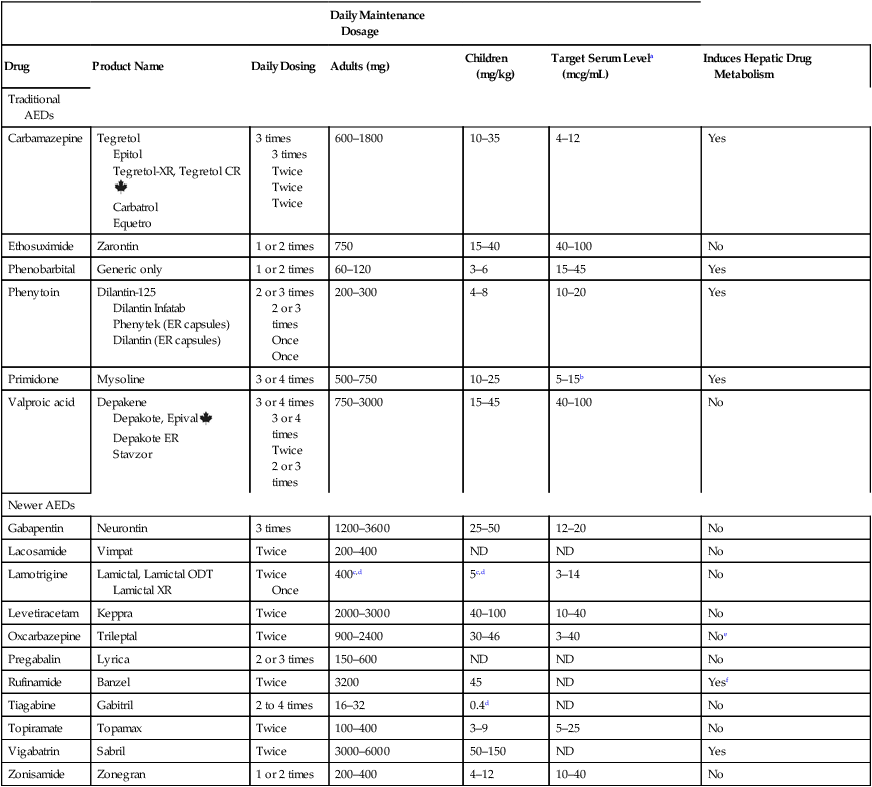
Monitoring plasma levels of AEDs is common. Safe and effective levels have been firmly established for most AEDs (Table 24–2). Monitoring these levels can help guide dosage adjustments.


TABLE 24–2 


Clinical Pharmacology of the Oral Antiepileptic Drugs (AEDs)


      Daily Maintenance Dosage    
Drug Product Name Daily Dosing Adults (mg) Children (mg/kg) Target Serum Levela (mcg/mL) Induces Hepatic Drug Metabolism
Traditional AEDs
Carbamazepine Tegretol
Epitol
Tegretol-XR, Tegretol CRimage
Carbatrol
Equetro
3 times
3 times
Twice
Twice
Twice
600–1800 10–35 4–12 Yes
Ethosuximide Zarontin 1 or 2 times 750 15–40 40–100 No
Phenobarbital Generic only 1 or 2 times 60–120 3–6 15–45 Yes
Phenytoin Dilantin-125
Dilantin Infatab
Phenytek (ER capsules)
Dilantin (ER capsules)
2 or 3 times
2 or 3 times
Once
Once
200–300 4–8 10–20 Yes
Primidone Mysoline 3 or 4 times 500–750 10–25 5–15b Yes
Valproic acid Depakene
Depakote, Epivalimage
Depakote ER
Stavzor
3 or 4 times
3 or 4 times
Twice
2 or 3 times
750–3000 15–45 40–100 No
Newer AEDs
Gabapentin Neurontin 3 times 1200–3600 25–50 12–20 No
Lacosamide Vimpat Twice 200–400 ND ND No
Lamotrigine Lamictal, Lamictal ODT
Lamictal XR
Twice
Once
400c,d 5c,d 3–14 No
Levetiracetam Keppra Twice 2000–3000 40–100 10–40 No
Oxcarbazepine Trileptal Twice 900–2400 30–46 3–40 Noe
Pregabalin Lyrica 2 or 3 times 150–600 ND ND No
Rufinamide Banzel Twice 3200 45 ND Yesf
Tiagabine Gabitril 2 to 4 times 16–32 0.4d ND No
Topiramate Topamax Twice 100–400 3–9 5–25 No
Vigabatrin Sabril Twice 3000–6000 50–150 ND Yes
Zonisamide Zonegran 1 or 2 times 200–400 4–12 10–40 No

image


CR = extended-release, ER = extended-release, ODT = orally disintegrating tablet, XR = extended-release.


aND = not determined.


bTarget serum level is 5 to 15 mcg/mL for primidone itself, and 15 to 40 mcg/mL for phenobarbital derived from primidone.


cDosage must be decreased in patients taking valproic acid.


dDosage must be increased in patients taking drugs that induce hepatic drug-metabolizing enzymes.


eOxcarbazepine does not induce enzymes that metabolize AEDs, but does induce enzymes that metabolize other drugs.


fRufinamide produces mild induction of CYP3A4.


Monitoring plasma drug levels is especially helpful when treating major convulsive disorders (eg, tonic-clonic seizures). Since these seizures can be dangerous, and since delay of therapy may allow the condition to worsen, rapid control of seizures is desirable. However, because these seizures occur infrequently, a long time may be needed to establish control if clinical outcome is relied on as the only means of determining an effective dosage. By adjusting initial doses on the basis of plasma drug levels (rather than on the basis of seizure control), we can readily achieve drug levels that are likely to be effective, thereby increasing our chances of establishing control quickly.


Measurements of plasma drug levels are less important for determining effective dosages for absence seizures. Why? Because absence seizures occur very frequently (up to several hundred a day), and hence observation of the patient is the best means for establishing an effective dosage: if seizures stop, dosage is sufficient; if seizures continue, more drug is needed.


In addition to serving as a guide for dosage adjustment, knowledge of plasma drug levels can serve as an aid to (1) monitoring patient adherence, (2) determining the cause of lost seizure control, and (3) identifying causes of toxicity, especially in patients taking more than one drug.


Suicide risk with antiepileptic drugs


In 2008, the Food and Drug Administration (FDA) warned that all AEDs can increase suicidal thoughts and behavior. However, data gathered since 2008 suggest that the risk may be lower than previously believed, and may apply only to certain AEDs.


The FDA based its warning on data from 199 placebo-controlled studies involving 11 different AEDs taken by 43,892 patients being treated for epilepsy, psychiatric disorders, and various pain disorders. After analyzing these data, the FDA concluded that, compared with patients taking a placebo, patients taking AEDs had twice the risk of suicidal thoughts and behaviors (0.43% vs. 0.22%). The reason underlying the increased risk was unknown. Of note, risk was higher among patients taking AEDs for epilepsy than among patients taking these drugs for other conditions, such as migraine, neuropathic pain, or psychiatric illness. Although the analysis was limited to 11 drugs, the FDA applied their warning to all AEDs. However, in the FDA’s own analysis, the association between suicide and AED use had statistical significance with just two drugs: topiramate and lamotrigine. Furthermore, with two other drugs—valproic acid and carbamazepine—their analysis showed some protection against suicidality.


Since the FDA issued its warning, other large studies have been conducted to clarify the relationship between AEDs and suicidality. Unfortunately, these studies have yielded conflicting results. Nonetheless, they do suggest three things. First, only some AEDs—especially topiramate and lamotrigine—are likely to increase suicidality, not all AEDs as warned by the FDA. Second, the risk of suicidal behavior may be related more to the illness than the medication: By analyzing data on 5,130,795 patients, researchers in the United Kingdom found that AEDs produced a small increase in suicidal behavior in patients with depression, but did not increase suicidal behavior in patients with epilepsy or bipolar disorder. And third, even if AEDs do promote suicidality, AED-related suicide attempts and completed suicides are very rare.


Given the uncertainty regarding AEDs and suicidality, what should the clinician do? Because epilepsy itself carries a risk for suicide, and because patients with epilepsy often have depression and/or anxiety (which increase the risk of suicide), prudence dictates screening all patients for suicide risk, whether or not AEDs increase that risk. In addition, once treatment begins, all patients should be monitored for increased anxiety, agitation, mania, and hostility—signs that may indicate the emergence or worsening of depression, and an increased risk of suicidal thoughts or behavior. Patients, families, and caregivers should be alerted to these signs and advised to report them immediately. Finally, two AEDs—topiramate and lamotrigine—should be used with special caution, given their significant association with suicidality.


Classification of antiepileptic drugs


The AEDs can be grouped into two major categories: traditional AEDs and newer AEDs. The traditional group has six major members, the last of which—valproic acid—was approved in 1978. The group of newer AEDs has thirteen members, all of which were approved in 1993 or later. As summarized in Table 24–3, both groups have their advantages and disadvantages. For example, clinical experience with the older AEDs is more extensive than with the newer ones, and the older drugs cost less. Both facts make the older drugs attractive. However, the older AEDs also have drawbacks, including troublesome side effects and complex drug interactions. Of importance, drugs in both groups appear equally effective—although few direct comparisons have been made. The bottom line? Neither group is clearly superior to the other. Hence, when selecting an AED, drugs in both groups should be considered.


Traditional antiepileptic drugs


The traditional AEDs have been in use for many years. Antiseizure properties of phenobarbital, the oldest member of the group, were demonstrated in 1912. Even the youngest member—valproic acid—has been in use for over three decades. Because of this extensive clinical experience, the efficacy and therapeutic niche of the traditional AEDs are well established. As a result, these drugs are prescribed more widely than the newer AEDs. In the discussion below, we focus on six of the traditional AEDs: phenytoin, carbamazepine, valproic acid, ethosuximide, phenobarbital, and primidone. The group has other members, but they are less important.


Although familiarity makes the traditional AEDs appealing, these drugs do have drawbacks. In general, they are less well tolerated than the newer AEDs, and they pose a greater risk to the developing fetus. Furthermore, owing to effects on drug-metabolizing enzymes (either induction or inhibition), they have complex interactions with other drugs, including other AEDs.


Phenytoin


Phenytoin [Dilantin, Phenytek] is our most widely used AED, despite having tricky kinetics and troublesome side effects. The drug is active against partial seizures as well as primary generalized tonic-clonic seizures. Phenytoin is of historic importance in that it was the first drug to suppress seizures without depressing the entire CNS. Hence, phenytoin heralded the development of selective medications that could treat epilepsy while leaving most CNS functions undiminished.


Pharmacokinetics

Phenytoin has unusual pharmacokinetics that must be accounted for in therapy. Absorption varies substantially among patients. In addition, because of saturable kinetics, small changes in dosage can produce disproportionately large changes in serum drug levels. As a result, a dosage that is both effective and safe is difficult to establish.


Metabolism.

The capacity of the liver to metabolize phenytoin is very limited. As a result, the relationship between dosage and plasma levels of phenytoin is unusual. Doses of phenytoin needed to produce therapeutic effects are only slightly smaller than the doses needed to saturate the hepatic enzymes that metabolize phenytoin. Consequently, if phenytoin is administered in doses only slightly greater than those needed for therapeutic effects, the liver’s capacity to metabolize the drug will be overwhelmed, causing plasma levels of phenytoin to rise dramatically. This unusual relationship between dosage and plasma levels is illustrated in Figure 24–1A. As you can see, once plasma levels have reached the therapeutic range, small changes in dosage produce large changes in plasma levels. As a result, small increases in dosage can cause toxicity, and small decreases can cause therapeutic failure. This relationship makes it difficult to establish and maintain a dosage that is both safe and effective.


Figure 24–1B indicates the relationship between dosage and plasma levels that exists for most drugs. As indicated, this relationship is linear, in contrast to the nonlinear relationship that exists for phenytoin. Accordingly, for most drugs, if the patient is taking doses that produce plasma levels that are within the therapeutic range, small deviations from that dosage produce only small deviations in plasma drug levels. Because of this relationship, with most drugs it is relatively easy to maintain plasma levels that are safe and effective.


Because of saturation kinetics, the half-life of phenytoin varies with dosage. At low doses, the half-life is relatively short—about 8 hours. However, at higher doses, the half-life becomes prolonged—in some cases up to 60 hours. Why? Because, at higher doses, there is more drug present than the liver can process. As a result, metabolism is delayed, causing the half-life to increase.


Adverse effects


Dermatologic effects.

Between 2% and 5% of patients develop a morbilliform (measles-like) rash. Rarely, morbilliform rash progresses to much more severe reactions: Stevens-Johnson syndrome (SJS) or toxic epidermal necrolysis (TEN). According to a 2008 alert from the FDA, the risk of developing SJS/TEN may be increased by a genetic mutation known as human leukocyte antigen (HLA)-B*1502, which occurs almost exclusively in people of Asian descent. However, data in support of this association are preliminary. As discussed below, vulnerability to SJS/TEN in patients taking another AED—carbamazepine—is clearly linked to HLA-B*1502. Because of this clear association, and because of the possible association between phenytoin-induced SJS/TEN and HLA-B*1502, patients with the mutation should definitely avoid carbamazepine, and should probably avoid phenytoin too. At this time, the FDA recommends testing for HLA-B*1502 in Asian patients taking carbamazepine, but not yet for Asian patients taking phenytoin.


Effects in pregnancy.

Phenytoin is a teratogen in animals and humans. In animals, the drug can cause cleft palate, hydrocephalus, renal defects, and micromelia (small or shortened limbs). In humans, phenytoin can cause cleft palate, heart malformations, and fetal hydantoin syndrome, characterized by growth deficiency, motor or mental deficiency, microcephaly, craniofacial distortion, positional deformities of the limbs, hypoplasia of the nails and fingers, and impaired neurodevelopment. Because of these effects, phenytoin is classified in FDA Pregnancy Category D, and hence should be used during pregnancy only if the benefits of seizure control are deemed to outweigh the risk to the fetus.


Phenytoin can decrease synthesis of vitamin K–dependent clotting factors, and can thereby cause bleeding tendencies in newborns. The risk of neonatal bleeding can be decreased by giving the mother prophylactic vitamin K for 1 month prior to and during delivery, and to the infant immediately after delivery.


Cardiovascular effects.

When phenytoin is administered by IV injection (to treat SE), cardiac dysrhythmias and hypotension may result. These dangerous responses can be minimized by injecting phenytoin slowly and in dilute solution.


Drug interactions

Phenytoin interacts with a large number of drugs. The more important interactions are discussed below.


Preparations, dosage, and administration


Dosage.

Dosing is highly individualized. Initial doses are usually given twice daily. Once a maintenance dosage has been established, once-a-day dosing is often possible (using extended-release capsules). For adults, a typical initial dosage is 100 to 125 mg 3 times a day; maintenance dosages usually range between 300 and 400 mg/day. For children, a typical initial dosage is 2.5 mg/kg twice a day; maintenance dosages usually range between 4 and 8 mg/kg/day.


Plasma drug levels are often monitored as an aid to establishing dosage. The dosing objective is to produce levels between 10 and 20 mcg/mL. Levels below 10 mcg/mL are too low to control seizures; levels above 20 mcg/mL produce toxicity. Because phenytoin has a relatively narrow therapeutic range (between 10 and 20 mcg/mL), and because of the nonlinear relationship between phenytoin dosage and phenytoin plasma levels, once a safe and effective dosage has been established, the patient should adhere to it rigidly.


When treatment is discontinued, dosage should be reduced gradually. Abrupt withdrawal may precipitate seizures.


Jul 24, 2016 | Posted by in NURSING | Comments Off on Drugs for epilepsy

Full access? Get Clinical Tree

Get Clinical Tree app for offline access