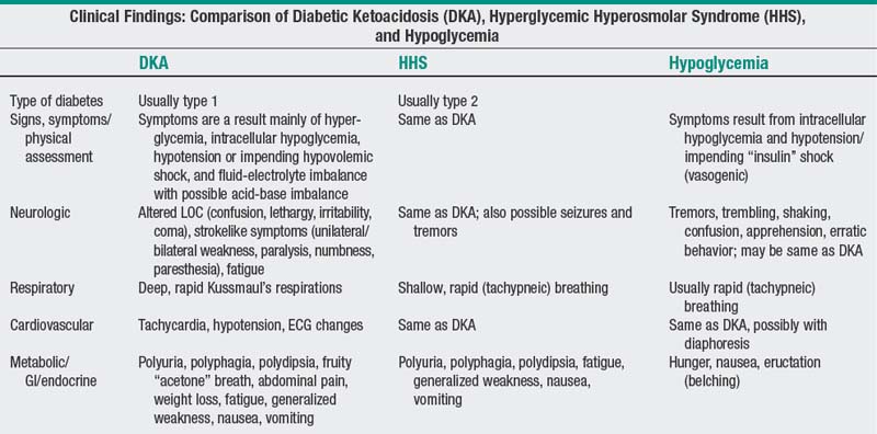
46 Diabetic ketoacidosis
Assessment/diagnostic tests
Nursing diagnoses:
Risk for shock
related to failure of regulatory mechanisms or decreased circulating volume occurring with hyperglycemia
| ASSESSMENT/INTERVENTIONS | RATIONALES |
|---|---|
| Assess for signs and symptoms of hypovolemic shock, including changes in VS q15min until patient remains stable for 1 hr. Notify health care provider promptly of significant findings. | Hyperglycemia acts as an osmotic diuretic, causing severe fluid and electrolyte losses that can lead to hypovolemic shock if untreated. HR greater than 120 bpm, BP less than 90/60 mm Hg or decreased 20 mm Hg or more from baseline, and CVP less than 2 mm Hg are signs of hypovolemia and necessitate notifying health care provider for timely intervention. |
| Assess for poor skin turgor, dry mucous membranes, sunken and soft eyeballs, tachycardia, and orthostatic hypotension. | These are physical indicators of hypovolemia. |
| Measure I&O accurately and weigh patient daily. Monitor urinary specific gravity and report findings of more than 1.020 in the presence of other indicators of dehydration. Report to health care provider if urine output is less than 30 mL/hr or less than 0.5mL/kg/hr for 2 consecutive hr. | Decreasing urinary output may signal diminishing intravascular fluid volume or impending renal failure. Loss of weight and output that exceeds intake may signal dehydration. However, loss of weight is unlikely in the setting of aggressive rehydration. To ensure accuracy, weight should be measured on the same scale if possible. |
| Administer intravenous (IV) fluids as prescribed. | This ensures adequate rehydration. Usually, normal saline or 0.45% saline is administered until plasma glucose falls to 200-300 mg/dL. After that, dextrose-containing solutions usually are given to prevent rebound hypoglycemia. Initially, IV fluids are administered rapidly (i.e., up to 2000 mL infused during the first 2 hr of treatment and 150-250 mL/hr thereafter until BP stabilizes). |
| Be alert to indicators of fluid overload; particularly in elders or in patients with a history of heart failure or renal failure. | Indicators of fluid overload (jugular vein distention, dyspnea, crackles, CVP more than 12 mm Hg) can occur with rapid infusion of fluids. |
| Administer insulin as prescribed. | Insulin usually is given by continuous IV infusion for rapid action and because poor tissue perfusion caused by dehydration sometimes makes the subcutaneous route less effective. Numerous protocols for IV insulin infusion are available, and vary widely in the dosing regimen. Safer, more effective protocols should take into consideration the patient’s level of insulin sensitivity and/or insulin resistance. The continuous infusion is administered through a separate IV tubing and controlled with an infusion control device. Computerized insulin dosing systems, dosing tables, and dosing algorithms are available. Dosage is adjusted based on serial glucose levels and resolution of ketosis. When formulas are used, the insulin sensitivity number is reflected as a variable multiplier that increases with higher levels of insulin resistance. Insulin analogues (i.e., NovoLog, HumaLog, Apidra) may be used in place of regular insulin to lower blood glucose levels. |
Before initiating treatment, flush the tubing with at least 30 mL of the insulin-containing IV solution. | Insulin, when added to IV solutions, may be absorbed by the container and plastic tubing. Flushing the tubing ensures that maximum adsorption of the insulin by the container and tubing has occurred before it is delivered to the patient. |
| Monitor laboratory results for abnormalities. Promptly report to health care provider serum K+ levels less than 3.5 mEq/L. Observe for clinical manifestations of the electrolyte, glucose, and acid-base imbalances associated with DKA as follows: | Before treatment there is risk of hyperkalemia from excess transport of intracellular K+ to extracellular spaces as a result of the acidosis. Na+ and Cl− are replaced with IV normal saline. K+ must be monitored and corrected carefully. After initiation of treatment, K+ returns to the intracellular compartment through accelerated transport into cells via insulin and following correction of acidosis, and therefore the patient is at risk for becoming hypokalemic. Use of phosphorus replacement is controversial, but if phosphorus levels remain low, potassium phosphate solutions can be used to assist with K+ and phosphate replacement. Proper rehydration and insulin dosing should correct ketoacidosis, and lactic acidosis resulting from hypoperfusion due to hypovolemia and/or hypovolemic shock. |
– Hypomagnesemia: Anorexia, nausea, vomiting, lethargy, weakness, personality changes, tetany, tremor or muscle fasciculations, seizures, confusion progressing to coma. – Hypoglycemia: Headache, impaired mentation, agitation, dizziness, nausea, pallor, tremors, tachycardia, diaphoresis. |
Nursing diagnosis:
Risk for infection
| ASSESSMENT/INTERVENTIONS | RATIONALES |
|---|---|
| Assess for evidence of infection. Monitor laboratory results for increased WBC count; culture purulent drainage as prescribed. | Infection is the most common cause of DKA in adults, whereas nonadherence to treatment regimen is more likely responsible for DKA in children and teenagers. Indicators of infection include fever, chills, pain with urination, vomiting, erythema and swelling around IV sites, and increased WBC count. |
| Use meticulous hand hygiene when caring for patient. | Patient is at increased risk for bacterial infection because of suppressed inflammatory response. |
| Manage invasive lines carefully. Schedule dressing changes according to agency policy. | Peripheral IV sites should be rotated at least q96h and dressings changed, depending on agency policy. Central lines should be discontinued as soon as feasible and when in place should be handled carefully. |
| Inspect insertion sites for erythema, swelling, or purulent drainage. Document the presence of any of these indicators, and notify health care provider. | These are signs of local infection that should be reported promptly for timely intervention. |
| Provide good skin care. | Intact skin is a first line of defense against infection. |
| Provide a pressure-relief mattress or pressure redistribution surface on patient’s bed. | These surfaces help prevent skin breakdown, which could lead to infection. |
| Use meticulous sterile technique when caring for or inserting indwelling urinary catheters. | This minimizes the risk of bacterial entry into the body. |
| Note: Limit use of indwelling urethral catheters to patients who are unable to void in a bedpan or when continuous assessment of urine output is essential. | There is increased risk of infection with indwelling catheters. Nationally recognized nurse-sensitive indicators recommend if an indwelling catheter is inserted, every effort is made to remove it within 48 hr. |
| Encourage hourly use of incentive spirometry while patient is awake, along with deep-breathing and coughing exercises. When patient stabilizes, enable patient to get out of bed, which also helps mobilize secretions and reduces the potential for deep vein thrombosis (DVT) or venous thromboembolus (VTE). | Deep inhalations with incentive spirometry along with deep breathing exercises expand alveoli and aid in mobilizing secretions to the airways. Coughing further mobilizes and clears the secretions. These exercises help prevent pulmonary infection. |
Only gold members can continue reading. Log In or Register to continue
Stay updated, free articles. Join our Telegram channel

Full access? Get Clinical Tree


