Craniocerebral Injuries


Craniocerebral Injuries


Karen S. March

Joanne V. Hickey


SCOPE OF THE PROBLEM

It is estimated that there are 1.7 million new cases of traumatic brain injury (TBI) annually in the United States; 2,75,000 of these will require hospital admission for treatment, approximately 52,000 people will die, and about 80,000 to 90,000 will have long-term disabilities.1 The incidence of TBI is highest in those of ages 0 to 4, 15 to 19, and greater than 75 years. Males are more likely to have head injury, although it is noted that females have a higher mortality rate. Motor vehicle collisions (17.3%), pedestrian struck by car (16.5%), falls (35.2%), assaults and violence (10%), and other or unknown (16%) are the major causes of TBI.2 Driving while under the influence of alcohol and/or drugs, lack of use of restraints among automobile occupants, and lack of use of helmets among motorcyclists are commonly associated with motor vehicle crashes. The number of alcohol-related TBIs in 2006 has dropped 36% since 1982 as a result of stricter laws regarding drinking and driving.3 Fatalities differ based on the cause of TBI, with firearm-related head injuries having a mortality rate of 91%.4

Disability after craniocerebral trauma has significant impact not only for the brain-injured person, but also for the family and society. Conservative estimates for permanent disability based on severity of head injury on presentation are 10% in mild, 66% in moderate, and 100% in severe injuries. The loss of human potential and the physical, emotional, psychosocial, and vocational impacts on the patient and family function are immeasurable and devastating. These impacts create the need for many health-related and community services.


OVERVIEW OF CRANIOCEREBRAL TRAUMA


Definitions and Classification

Craniocerebral trauma and TBI are general designations to denote injury to the skull, brain, or both that is of sufficient magnitude to interfere with normal function and to require treatment. Chart 16-1 provides a classification of scalp and craniocerebral injuries.

Clinically, TBI is often classified according to a severity injury index called the Glasgow Coma Scale (GCS) (Fig. 16-1). Alternatively, craniocerebral trauma can be classified according to location.


  • Scalp injuries: this may include contusion, abrasion, laceration, and subgaleal hematoma


  • Skull fractures: further classified according to the following.


    • Type: include linear skull fracture, comminuted skull fracture, and depressed skull fracture


    • Location: based on the anatomic location of the fractures such as frontal bone fracture, temporal skull fracture, and basilar skull fracture


  • Brain injuries: classified as follows.


    • Focal injuries: include contusion, laceration, and hemorrhage. The latter is further classified as epidural hematoma (EDH), subdural hematoma (SDH), subarachnoid hemorrhage, and intracerebral hematoma (ICH)


    • Diffuse injuries: include concussion and diffuse axonal injury (DAI)


CHART 16-1 Classification of Scalp and Craniocerebral Trauma
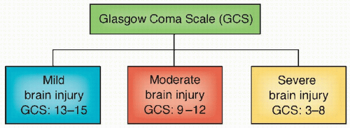


image


Anatomic Correlations

The cranial vault can be envisioned as a solid container with only one major opening at the base of the skull, the foramen magnum. The cranial vault is separated by bony buttresses into three compartments called fossae (anterior fossa, middle fossa, and posterior fossa). The internal plate of the intracranial vault varies in that it is smooth in some areas such as the occipital bone and highly irregular in other areas such as the frontal area. Some blood vessels lie close to the bone, such as the middle meningeal artery, while other blood vessels go through bony openings to get into the skull. See Chapter 5 for neuroanatomy review.


PRIMARY BRAIN INJURY

TBI results from a mechanical force or load that sets the head in motion causing cellular damage.6 Such mechanical forces are differentiated as impact, strain, or compression. Impact loading involves direct contact of an object to the head, which results in skull deformation and/or fractures. Strain is the result of blunt, acceleration, deceleration, or rotational forces applied to the brain tissue that tear or shear tissue and vessels and disrupt cells. Compression are prolonged static forces applied to brain tissue that
distort tissue, alter perfusion, and result in increased intracranial pressure (ICP). Head motions caused by these mechanical forces result in contact phenomena injuries and head motions of accelerations and decelerations (Fig. 16-3).7


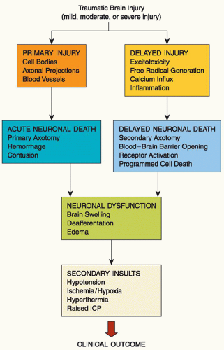
Figure 16-2 ▪ Key primary and secondary pathologic events with traumatic brain injury. ICP, intracranial pressure. (From: Dietrich, W. D. (2000). Trauma of the nervous system: Basic neuroscience of neurotrauma. In W. G. Bradley, R. B. Daroff, G. M. Fenichel, & C. D. Marsden (Eds.). Neurology in clinical practice (3rd ed., pp. 1045-1054) Boston: Butterworth & Heinemann.)


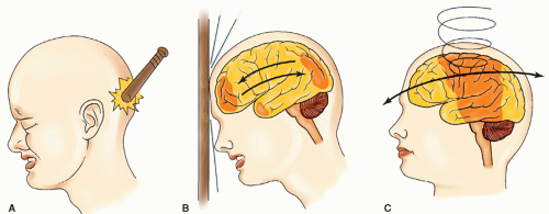
Figure 16-3 ▪ Mechanisms of injury: (A) contact, (B) acceleration-deceleration, and (C) rotational acceleration-deceleration.
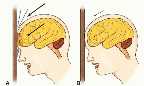


Figure 16-4A: High-velocity contact impact on the brain and skull. B: A low-velocity contact impact damages only the skull.


  • Acceleration injuries are the result of a moving object striking the head. Deceleration injuries result from the moving head striking an immobile object. The local effects of such forces are scalp laceration, skull fracture, extradural hematoma, contusions, lacerations, and intracerebral hemorrhage.8 The velocity (low or high) of the impact determines whether the injury is restricted to the scalp or skull (low velocity) or includes the brain (high velocity) (Fig. 16-4).


  • Acceleration-deceleration injuries are caused by abrupt changes in the velocity of the brain’s principally rapid forward movement followed by an abrupt stop (inertia) within the cranial vault. It is produced by head motion at the instant after injury and causes strain on cerebral tissue.9 These strains often operate simultaneously or in rapid succession to produce injury by compression (pushing together of tissue), tension (traction on tissue), or shearing (opposite but parallel sliding motion of the planes of an object).

A special type of acceleration-deceleration motion is rotation (sometimes called angular acceleration); it is common at sites
where the brain tissue hits bony buttresses in the cranial vault. The effects of linear acceleration of the head are much less significant than those resulting from rotational forces. The acceleration-deceleration mechanism is responsible for two important types of injury encountered in blunt TBI, acute SDH and DAI.

Whether the head is fixed or free, a certain amount of acceleration-deceleration always occurs at the time of impact to the skull. The difference in density between the skull (a solid substance) and the brain (a semisolid substance) causes the skull to move faster than its intracranial contents. The brain, which is located within the rigid skull and compartmentalized by the dura and bony buttresses, responds to the force exerted on the skull by gliding forward and then rotating within the compartment. The rotational force produces distortion of the brain as well as tension, stretching, and shearing of involved tissue. Shearing may either result in coup (below the area of impact) and/or contrecoup (opposite of coup area) injuries.10

The maximal amount of injury is usually found at the frontal and temporal poles and on the inferior surfaces of the frontal and temporal lobes where brain tissue comes in contact with bony protuberances at the base of the skull. Shearing or sliding of cerebral tissue over another portion implies stresses on two different planes. With rotational acceleration, the stress of shearing is directed toward areas where tough, fibrous tissue and cerebral tissue meet. These high-risk areas include the crista galli, the sphenoid wing, the margins of the tentorium or falx, and the foramen magnum. The degree of injury will depend on the extent and direction of the angular acceleration. Rotational movement in the brain can cause damage to axons even without gross visible lesions.11

Blast injuries occur when individuals are exposed to explosive forces. Blast waves result in changes in atmospheric pressure with waves of over (increased pressure) and under (decreased pressure) pressurization as gases expand and contract putting stress and strain on brain tissues. It is thought that cerebral damage may also occur through pressure waves generated through the cerebrospinal fluid. Animal studies suggest that energy waves result in cortical neuronal and diffuse white injury with changes in myelin and axonal structures. This damage is predominately seen in the hippocampus and brainstem.12


SECONDARY BRAIN INJURY

As discussed above, primary injury is the immediate response to the initial impact of mechanical force to the head, which results in cellular damage including neurons and glial cells, which further leads to axonal dysfunction. By comparison, secondary injury is a delayed, physiologic response to the primary injury. Following the primary head injury there is a massive neuronal depolarization that occurs which leads to a number of neuropathological changes, including the activation of biochemical, metabolic, and inflammatory cascades, which may lead to ischemia, cerebral edema, inflammation, and neuronal death.13 Cerebral pressure autoregulation is altered resulting in the brain’s increased susceptibility to systemic blood pressure changes and other physiologic insults. Table 16-1 describes the intracranial and systemic insults that lead to secondary brain injury. The deleterious roles of these physiologic insults on TBI will be discussed later in this chapter in the management section.

Based on experimental models, several theories have been generated to explain the cause and mechanisms associated with secondary injury in TBI. Among the major theories that have been the focus of several clinical trials are neurotoxicity secondary to release of excitatory neurotransmitters (EENs) that alters the cellular ionic homeostasis, membrane dysfunction due to the oxidative stress-free radical cascade, and inflammation.15 Fig. 16-5 illustrates this ischemic cascade.


TABLE 16-1 MRI FINDINGS AND DEFICITS FOR DIFFUSE AXONAL INJURY AND mTBI


MRI FINDINGS


DEFICITS


Genu of corpus callosum affected


More common in mild traumatic brain injury


Splenium of corpus callosum affected


More common in severe traumatic brain injury


When mid-portion of genu of corpus callosum affected


Deficits with ambulation, balance, motor planning, and executive and cognitive functions


When anterior portion of genu of corpus callosum affected


Asynchronous motor coordination between sides, planning, and mental flexibility deficits


When body and splenium of corpus callosum affected


Tactile deficits in which patient unaware of deficits because deficits compensated by visual control


From: Matsukawa, H., Shinoda, M., Fujii, M., Takahashi, O., Yamamoto, D., Murakata, A., et al. (2011). Genu of corpus callosum as a prognostic factor in diffuse axonal injury. Journal of Neurosurgery, 115(5), 1019-1024. doi: 10.3171/2011.6.JNS11513.


Neurotoxicity. The release of excitatory amino acids (EAAs) such as glutamate and aspartate is widespread after trauma, resulting in cell swelling, vacuolization, and cell death.16 Through activation of EAA receptors (e.g., glutamate and aspartate), there is an abnormal intracellular calcium influx. In addition, extensive membrane depolarization, induced by trauma, allows for a nonselective opening of the voltage-sensitive calcium channels and an abnormal accumulation of calcium within neurons and glia. The calcium shifts are associated with activation of intracellular lipolytic and proteolytic enzymes, protein kinases (calpains), protein phosphatases, dissolution of microtubules, expression of neurotropic factors, altered gene expression, and activation of cell death genes.17, 18, 19, 20

In response to injury there is often failure of aerobic glycolysis, phosphocreatine production, activation of high-energy cellular functions, and production of adenosine triphosphate (ATP). Failure of aerobic glycolysis increases lactate production and decreases intracellular pH, resulting in cellular acidosis. With failure of ATP production, the sodium-potassium pump fails, sodium enters the cell (normally there is higher intracellular concentrations of potassium [K+] and higher extracellular concentrations of sodium [Na+]), water follows resulting in cytotoxic edema.21

A major final common pathway for cell death is loss of calcium homeostasis.22 Loss of calcium homeostasis inhibits mitochondrial function. This, in turn, leads to increased breakdown of the cell membrane, and the production of toxins (i.e., eicosanoid, platelet-activating factor, and free radicals). Concurrently, following trauma, there is immediate severe cellular energy failure causing a strikingly increased level of extracellular EENs. These EENs include the EAAs, glutamate and aspartate, and the amine acetylcholine. The source
of EAAs is believed to be injured, energy-depleted, and depolarized neural cells (neurons and glia) that release their glutamate. Escalating levels of glutamate and aspartate stimulate specific EAA receptors that normally mediate excitatory synaptic transmission between neurons.

Three ion channels are known to be activated by glutamate and are named for their pharmacologic similarity. These ion channels are kainate receptor (↑ Na+ influx and K+ efflux), adenosine monophosphate (AMP) A receptor (α-amino-3-hydroxy-5-methyl-4-isoxazole propionic acid) (↑ Na+ influx and K+ efflux), and N-methyl-D-aspartate (NMDA) receptor (↑ Na+, Ca2+). Excessive stimulation of glutamate receptors opens ionic channels, which causes sodium-mediated cellular swelling and calcium-mediated neuronal disintegration from membrane lipid hydrolysis and protease activation.23 Thus, overstimulated EAA receptors have been implicated as a final common pathway of neurotoxicity in numerous central nervous system (CNS) problems, including traumatic injury.24

Oxidative Stress-Free Radical Cascade. High levels of intracellular calcium, activation of phospholipases, breakdown of the cell wall, and the formation of EAA lead to the formation of reactive oxygen species or free radicals (superoxide, hydroxyl, hydrogen peroxide, singlet oxygen, and nitrous oxide). These free radicals damage the cell membrane, mitochondria, proteins, and DNA.25

The blood and breakdown products within the brain following tissue injury become another source of oxygen-free radicals. Free iron from hemoglobin breakdown can transfer an electron to oxygen-forming superoxide molecules or to hydrogen peroxide to form hydroxyl. These free radicals can overwhelm cytoplasm defenses and begin to oxidize cellular membrane by lipid peroxidation. Through lipid peroxidation, electrons are transferred to unsaturated fatty acids, which form free radicals called lipid peroxyl or alkoxy moieties.26 Ongoing lipid peroxidation causes breakdown of the cellular membrane and oxidation of membrane lipoproteins. The process also spreads to adjacent cells and perpetuates cell death and edema.27

Inflammation. Although the inflammatory cascade which is activated within minutes of primary injury following TBI can contribute to further injury some have neuroprotective properties. Cytokines are major players of a process that includes expression of adhesion molecules, cellular infiltration, and further release of more inflammatory cells and growth factors leading to either cell regeneration or cell death.28 This dual role of cytokines in the pathophysiology of TBI has been widely studied and conflicting results have been found on whether or not cerebral inflammatory cascade following TBI is neuroprotective or contributory to neuronal and glial cell damage. Inflammatory mediators such as tumor necrosis factor (TNF), interleukins (ILs) (IL-1, IL-6, IL-8, IL-10, and IL-12), and nerve growth factor may be neuroprotective.29, 30, 31, 32, 33 Specifically, TNF has been associated with cerebral edema, blood-brain barrier (BBB) disruption, and neuronal cell death in the acute phase of TBI. Neutrophil and macrophage infiltration is also believed to exacerbate post-traumatic altered homeostasis of the brain and BBB disruption. On the
other hand, IL-6 demonstrates neuroprotective function by preventing TNF synthesis, promoting nerve growth factor and neuronal regeneration, and neutralizing NMDA-mediated toxicity. Another product of inflammatory cells is nitric oxide. This is synthesized by neuronal and endothelial cells and can be a potent vasodilator and thus improve perfusion, downregulate EAA, and inhibit cell death.34


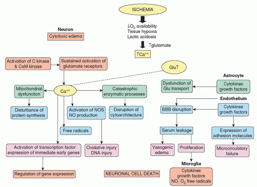
Figure 16-5 ▪ Pathophysiological changes with ischemia (need permission).


PRIMARY INJURY: DIAGNOSIS AND MANAGEMENT


Scalp Lacerations

The velocity and characteristics of the impacting contact object determine the extent of scalp injury. Injuries to the scalp can be classified as follows.


  • Abrasion: the top layer of the scalp is scraped away; this is a minor injury that may cause slight bleeding. The area is cleaned and possibly dressed, and no other treatment is required.


  • Contusion: the scalp is bruised with possible effusion of blood into the subcutaneous layer without a break in the integrity of the skin; there is no specific treatment.


  • Laceration: the scalp is torn and may bleed profusely; suturing may be necessary.


  • Subgaleal hematoma: a hematoma in the subgaleal layer of the scalp occurs and will usually absorb on its own.

A scalp contusion may benefit from the application of ice initially to prevent a hematoma from forming. Skull films may be ordered to rule out a skull fracture. Depending on other signs and symptoms, a computed tomography (CT) scan rather than a skull film may be ordered to rule out underlying skull or brain injury. Scalp lacerations often require suturing and aseptic management.


Skull Fracture

The mechanism of skull injury is direct contact. Factors that determine the degree of injury include the skull’s thickness at the point of impact and the weight, velocity, and angle of impact of the intruding object. At impact, several actions are set in motion in split-second sequence. At the point of impact, there is a relative indentation that may be temporary or permanent, depending on velocity. Stress waves are set into motion, radiating throughout the entire skull. With high-velocity impact, a depressed skull fracture with or without a dual tear and cerebral laceration can occur. With low-velocity impact, the area of indentation rebounds outward and may result in no fracture, a linear fracture, or a comminuted fracture (Fig. 16-4). The fracture line extends from the point of impact and is directed by thicker bony areas toward the base of the skull.


Classification

Skull fractures are classified as follows.


  • Linear: a singular fracture line occurring to the skull, which could be displaced or nondisplaced


  • Comminuted: the skull is splintered or shattered into pieces


  • Depressed: a fracture of the skull in which a fragment is depressed more than half the width of the bone; the scalp and/or dura may or may not be torn


  • Open depressed fracture: also known as compound skull fracture; is an opening of the skull as a result of comminuted depressed skull fractures and tearing of the dura mater and the scalp


  • Basal skull fracture: often arises from extension of a linear fracture into the base of the skull. The frontal and temporal bones are usually affected so that the fracture involves the anterior or middle fossa. It is important to distinguish between fractures of the cranial vault and those of the base of the skull. Although the mechanism by which the fractures arise is similar, the consequences of basilar fractures are more serious than those of cranial vault fractures. A feature of basal skull fractures is the frequency with which they traverse the paranasal air sinuses (frontal, maxillary, or ethmoid) of the frontal bone or the air sinuses located in the petrous portion of the temporal bone. The fragility of the bones in these areas and the intimate adherence of delicate dura account for the frequency of lesions in these areas and the consequent leakage of CSF through the dural tear manifested either by rhinorrhea (from the nose) or otorrhea (from the ear). Continued leakage of CSF can lead to serious infections. Such patients are at high risk for meningitis, abscess formation, and osteomyelitis from organisms gaining entry by way of the ear, nose, or paranasal sinuses through the dural tear.

A CT is used to diagnose a skull fracture. The ease with which a diagnosis of skull fracture is made depends on the site of the fracture. If a fracture is found on CT, there is always the question of associated brain injury, and magnetic resonance imaging (MRI) provides better resolution and clearer pictures at the fracture-cerebral site.

Most basal skull fractures are extensions of fractures of the cranial vault. A CT scan may not be sensitive enough to clearly show a basal skull fracture. A CT scan and clinical criteria are used to make a diagnosis. For example, a linear fracture of the parietal bone may be readily evident on a regular CT scan, whereas thin-slice CT may be necessary to find a basal skull fracture. If the paranasal sinuses are fractured, air may be evident in the frontal and maxillary sinuses on x-ray studies. With fracture of the temporal bone, the mastoid sinuses may be opaque. Common findings of anterior fossa fractures (fractures of the paranasal sinuses) include the following.


  • Rhinorrhea (drainage of CSF, blood, or both from the nose)


  • Subconjunctival hemorrhage of the eye


  • Periorbital ecchymosis (raccoon’s eyes)

With middle fossa (fractures associated with fracture of the temporal petrous bone) and posterior fossa fracture, findings include the following.


  • Otorrhea (drainage of CSF, blood, or both from the ear)


  • Hemotympanum (blood behind the tympanic membrane)


  • Battle’s sign (ecchymosis over mastoid bone that develops 12 to 24 hours after injury)


  • Conductive hearing loss (may be associated with signs of vestibular dysfunction, such as vertigo, nausea, and nystagmus)


  • Possible facial nerve palsy (Bell’s palsy) that appears 5 to 7 days after injury

Treatment of skull fractures depends on the type of fracture. Generally, linear skull fractures do not require special medical management other than observation for underlying cerebral injury. A depressed skull fracture with an open scalp, skull, and dura mater requires surgery to debride, elevate, and remove bone fragments from the wound. If the fragments are extensive, a craniectomy may
be necessary. Surgical elevation of depressed skull fractures may be necessary with greater than 8- to 10-mm depression, neurological deficit, CSF leak, and presence or absence of open depressed fracture.35 For cosmetic and brain protective purposes, a cranioplasty with insertion of a bone or artificial graft may follow immediately or be postponed for a few months (approximately 3 to 6 months) if brain swelling is present.

Use of prophylactic antibiotics with basal skull fractures is controversial; many argue that prophylactic use allows for proliferation of other virulent organisms. Most CSF leaks resolve spontaneously within 7 to 10 days. To aid resolution of leakage lasting more than 4 to 5 days, a lumbar catheter for continual drainage of CSF may be inserted.36 If leakage of CSF continues, a craniotomy may be necessary to repair the tear surgically or to repair the leakage with grafts.

Other complications associated with basal skull fracture include cerebrovascular injury such as internal carotid artery (ICA) injury at the point of entry to the cranial vault through the foramen at the base of the skull. Fracture in or around the foramen can result in cerebral hemorrhage from laceration, thrombosis, development of a traumatic aneurysm, a carotid-cavernous sinus fistula, or carotid-cavernous sinus compression. A carotid-cavernous sinus fistula is characterized by chemosis, bruit, and pulsating exophthalmos. The oculomotor, trochlear, trigeminal, and abducens cranial nerves pass through the cavernous sinus. Compression of the sinus may be evident because of ophthalmoplegia or trigeminal dysfunction. Another potential complication is trapping portions of the frontal arachnoid and dura between the fracture edges, thus creating a permanent route for leakage of CSF. Radiographic and surgical identification of the exact area of the dural tear is extremely difficult; yet such identification is necessary to facilitate surgical repair.


Traumatic Brain Injuries

Injuries to the brain can be focal or diffuse. Focal injuries include contusions and hematomas. Concussions, diffuse axonal injuries, and traumatic subarachnoid hemorrhage (TSAH) are the major diffuse injuries. TBI could also be classified as penetrating or nonpenetrating. Common penetrating injuries are caused by gunshots and impalement and may result in contusions, hematoma, laceration, and cerebral edema. In all cases, the diagnosis of TBI is made with a noncontrast CT scan. In some instances, a contrast CT or an MRI may be requested after a noncontrast CT scan, but neither a contrast CT scan nor an MRI is required on an emergency basis. CT scan is more sensitive in detecting hematoma while MRI is superior in identifying DAI. In severe TBI, a follow-up CT is often ordered 3 to 5 days postinjury or as necessary to follow progress and as needed for investigation of any neurological changes.


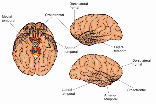
Figure 16-6 ▪ Cerebral contusions. The most frequently involved areas of the brain in cerebral contusions are the orbitofrontal and anterior-temporal regions. These are the areas that come in direct contact with the irregular bony surfaces in the inside frontal skull area.

Nearly 50% of all multi-trauma patients have a head injury. Patients with a head injury often have concurrent cervical spine, maxillofacial, or cervical vascular injuries.


Focal Cerebral Injuries: Contusions, Lacerations, and Hematomas

Cerebral Contusions. The anterior and middle fossae at the base of the skull have irregular, bony surfaces that are capable of contusing or lacerating the brain on impact. The distribution of contusions in these particular areas is explained by the movement of the brain within the skull (Fig. 16-6). The frontal and temporal poles are vulnerable because of the relative restraint created by the sphenoidal ridges and other bony irregularities of the base of the skull. Less common sites of injury are the inferolateral angles of the occipital lobes, the medial surfaces of the hemispheres, and the corpus callosum. The firm falx cerebri and tentorium cerebelli not only restrict movement of the brain, but also contribute to contusion or laceration during impact as cerebral tissue impacts on these hard surfaces. This is particularly true when the rotational force of acceleration-deceleration is applied to the frontal-temporal area. Injury to the inferolateral angles of the occipital lobe and, less commonly, the inferior surface of the cerebellar and cerebral peduncles can result from impact by the tentorium. A blow to the vertex of the head may cause cerebellar, cerebellar tonsillar, and brainstem contusions initiated by the downward thrust of the brain toward the foramen magnum.

A cerebral contusion is a bruising of the surface of the cortex to subcortical white matter (cerebral parenchyma). Contusions may occur from blunt trauma (direct contact), a depressed skull
fracture, a penetrating wound, or an acceleration-deceleration closed injury. With acceleration-deceleration, the sites of injury are generally predictable and are located where the brain has an impact on the bony protuberances within the skull (Fig. 16-6). These areas include the frontal poles, frontal-orbital areas, frontal-temporal junction around the Sylvian fissure (where the brain is close to the lesser sphenoidal wings), and temporal poles (the inferior and lateral surfaces of the temporal lobes where there is a shelf-like separation between the anterior and middle fossae).

The clinical effect of a contusion depends on its size and related cerebral edema. Small, unilateral, frontal lesions may be asymptomatic, whereas larger lesions may result in neurological deficits.

Contusions can cause secondary mass effect from edema resulting in increased ICP, and possible herniation syndromes. For patients with cerebral contusions, no surgical interventions are indicated and patients are managed medically with emphasis given toward prevention of secondary injuries such as ICP management.

Classification. Contusions are classified as gliding or surface contusions.6 Gliding contusions result from rotational motion and are likely associated with DAI. Usual areas involved are the parasagittal regions of the brain with bilateral and symmetric focal hemorrhages of the cortex and surrounding white matter. Surface contusions, on the other hand, are injuries caused by contact forces and include the following.37


  • Fracture contusions occur at the sites of fractures and are particularly severe in the frontal lobes when associated with fractures in the anterior fossa.


  • Coup contusions are found directly under the areas of impact (contact).


  • Contrecoup contusions, by contrast, are cerebral injuries at the opposite pole of direct contact. A contrecoup injury is most often a contusion, but occasionally it may be a laceration. Both coup and contrecoup injuries are caused by the rapid acceleration-deceleration of the semisolid brain within the rigid cranial vault.


  • Herniation contusions occur at the time of injury at the point of contact where the medial part of the temporal lobe produces an impact at the edge of the tentorium or the cerebellar tonsils against the foramen magnum.

Cerebral Lacerations. A cerebral laceration refers to a traumatic tearing of the cerebral tissue. It is related to high-impact injuries and is treated in the same manner as a cerebral contusion is managed. Laceration may also be associated with a depressed skull fracture or other penetrating injuries.

Intracranial Hemorrhage/Hematomas. Traumatic intracranial hemorrhage is a common complication of TBI. Although bleeding may begin immediately after injury, its presence may not be clinically apparent until sufficient blood accumulates to cause signs and symptoms of a space-occupying lesion and mass effect. The interval between bleeding and the appearance of clinical symptoms may be minutes or weeks, depending on the site and rate of bleeding. Intracranial hemorrhage may be an occult development in a patient who has sustained a seemingly minor TBI in which consciousness has been maintained or quickly restored. Other patients with hemorrhage may be unconscious from the moment of impact.

The major types of bleeding associated with head trauma are EDH, SDH, and ICH. Other bleeding includes TSAH and intraventricular hemorrhage (IVH). Each lesion is a distinct clinical entity, but two or more types of hemorrhagic lesions can coexist.

Epidural Hematoma. EDH, also known as an extradural hematoma, refers to bleeding into the potential space between the inner table of the skull (inner periosteum) and the dura mater. It accounts for about 2.7% to 4% of traumatic head lesions. EDHs are seen most often in 20- to 30-year-old individuals or 6- to 10-year-old children. Classically, EDHs are arterial in origin, occurring from a tear of an artery in a patient who has sustained a skull fracture. The most common location for an EDH is a fracture to the thin, squamous portion of the temporal bone (90%), under which is located the middle meningeal artery. Therefore, common locations of the bleed are the temporal and temporoparietal regions, but may also occur in the frontal area. A more recent study found more EDH may be venous, the result of torn dural venous sinuses particularly in the parieto-occipital region or posterior fossa. As the bleed expands, it gradually strips away the dura from the inner table of the skull, and a large, ovoid mass develops, creating pressure on the underlying brain and causing a mass effect.

Clinically, the “classic” description of an EDH was that of a momentary unconsciousness followed by a lucid period lasting for minutes to several hours. EDH is often referred to as the “talk and die syndrome” because the patient may present with a lucid period followed by a rapid deterioration in the level of consciousness (LOC) from drowsiness to lethargy to coma as a mass effect and herniation developed. Only about 10% to 27% of patients with EDH present with the above classic manifestation. Other findings include ipsilateral pupillary dilation, contralateral hemiparesis, headache, vomiting, seizures, and hemi-hyperreflexia with a unilateral Babinski sign.35

In 18% to 44% of patients, a unilateral dilated ipsilateral pupil without loss of consciousness is found. Bradycardia and respiratory distress are late findings. If untreated, neurological deterioration may occur from drowsiness to lethargy and then to coma as mass effect and herniation developed.

EDHs are commonly identified through CT scan of the head (without contrast), which appears as a biconvex or lenticular high-density lesion adjacent to the skull (Fig. 16-7). Mass effect is frequently associated with this bleed, which can be seen radiographically. Lesions not noted on initial CT scan but on subsequent CT scans are known as delayed EDHs.


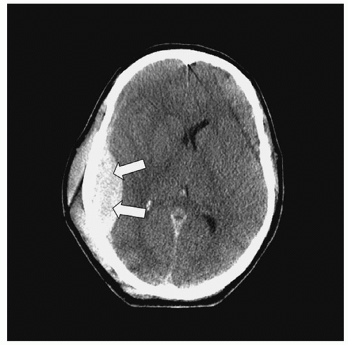
Figure 16-7 ▪ Computed tomography scan: epidural hematoma.


Immediate diagnosis and surgical evacuation of the hematoma are associated with lower mortality rate. Prognosis is associated with the severity and the duration of the brain compression caused by the EDH. Mortality is about 10%. Therefore, early diagnosis and management are important.38 Surgical indication includes symptomatic EDH and/or acute, greater than 30 cc bleed. Presence of coagulopathy associated with a hematoma should also be identified and optimally corrected to promote hemostasis and prevent rebleeding. Hematomas of less than 30 cc with less than 15-mm thickness and a midline shift of less than 5 mm in a patient who is conscious without focal deficits can be monitored with serial CT scans and close neurologic observation.39


Subdural Hematoma. An SDH refers to bleeding between the dura mater and arachnoid or pial layer. Approximately 12% to 29% of patients with post-traumatic intracranial lesions have SDHs. Most SDHs are caused by tearing of the bridging veins located over the convexity of the brain. Other causes include tearing of small cortical arteries, cerebral contusions, and acute bleeding into chronic SDHs. Bilateral SDHs are not uncommon. SDHs are categorized based on the interval between injury and the appearance of signs and symptoms. Although there are no uniform definitions, three classes with approximate time intervals are recognized.


  • Acute: up to 48 to 72 hours. This consists of clotted blood that is hyperdense and crescent shaped on CT.


  • Subacute: 2 to 3 days to about 2 weeks. The clot now lyses, and blood products and fluid are present. Clot will be seen isodense on CT scan.


  • Chronic: longer than 2 weeks to several months. The clot is a fluid mass and is hypodense on CT.

Clinical presentations are usually due to brain compression and vary according to the location of the lesion and the interval between injury and presentation of symptoms. Common signs and symptoms associated with an acute SDH include gradual or rapid deterioration of the LOC from drowsiness, slow cerebration, and confusion to coma; pupillary changes; and hemiparesis or hemiplegia. Subacute SDHs are associated with less severe underlying compression. Signs and symptoms of subacute SDHs correspond closely to those of the acute SDH. Chronic SDHs can develop from seemingly minor TBIs. The time elapsed between injury and the development of symptoms may be months, so the initial injury itself may not be recalled. The lesion becomes encased within a membrane that is easily separated from the arachnoid and dura. The SDH slowly enlarges, probably because of repeated small bleeding, until a mass effect occurs. The most common symptoms of a chronic SDH include headache (progressing in severity), slow cerebration, confusion, drowsiness, and possibly seizure. Papilledema, sluggish ipsilateral pupillary response, and hemiparesis may later develop.


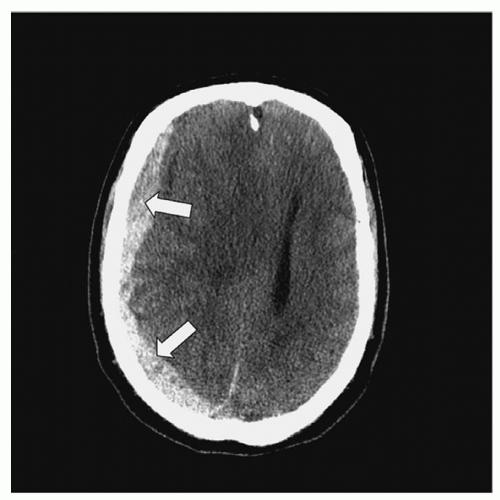
Figure 16-8 ▪ Computed tomography: subdural hematoma.

Elderly patients with cerebral atrophy associated with the normal aging process are prone to develop SDH because of traction on fragile bridging veins. This is particularly true with tear and rupture in the elderly with cerebral trauma. Atrophy also provides more free space into which bleeding can occur before symptoms are evident from a mass effect. With chronic SDH, the development of symptoms can be subtle because of gradual spatial compensation. Patients who have had long-term alcohol abuse with related cerebral atrophy and impaired blood clotting resulting from altered liver function and/or use of anticoagulants are also especially prone to SDH.

A CT scan of the head will reveal a crescentic hyperdense lesion with edema/mass effect in acute SDH. Unlike EDH, CT scan findings of acute SDH appear concave over the brain surface and more diffuse (Fig. 16-8). The appearance of subacute and chronic SDH, as discussed earlier, varies according to density on CT scan.

Immediate surgical evacuation of the clot is recommended for symptomatic SDHs that are greater than 10 mm in thickness or greater than 5 mm midline shift. Factors that affect outcome include initial GCS score, pupillary status, time interval between trauma and treatment, clot size, mass effect, and presence of other traumatic lesions.40 Low GCS score (<7), the presence of pupillary abnormalities, delay in treatment (>4 hours after injury), larger hematoma volume (>100 mL), midline shifts >1.5 cm, and associated cerebral contusions and/or TSAH are associated with poor functional survival and higher mortality rate. Mortality rates for patients requiring surgery are between 40% and 60%.


Intracerebral Hematoma. An ICH refers to bleeding into the brain parenchyma (Fig. 16-9) resulting from contusions or blood vessel injury. Approximately 8.2% of all TBI and 13% to 35% of all severe TBI cases are due to ICH.41 Traumatic parenchymal lesion vary with some evolving from contusions, others evolve over time (delayed traumatic ICH or DITCH); thus surgical intervention requires careful timing. Common locations are below the surface of the cortex in the white matter most commonly in the frontal and temporal lobes while others are deeper in the thalamus, basal ganglia, or parasagittal white matter. Manifestations include headache; altered mental status, which can vary from confusion to a reduced LOC or coma; contralateral hemiplegia; and ipsilateral pupil dilation.42 Clinically, ICHs behave as expanding, space-occupying lesions and consequently cause increased ICP, which could result in herniation. ICHs are seen on CT scan as high-density lesions in the brain parenchyma.

Similarly to EDH and SDH, the size of the ICH determines the best treatment approach. Considerable controversy exists regarding the indications for surgery in ICH. This is a complex decision based on the patient’s neurological condition, size and location of the hematoma, patient’s age, and patient/family wishes. Surgery is recommended for patients with a GCS 6 to 8 with frontal or temporal contusion with a volume greater than 20 cc and with a midline shift of more than 5-mm or compressed cisterns; any lesion with a volume greater than 50 cc; or a lesion that results in refractory increased ICP. Procedures include lobectomies and decompressive craniectomies. Deeper lesions which are stable are often managed medically with supportive care and management of increased ICP.


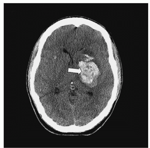
Figure 16-9 ▪ Computed tomography scan: intracerebral hematoma.


Traumatic Subarachnoid Hemorrhage. It is estimated that between 33% and 60% of TBI patients may have a TSAH which is associated with a two-fold increased risk of death. TSAH is the accumulation of blood in the subarachnoid space and may be associated with contusions and SDH. Increased density spread thinly over the convexity causing fullness of the sulci and basal cisterns are usually noted on noncontrast CT scan of the head. This CT scan differs from the scan of aneurysmal SAH where the blood is found predominately in basal regions. The bleeding is thought to be the result of acceleration/deceleration forces that cause tearing in the pia mater on the roof of the ventricles. The distribution of the blood increases the risk of the patient developing communication hydrocephalus. Development of delayed ischemic events from vasospasm (2% to 41%) is another risk and is associated with a poor outcome. One study found that fever at admission and a contusion on CT scan were risk factors for the development of post-traumatic vasospasm.43 Onset of symptoms varies according to location and the rate of bleeding. Clinical presentation includes headache, reduced LOC, nuchal rigidity, hemiplegia, and/or ipsilateral papillary abnormalities. Cerebral angiography is not indicated but may be done in cases when there is unclear history of trauma to exclude the presence of cerebral aneurysm.44

Management is usually directed at supportive care and follows the treatment for aneurysmal subarachnoid hemorrhage A Cochrane review on the use of calcium channel blockers (e.g., nimodipine) has shown little to no benefit.45 A cerebral angiogram to exclude vasospasm may be indicated in cases of delayed focal neurological deficits following TSAH.

Intraventricular Hematoma. IVH (Fig. 16-10) occurs secondary to TSAH or as an extension from an ICH and could be suggestive of severe head injury. It is reported in approximately 10% of severe head injuries.46 Signs and symptoms include altered LOC, hemiparesis, ipsilateral pupil dilation, and intracranial hypertension.
Management consists of drainage of CSF with a ventriculostomy, ICP management, and prevention of secondary brain injuries.


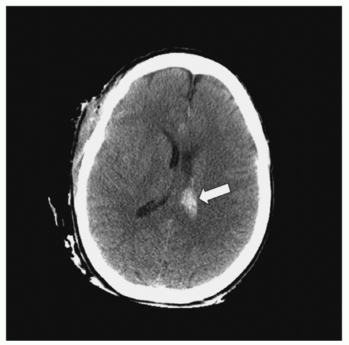
Figure 16-10 ▪ Computed tomography scan: intraventricular hematoma.


Diffuse Cerebral Injuries: Diffuse Axonal Injuries

Diffuse Axonal Injury. DAI or traumatic axonal injury (TAI) is a primary TBI associated with rotational acceleration-deceleration forces during which shearing forces damage nerve fibers at the moment of injury (Fig. 16-11). Because of the difference in acceleration gradient on certain areas of the brain during primary injury, shearing forces at the white-gray junction, corpus callosum, brainstem, and sometimes cerebellum produce diffuse tearing of axons and small blood vessels.47, 48 Until recently, it was believed that DAI was most commonly associated with noncontact injuries. A recent study found that more patients with near-sided side impact crashes in which the head makes contact with a hard surface, sustained DAI than frontal and far-sided impact crashes.49 DAI accounts for approximately 13.9% to 72% of primary brain injuries and 35% of deaths from all TBIs.37, 50, 51 It is characterized by distinct gross and microscopic findings including axonal swellings that are widely distributed in the cerebral hemispheric white matter, corpus callosum, and upper brainstem; gross hemorrhagic lesions of the corpus callosum; and gross hemorrhagic lesions involving one or both dorsolateral quadrants of the rostral brainstem.52


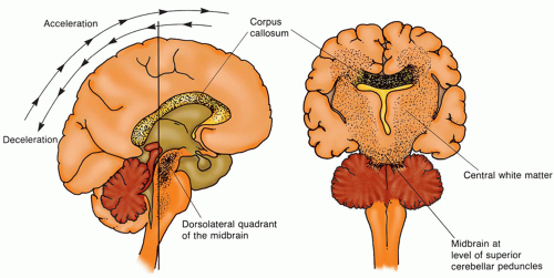
Figure 16-11 ▪ Diffuse axonal injury. Diffuse axonal injury results from acceleration-deceleration and shearing force on the brain. Depending on the severity of the injury, the areas of the brain most often affected are the corpus callosum, the dorsolateral area of the midbrain, and the parasagittal white matter.


TABLE 16-2 CAUSES OF SECONDARY BRAIN INJURIES


INTRACRANIAL CAUSES


SYSTEMIC CAUSES


  • Intracranial hypertension


  • Cerebral edema/hematoma


  • Hydrocephalus


  • Seizures


  • Cerebral vasospasm


  • Infection


  • Hypotension


  • Hypoxemia


  • Hypercarbia/hypocarbia


  • Hyperthermia


  • Hyponatremia


  • Hyperglycemia/hypoglycemia


The angular acceleration-deceleration force produces axonal shearing (primary axotomy) alone or together with tearing of blood vessels that cause microscopic petechial hemorrhage. In animal models, primary axotomy, in which axons are sheared, severed, or disconnected, is uncommon.53, 54 In transected axons, axoplasmic transport from the cell body to the axon terminal continues. However, the transported material cannot pass the damaged area, where it accumulates, causing the axon to swell.55 Most axons undergo a series of changes that result in secondary axotomy. Although the timeline for neuronal death varies, primary axotomy usually occurs in approximately 1 hour, whereas secondary axotomy is more protracted. Table 16-2 illustrates the proposed sequence of primary
and secondary axotomy changes due to injury.56 Table 16-3 provides a grading system DAI that occurs with acceleration-deceleration forces to the brain. The grading system is based on the distribution of pathologic findings.36


TABLE 16-3 SEQUENCE OF PRIMARY AND SECONDARY AXOTOMY CHANGES


PRIMARY (<1 HR) AXOTOMY CHANGES


SECONDARY (>4 HRS) AXOTOMY CHANGES


Axolemma permeability increases (axotomy)


Activation of intracellular leukocytic & proteolytic enzymes, including protein kinase, occurs (axotomy)


An influx of calcium occurs (axotomy)


Changes occur in phosphorylation


Microtubules are lost


Change occurs in the structure of neurofilament


Mitochondria swell


Loss of axonal transport occurs


Focal loss of axonal transport occurs


Axonal swelling occurs in 2 to 4 hrs Neurofilament proteolysis occurs


Adapted from: Maxwell, W. L., Povlishock, J. T., & Graham, D. I. (1997). A mechanistic analysis of non-disruptive axonal injury. Journal of Neurotrauma, 14, 419-440.


Recent research has increased understanding of patient outcomes from DAI. Diffuse injury to axons, which are apparent microscopically throughout the white matter of the cerebral hemispheres, the cerebellum, and the brainstem, has three presentation forms related to the length of the patient’s survival.37 In short-term survival (days), there are a large number of axonal bulbs throughout the white matter. It takes about 15 to 18 hours for the axonal bulbs to appear in humans.57 In intermediate survival (weeks), there are large numbers of microglia clusters throughout the white matter. In long-term survival (e.g., vegetative state), there is long-tract degeneration of the Wallerian type in these areas. In these survivors, the degenerative effects are finally replaced by diffuse white matter gliosis. Gliosis is the chief finding in long-term survivors and is related to severe and permanent disabilities.58, 59

A growing body of knowledge regarding location of DAI lesion on MRI is helping with prognosticating patient outcomes. The number and location of lesions help determine severity of injury and type of neurologic deficits which can be expected. Examples of these data can be found in Table 16-4.14

DAI is characterized clinically by functional cerebral failure, which may range from confusion to coma and death. It is also classified clinically as mild, moderate, or severe based on coma duration and brainstem signs.60, 61


  • Mild DAI: coma lasting 6 to 24 hours with the patient beginning to follow commands by 24 hours. Outcome: death is uncommon, but cognitive and neurological deficits are common.


  • Moderate DAI: coma lasting longer than 24 hours, but without prominent brainstem signs. This is a common presentation (about 45% of all patients). Outcome: incomplete recovery in those who survive.


  • Severe DAI: coma is prolonged and associated with prominent brainstem signs (e.g., decortication, decerebration). This presentation is seen in about 36% of all DAI patients. Outcome: death or severe disability.


TABLE 16-4 GRADING SYSTEM FOR DIFFUSE AXONAL INJURY (DAI) BASED ON THE DISTRIBUTION OF PATHOLOGIC FINDINGS


  • DAI grade 1 (mild): there is histologic evidence of axonal damage in the white matter injury primarily of the cerebral hemispheres, referred to as lobar. Cinically, coma >24 hrs, followed by mild to moderate memory impairment and mild to moderate disabilities.


  • DAI grade 2: there are focal lesions of the corpus callosum with or without lobar lesions. Clinically, coma >24 hrs followed by confusion and amnesia. Mild to severe memory loss, behavioral, and cognitive deficits.


  • DAI grade 3: lesions of the dorsolateral quadrant of the rostral brainstem with or without lobar or corpus callosal injury. Clinically, coma long lasting with flexor and extensor posturing. Cognitive, memory, speech, sensorimotor, and behavioral deficits.


From: Greenberg, M. S. (2010). Handbook of neurosurgery (7th ed., p. 853). New York: Thieme Publishers.


The clinical diagnosis of DAI is based on immediate onset of coma in a patient who has significant cerebral trauma and no intracranial lesion noted on CT scan. In many cases, an MRI may demonstrate lesions not evident on a CT. The treatment is supportive care, specifically to the unconscious patient, which will be further discussed in this chapter. DAI is associated with significant disability or death whether or not there is a coexisting traumatic lesion such as contusion or hematoma.

Concussions. Concussion or mild TBI (mTBI) is the most common TBI. A cerebral concussion is defined as an injury caused by a blunt acceleration-deceleration force (with shearing stress on the reticular formation) resulting in a brief loss of consciousness (or no loss of consciousness), and/or brief retrograde amnesia, no focal neurologic deficits, and no significant CT findings. Though the presenting symptoms may seem minor (headache, drowsiness, confusion, dizziness, irritability, giddiness, visual disturbances [seeing stars], and gait disturbances), these patients may be vulnerable to other sequelae (see Mild TBI section).

Concussions are classified as mild or classic, based on the degree of symptoms, particularly those of unconsciousness and memory loss. Mild concussion is defined as temporary neurological dysfunction without loss of consciousness or memory. By contrast, a moderate or classic concussion includes temporary neurological dysfunction, unconsciousness, and memory loss. Recovery of consciousness varies from minutes to hours. Some patients will develop a postconcussion syndrome (described later in this section).


The diagnosis of concussion is based on the patient’s history and neurological examination. CT scan or MRI is usually unremarkable, although radiologic testing is still recommended in cases where concussion is suspected to exclude any other traumatic lesion and confirm absence of any focal lesion on a CT scan, if a CT scan is done.
Depending on the severity of the concussion, a short-stay admission may be done to observe the patient’s neurological status. In other cases, a patient may be discharged home under the supervision of a responsible adult. Specific verbal and written instructions about what signs and symptoms to look for and report immediately to the physician should be provided. The patient should also be cautioned in avoiding alcohol, illicit drugs, and/or other substances such as narcotic analgesics or sedatives that may mask any signs of neurological dysfunction.62 For any persistent and/or worsening signs and symptoms, follow-up is imperative.


Brain Injuries Caused by Gunshots and Impalement

Gunshot wounds to the head are responsible for most penetrating brain injuries, and they account for approximately 35% of deaths from brain injury in people younger than 45 years of age.63 The wound created by the bullet depends on ballistics (i.e., the size, shape, velocity, and direction of the bullet). As the bullet propels forward and penetrates the skull, it compresses the air in front of it, thereby increasing the destruction of brain tissue locally and remotely.64 The major effects of missile injuries are cerebral contusions and lacerations, focal tissue necrosis, hemorrhage from tearing of blood vessels, and focal or generalized cerebral edema. Hemorrhage and edema may produce increased ICP and possible herniation associated with rapid expansion during impact and in response to a space-occupying lesion. The severity of injury sustained depends on the structures involved and herniation. Cerebral abscesses are postinjury concerns because of the microorganisms that have been carried into the brain from surface debris of skin, hair, and bone fragments. Other late complications include traumatic aneurysms, seizures, and fragment migration.

Missile injuries are classified as follows.


  • Tangential injuries, in which the missile does not enter the cranial cavity but produces a scalp laceration, facial injury, comminuted skull fracture, meningeal tear, or cerebral contusion-laceration.


  • Penetrating injuries, in which the missile enters the cranial cavity but does not pass through it, resulting in the presence of metal, bone fragments, hair, and skin within the brain. There is direct injury to cerebral tissue in the path of the bullet as well as injury to tissue from the high-pressure waves created by the high-velocity bullet. This high pressure can cause coup and contrecoup injuries, spikes in ICP, and herniation.


  • Through-and-through injuries, in which the missile perforates the cranial contents and leaves through an exit wound. There is one tract created from a missile entering the brain, although several tracts are possible if the bullet ricochets off bony structures within the cranial vault.

The history and evidence of entrance and possible exit gunshot wounds indicate probable injury. A CT scan determines the amount of injury and identifies bone fragments and the location of bullets, if retained. Emergency surgical intervention is common for salvageable patients through evacuation of hematomas, such as EDH or SDH; to debride the wound and remove the bullet and necrotic tissue; and to treat other system injuries caused by bullets or trauma. Survivors usually require ICU management for intracranial hypertension and system complications associated with severe TBI as well of injuries to other body systems.

Penetrating injuries can also be the result of objects such as knives, sticks, scissors, nails, and the like piercing areas where the bone is thin such as the orbit. These stabbing type of injuries produce a localized laceration of the parenchyma and blood vessels rather than more extensive injury seen with gunshot wounds.


OTHER INJURIES RELATED TO TRAUMATIC BRAIN INJURY


Facial Fractures

Facial fractures commonly coexist with TBI. Injuries may involve the soft tissue (contusions, lacerations), the facial bones (fractures), or both. The facial bones include most of the paranasal sinuses and the primary receptor organs for the senses of vision, hearing, taste, and smell. Facial injuries can result in disfigurement, motor and sensory dysfunction, and deficits in communication. Cerebral spinal fluid (CSF) leaks often accompany facial fractures. Table 16-5 summarizes common facial fractures. A CT scan is used to diagnose a facial fracture that can lead to injury of the eye. Orbital fractures are often undiagnosed or are confused with cranial nerve injury.

Some facial fractures require surgical repair and reconstruction for cosmetic effects; therefore, appropriate consultation with oral maxillofacial or plastic surgery is necessary. Patients with a significant CSF leak may have a ventricular or lumbar catheter inserted to decrease the uncontrolled leakage and in some cases surgical repair of the dura may need to be performed. A series of surgical procedures may be necessary to achieve the desired outcome. Timing of surgery depends on stabilization and management of other higherpriority needs in a multitrauma patient. Some studies have shown that early craniofacial repair can be performed safely with appropriate general surgical and neurosurgical support in selected patients, thus avoiding costly delays and complications.68


Cranial Nerve Injuries Associated with Skull Fractures

Specific cranial nerves tend to be compressed or injured because of anatomic location, transection, and attachments. Frontal bone fractures are associated with olfactory nerve (most vulnerable) and sometimes optic nerve injury. Cribriform plate fractures often produce anosmia because of injury of the olfactory nerve. Orbital plate fractures may affect the optic and oculomotor nerves, resulting in loss of vision and impaired eye movement. The orbits are created by the fusion of many bones. Any of these bones may become fractured in a TBI. Isolated lesions of the trochlear or abducens nerves are rare. Temporal bone fractures often result in facial nerve paralysis, the most commonly injured motor cranial nerve. Auditory nerve dysfunction of the cochlear or vestibular branches is seen with less frequency. Rare cases of palsies to the lower cranial nerve (glossopharyngeal, vagus, spinal accessory, and hypoglossal nerves) had also been reported with basal skull fractures, specifically those with occipital condyle fractures.69 Treatment varies depending on the cranial nerve affected but may include surgical decompression, steroid therapy for optic nerve injuries, and supportive measures, such as the use of eye lubricant or patch in patients with an impaired corneal reflex or placement of a feeding tube for patients with dysphagia.


MANAGEMENT OF THE TRAUMATIC BRAIN INJURY PATIENT: THE CONTINUUM OF CARE


Severity of Injury

Because TBI is a very heterogeneric disease it has been very difficult to come up with clear universally accepted definitions. Also much
of the focus of care has been on the most severe rather than the largest group of patients the “walking wounded.”


TABLE 16-5 COMMON FACIAL FRACTURES


BONE


FRACTURE


SIGNS AND SYMPTOMS


Mandible


  • Only movable bone of the face


  • Composed of lower jaw and ramus


  • Lower jaw or chin, portion that contains the teeth


  • Ramus portion vertical with condyloid processes that fit into the temporomandibular joint


  • Most frequently fractured facial bone


  • Because of its arched shape, fractures common in two places


  • Malalignment of the teeth


  • Pain


  • Bruising and laceration over the fracture site


  • Ecchymosis in floor of the mouth


  • Palpation of a “shelf” defect in the inferior border


  • Inability to palpate condylar movement when the little finger is placed in the external ear canal and the jaw is opened


Maxilla


Midface Fractures


Midface Fractures


  • Holds upper teeth


  • Includes the palate


  • Forms a portion of the floor of the orbits


  • Forms part of floor and outer wall of the nasal fossa


  • Meets temporal and zygoma bones laterally


  • Contains maxillary sinus


  • Involved in midface fractures (involves the maxillae, naso-orbital bones, and zygomatic bones)


  • Midface fractures classified using the system devised by René Le Fort.


  • Le Fort I fracture: horizontal detachment of the maxilla at the nasal floor; leaves maxillary alveolar ridge of the hard palate mobile


  • Le Fort II fracture: pyramid-shaped fracture of the central part of the face; includes transverse fractures across the medial maxillae and nasal bones, medial half of the infraorbital rim, and medial part of the orbit and orbital floor


  • Le Fort III fracture: separates the cranial and facial bones; includes a Le Fort II fracture along with fractures of both zygomatic bones so that the fracture line cuts through both orbits transversely


  • Distortion of facial symmetry (elongated face, flattened naso-orbital area)


  • Possible pushing of the upper and lower molar teeth together


  • Inability to close jaw


  • Pain


  • Edema


  • Ecchymosis of buccal mucosa in the lateral portions


  • Abnormal movement (free-floating maxillary segment)


  • Possible respiratory obstruction


  • Hemorrhage


Zygoma


  • Forms the prominence of cheek bone


  • Forms part of outer wall and floor of orbit


  • Part of temporal and zygomatic fossa


  • Often involved in midface fractures


  • Fractures of the zygoma often called tripod fractures because of their shape


  • Fracture of the zygoma always involves the orbits


  • Flatness of the cheek


  • Loss of sensation on the side of the face of the fracture


  • Diplopia


  • Ophthalmoplegia


Nasal


  • Forms bridge of nose


  • Forms part of upper inner orbit


  • May occur alone or in conjunction with orbital or Le Fort fractures


  • Ecchymosis and edema of the dorsum of the nose


  • Nosebleed


  • Laceration


Orbital


Blow-Out Fractures


  • Seven facial and cranial bones that form the orbits (frontal, maxillary, zygomatic, lacrimal, sphenoid, ethmoid, and palatine)


  • Fracture as Le Fort fractures or, less frequently, as orbital blow-out fracture


  • Blow-out fractures result from spike in intraorbital pressure caused by a blunt object (fist, baseball) directed at the globe; spike in pressure, fracture at the weakest point—the orbital floor or medial wall; the orbital contents may protrude into the maxillary sinus


  • Sinking of globe


  • Diplopia (secondary to injury of the extraocular muscles)


  • Ophthalmoplegia


  • Possible blindness (secondary to detached retina)


  • Edema


  • Ecchymosis of the eyelid


  • Conjunctival hemorrhage


  • Paresthesia


From: Bertz, J. (1981). Maxillofacial injuries. Clinical Symposia, 33(4), 1-32; Black, J., & Arnold, P. G. (1982). Facial fractures. American Journal of Nursing, 82(7), 1086; and Lower, J. (1986). Maxillofacial trauma. Nursing Clinics of North America, 21(4), 611-628.


National Institute of Neurological Disorders and Stroke (NINDS) has defined mTBI as a patient who suffers a brief (seconds or minutes) or no loss of consciousness and symptoms such as headache, confusion, dizziness, visual symptoms, auditory problems (tinnitus), fatigue, memory, concentration, attention, or thinking to name a few. For moderate or severe injury the headache may worsen and not go away, have repeated episodes of nausea and vomiting, decreasing LOC, seizures, restlessness, confusion, pupillary changes, and other focal neurologic changes.70

The National Traumatic Data Coma Bank describes severity of TBI by GCS: mild GCS 13 to 15; moderate GCS 9 to 12; and severe GCS 3 to 8. A more recent study conducted by Andriessen et al.
in 2011 defined an mTBI as a GCS of 14 to 15, moderate as GCS of 9 to 13, and severe as 8 or less. The authors also incorporated length of coma into their definitions; severe is a coma of more than 6 hours; moderate coma as less than 6 hours or GCS never less than 8.71

Mild TBI (GCS: 14 to 15) is a transient event in which there may be a dazed appearance, unsteady gait, and short-term confusion after a blow to the head. The patient feels well after a few minutes; most patients have full recovery without problems. Some will have post-traumatic amnesia and postconcussion syndrome. The WHO defines mTBI as injury with one or more of the following characteristics: confusion or disorientation, loss of consciousness for up to 30 minutes, post-traumatic amnesia for less than 24 hours, and/or other transient neurological abnormalities (focal signs, seizure, and intracranial lesion not requiring surgery, GCS score of 13 to 15 after 30 minutes postinjury or upon presentation for health care). All symptoms of mTBI are unrelated to drugs, alcohol, medications, caused by other injuries or their treatment (systemic injuries, facial injuries, intubation), caused by other problems (psychological trauma, language barrier or coexisting medical conditions) or caused by penetrating head injury.

While the American Congress of Rehabilitation Medicine Special Interest Group on Mild Traumatic Brain Injury defines mTBI as “A traumatically induced physiological disruption of brain function, as manifested by at least one of the following: any loss of consciousness; any loss of memory for events immediately before or after the accident; any alteration in mental state at the time of the accident (e.g., feeling dazed, disoriented, or confused); and focal neurologic deficit(s) that may or may not be transient but where the severity of the injury does not exceed the following: loss of consciousness of approximately 30 minutes or less, after 30 minutes, an initial GCS score of 13 to 15, and post-traumatic amnesia not greater than 24 hours.”72

Sports or recreational trauma are a common mechanism for those who suffer a concussion. Sport concussion has gained a lot of attention in the last decade with a number of high-profile figures suffering the consequences of a concussion. Several issues have been identified: second impact syndrome, repeat or multiple concussions and their relationship to long-term cognitive issues, and traumatic encephalopathy.

Research has shown that mildly injured brain is more vulnerable if injured again before it has recovered and may result in fatal cerebral edema. This syndrome is referred to as second impact syndrome and has led to the development of guidelines for return to play after a concussion. Most concussion resolve (80% to 90%) within a week to 10 days; however, the young brain may be longer. In sports, a player may show only vague symptoms to the untrained eye and want to return to play. In November 2009, a consensus conference was convened to draft global recommendations on how to handle this dilemma. A number of guidelines have been drafted by a variety of organizations and are similar. Table 16-6 summarizes the on-field or sideline recommendations from this conference. Table 16-7 is the graduated return to play recommendations.73

Long-term neuropsychiatric issue may be associated with repeated concussions, even micro- or sub-concussions that are not associated with removal from the game. These issues include depression, cognitive changes, dementia, and possibly even traumatic encephalopathy. Dementia pugilistica, now called chronic traumatic encephalopathy, is a syndrome of progressive neurodegeneration with signs similar to Alzheimer’s resulting from exposure to repetitive subconcussive head impacts. It was first described in retired boxers and is associated with personality changes, memory impairment, parkinsonism, and speech and gait abnormalities. Gross cerebral and medial temporal lobe atrophy and extensive τ-immunoreactive neurofibrillary tangle deposition are classic pathologic changes found in these patients.74, 75


TABLE 16-6 RECOMMENDATIONS FOR EVALUATION OF SPORT CONCUSSION


On-field or Sideline Evaluation of Acute Concussion


When a player shows any features of a concussion.


  1. the player should be medically evaluated onsite using standard emergency management principles and particular attention should be given to excluding a cervical spine injury.


  2. the appropriate disposition of the player must be determined by the treating health care provider in a timely manner. If no health care provider is available, the player should be safely removed from practice or play and urgent referral to a physician arranged.


  3. once the first-aid issues are addressed, an assessment of the concussive injury should be made using the SCAT2.


  4. The player should not be left alone following the injury and serial monitoring for deterioration is essential over the initial few hours following injury.


  5. A player with diagnosed concussion should generally not be allowed to RTP on the day of injury. Occasionally in adult athletes, there may be RTP on the same day as the injury.


Evaluation in Emergency Room or Office by Medical Personnel


An athlete with concussion may be evaluated in the emergency room or doctor’s office as a point of first contact following injury or may have been referred from another care provider. In addition to the points outlined above, the key features of this exam should include the following.


  1. A medical assessment encompassing a comprehensive history and detailed neurological examination including a thorough assessment of mental status, cognitive functioning, and gait and balance.


  2. A determination of the clinical status of the patient including whether there has been improvement or deterioration since the time of injury. This may involve seeking additional information from parents, coaches, teammates, and eyewitnesses to the injury.


  3. A determination of the need for emergent neuroimaging in order to exclude a more severe brain injury involving a structural abnormality.


From: McCrory, P., Meeuwisse, W., Johnston, K., Dvorak, J., Aubry, M., Molloy, M., et al. (2009). Consensus statement on Concussion in Sport 3rd International Conference on Concussion in Sport held in Zurich, November 2008. Clinical Journal of Sport Medicine, 19(3), 185-200. doi: 10.1097/JSM.0b013e3181a501db, with permission.


Mild TBI or concussion plagues soldiers who are exposed indirectly to blasts and explosions. Sequelae leave them and their fellow soldiers vulnerable to the consequence of subtle cognitive issues that put everyone at risk. As a result the military has developed guidelines similar to those used for sports injury aided medical personnel in evaluating whether they are safe to return to active duty.76

The American College of Emergency Physicians (ACEP)/Centers for Disease Control and Prevention (CDC) Panel revised the Clinical Policy: Neuroimaging and Decisionmaking in Adult Mild Traumatic Brain Injury in the Acute Setting in 2008. They recommended a noncontrast head CT in patients with a loss of consciousness or posttraumatic amnesia if they have one or more of the following present: headache, vomiting, age greater than 60 years, drug or alcohol intoxication, deficits in short-term memory, physical evidence of trauma above the clavicle, posttraumatic seizure, GCS score less than 15, focal neurologic deficit, or coagulopathy. They also recommend a noncontrast head CT in patients with NO loss of consciousness or posttraumatic amnesia if there is a focal neurologic deficit, vomiting, severe headache, age 65 years or greater, physical signs of a basilar skull fracture, GCS score less than 15, coagulopathy, or a dangerous mechanism of injury (ejected from vehicle, fall, struck by
car). MRI was not recommended in the acute emergency setting. Patients with no isolated mTBI and a negative CT can be safely discharged from the emergency department with information about post-concussive symptoms.77 Recommendations for the evaluation of patient’s following sports concussion emphasis that MRI is more sensitive than CT at detecting axonal injury. Other techniques that may be beneficial are diffusion weighted MRI, functional MRI, MRI spectroscopy, SPECT scan or PET.78


TABLE 16-7 GRADUATED RETURN TO PLAY PROTOCOL


REHABILITATION STAGE


FUNCTIONAL EXERCISE AT EACH STAGE OF REHABILITATION


OBJECTIVE OF EACH STAGE


1. No activity


Complete physical and cognitive rest


Recovery


2. Light aerobic activity


Walking, swimming, or stationary cycling keeping intensity at less than 70% of maximum predicted heart rate


No resistance training


Increase heart rate


3. Sport-specific exercise


Skating drills in ice hockey; running drills in soccer


No head impact activities


Add movement


4. Noncontact training drills


Progression to more complex training drills (e.g., passing drills in football and hockey)


May start progressive resistance training


Exercise Coordination Cognitive load


5. Full contact practice


Following medical clearance, participate in normal training activities


Restore confidence


Assessment of functional skills by coaching staff


6. Return to play


Normal game play


From: McCrory, P., Meeuwisse, W., Johnston, K., Dvorak, J., Aubry, M., Molloy, M., et al. (2009). Consensus statement on Concussion in Sport 3rd International Conference on Concussion in Sport held in Zurich, November 2008. Clinical Journal of Sport Medicine, 19(3), 185-200. doi: 10.1097/JSM.0b013e3181a501db, with permission.

Only gold members can continue reading. Log In or Register to continue

Stay updated, free articles. Join our Telegram channel

Jul 14, 2016 | Posted by in NURSING | Comments Off on Craniocerebral Injuries

Full access? Get Clinical Tree

Get Clinical Tree app for offline access