M. Linda Workman
Cancer Development
Learning Outcomes
Safe and Effective Care Environment
Physiological Integrity
3 Explain why causes of specific cancers can be hard to establish.
4 Use knowledge of basic biology to understand how normal cells can become malignant.
5 Distinguish the features of normal cells from those of benign tumors and cancer cells.
6 Discuss the roles of oncogenes and suppressor genes in cancer development.
7 Compare the cancer development processes of initiation and promotion.
8 Interpret cancer grading, ploidy, and staging reports.
9 Explain how cancer metastasis occurs and the common sites of distant metastasis.
10 Discuss the role of immunity in protection against cancer.

http://evolve.elsevier.com/Iggy/
Animation: Metastatic Spread of Breast Cancer
Audio Glossary
Key Points
Review Questions for the NCLEX® Examination
Most common types of altered cell growth, such as moles or skin tags, are benign (harmless) and do not require intervention. Malignant cell growth, or cancer, however, is serious and, without intervention, leads to death. Cancer is a common health problem in the United States and Canada. Nearly 1.5 million people are newly diagnosed with cancer each year (American Cancer Society [ACS], 2011; Canadian Cancer Society, 2010). Some types of cancer can be prevented; others have better cure rates if diagnosed early. As a nurse, you can have a vital impact in educating the public about cancer prevention and early detection methods.
Although cancer is common today, it is not a new disorder. Some types of cancer are more common today, especially among more affluent societies, than in centuries past. Two reasons for this increase are the longer life expectancy of people in more affluent countries and increased exposure to substances that cause cancer.
Cancer will occur in about 1 of every 3 people currently living in North America (ACS, 2011; Canadian Cancer Society, 2010), although cancer risk differs for each person. More than 10 million Americans with a history of cancer are alive today, nearly 5 million of whom are considered cured (ACS, 2011).
Pathophysiology
Growth of cells and tissues is expected during infancy and childhood, and many human body cells continue to “grow” by mitosis (cell division) long after maturation is complete. Such cells are located in tissues in which constant damage or wear is likely and continued cell growth is needed to replace dead tissues. Cells of the skin, hair, mucous membranes, bone marrow, and linings of organs such as the lungs, stomach, intestines, bladder, and uterus, among others have the ability to divide throughout a person’s life span. The growth of these cells is well controlled, ensuring that only the right number of cells is always present in any tissue or organ.
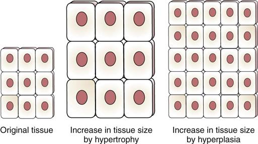
Some tissues and organs stop growing by cell division after development is complete. For example, heart muscle cells no longer divide after fetal life; the number of heart muscle cells is fixed at birth. The size of the heart increases as the person grows because each cell gets larger, but the number of heart muscle cells does not increase. Growth that causes tissue to increase in size by enlarging each cell is hypertrophy. Growth that causes tissue to increase in size by increasing the number of cells is hyperplasia (Fig. 23-1).
Any new or continued cell growth not needed for normal development or replacement of dead and damaged tissues is called neoplasia. This cell growth is always abnormal even if it causes no harm. Whether the new cells are benign or cancerous, neoplastic cells develop from normal cells (parent cells). Thus cancer cells were once normal cells but changed to no longer look, grow, or function normally. The strict processes controlling normal growth and function have been lost. To understand how cancer cells grow, it is helpful to first understand the regulation and function of normal cells.
Biology of Normal Cells
Many different normal cells work together to make the whole person function at an optimal level. For optimal function, each cell must perform in a predictable manner.
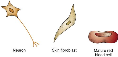
Specific morphology is the feature in which each normal cell type has a distinct and recognizable appearance, size, and shape, as shown in Fig. 23-2.
A small nuclear-to-cytoplasmic ratio means that the nucleus of a normal cell does not take up much space inside the cell. As shown in Fig. 23-2, the size of the normal cell nucleus is small compared with the size of the rest of the cell, including the cytoplasm.
Differentiated function means that every normal cell has at least one special function it performs to contribute to whole-body function. For example, skin cells make keratin, liver cells make bile, cardiac muscle cells contract, nerve cells conduct impulses, and red blood cells make hemoglobin.
Tight adherence occurs because normal cells make proteins that protrude from the membranes, allowing cells to bind closely and tightly together. One such protein is fibronectin, which keeps most normal tissues bound tightly to each other. Exceptions are blood cells. Red blood cells and white blood cells produce no fibronectin and do not usually adhere together.
Nonmigratory means that normal cells do not wander throughout the body (except for blood cells). This occurs in normal cells because they are tightly bound together, which prevents cell wandering from one tissue into the next.
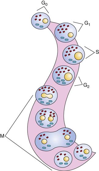
Orderly and well-regulated growth is a very important feature of normal cells. They divide (undergo mitosis) for only two reasons: (1) to develop normal tissue or (2) to replace lost or damaged normal tissue. Even when they are capable of mitosis, normal cells divide only when body conditions, including the need for more cells, adequate space, and sufficient nutrients and other resources, are just right. Cell division (mitosis), occurring in a well-recognized pattern, is described by the cell cycle. Fig. 23-3 shows the phases of the cell cycle. The amount of time needed for one cell to divide completely into two cells is the generation time. For normal cells, the generation time varies by tissue and organ type.
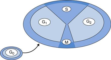
Living cells not actively reproducing are in a reproductive resting state termed G0. During the G0 period, cells actively carry out their functions but do not divide. Normal cells spend most of their lives in the G0 state rather than in a reproductive state.
Mitosis makes one cell divide into two cells. These two new cells are identical to each other and to the cell that began mitosis. The steps of entering and completing the cell cycle are tightly controlled. Much of this control is regulated by proteins produced by “suppressor genes.”
Whether a cell enters and completes the cell cycle to form two new cells depends on the presence and absence of specific proteins. Proteins that promote cells to enter and complete cell division are produced by oncogenes and are known as cyclins. When cyclins are activated, they allow a cell to leave G0 and enter the cycle. These activated cyclins then drive the cell to progress through the different phases of the cell cycle and divide. Proteins produced by suppressor genes regulate the amount of cyclins present and ensure that cell division occurs only when it is needed. Thus normal cell division is a balance between the proteins that promote cell division (cyclins) and the proteins that limit cell division (suppressor gene products).
Fig. 23-4 shows the activities of the phases of the cell cycle (described below):
Contact inhibition is the stopping of further rounds of cell division when the dividing cell is completely surrounded and touched (contacted) by other cells. Of the normal cells that can divide, each cell divides only when some of its surface is not in direct contact with another cell. Once a normal cell is in direct contact on all surface areas with other cells, it no longer undergoes mitosis. Thus normal cell division is contact inhibited.
Apoptosis is programmed cell death. Not only do normal cells have to divide only when needed and perform their specific differentiated functions, some cells also have to die at the appropriate time to ensure optimum body function. Thus normal cells have a finite life span. With each round of cell division, the telomeric DNA at the ends of the cell’s chromosomes shortens (see Chapter 6). When this DNA is gone, the cell responds to signals for apoptosis. The purpose of apoptosis is to ensure that each organ has an adequate number of cells at their functional peak.
Normal chromosomes (euploidy) is a feature of most normal human cells. These cells have 23 pairs of chromosomes, the correct number for human beings.
Biology of Abnormal Cells
Body cells are exposed to a variety of conditions that can alter how the cells grow or function. When either cell growth or cell function is changed, the cells are considered abnormal. Table 23-1 compares features of normal, benign tumor, and cancer (malignant) cells.
TABLE 23-1
CHARACTERISTICS OF NORMAL AND ABNORMAL CELLS
| CHARACTERISTIC | NORMAL CELL | BENIGN TUMOR CELL | MALIGNANT CELL |
| Cell division | None or slow | Continuous or inappropriate | Rapid or continuous |
| Appearance | Specific morphologic features | Specific morphologic features | Anaplastic |
| Nuclear-to-cytoplasmic ratio | Small | Small | Large |
| Differentiated functions | Many | Many | Some or none |
| Adherence | Tight | Tight | Loose |
| Migratory | No | No | Yes |
| Growth | Well regulated | Expansion | Invasion |
| Chromosomes | Diploid (euploid) | Diploid (euploid) | Aneuploid* |
| Mitotic index | Low | Low | High* |
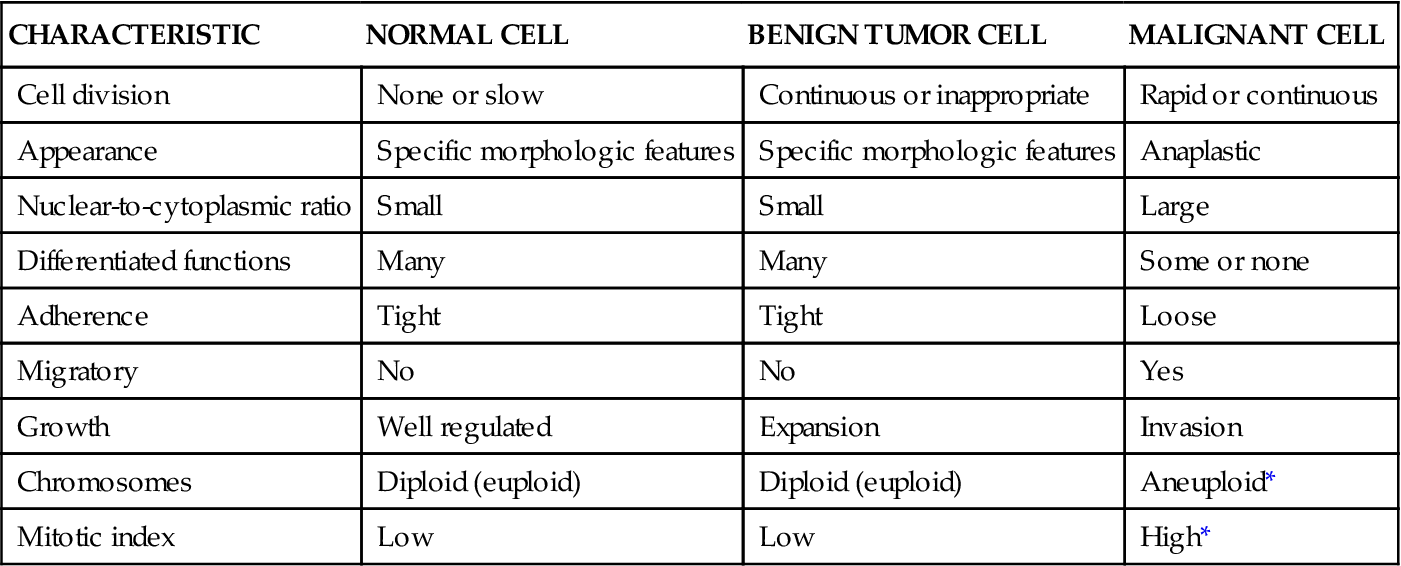
Features of Benign Tumor Cells
Benign tumor cells are normal cells growing in the wrong place or at the wrong time. Examples include moles, uterine fibroid tumors, skin tags, endometriosis, and nasal polyps. Benign tumor cells have these characteristics:
Features of Cancer Cells
Cancer (malignant) cells are abnormal, serve no useful function, and are harmful to normal body tissues. Cancers commonly have these features:
Cancer Development
Carcinogenesis/Oncogenesis
Carcinogenesis and oncogenesis are other names for cancer development. Table 23-2 lists key concepts about cancer development. The process of changing a normal cell into a cancer cell is called malignant transformation, occurring through the steps of initiation, promotion, progression, and metastasis.
TABLE 23-2
KEY CONCEPTS RELATED TO CANCER DEVELOPMENT

Initiation is the first step in carcinogenesis. Normal cells can become cancer cells if their genes promoting cell division, oncogenes, are turned on excessively (overexpressed). Initiation is a change in gene expression caused by anything that can penetrate a cell, get into the nucleus, and damage the DNA. Such changes can activate oncogenes that should have only limited expression and can damage suppressor genes, which normally limit oncogene activity. Thus initiation leads to excessive cell division through DNA damage that results in either loss of suppressor gene function or enhancement of oncogene function.
Initiation is an irreversible event that can lead to cancer development. After initiation, a cell can become a cancer cell if the cellular changes that occurred during initiation continue and cell division is not impaired. A cancer cell is not a health threat unless it can divide. If it cannot divide, it cannot form a tumor. If growth conditions are right, however, widespread metastatic disease can develop from just one cancer cell.
Substances that change the activity of a cell’s genes so that the cell becomes a cancer cell are carcinogens. Carcinogens may be chemicals, physical agents, or viruses. There are more than 50 substances known to cause cancer and about another two hundred that are suspected to be carcinogens. The National Toxicology Program’s website lists these substances (http://ntp.niehs.nih.gov/; U.S. Department of Health and Human Services, 2011). Chapters presenting the care of patients with specific cancers discuss specific carcinogens (when known) within the Etiology sections.
Promotion is the enhancement of growth of an initiated cell. Promoters are substances that promote or enhance growth of the initiated cancer cell. Once a normal cell has been initiated by a carcinogen and is a cancer cell, it can become a tumor if its growth is enhanced. Many normal hormones and body proteins, like insulin and estrogen, can act as promoters and make altered cells divide more frequently. The time between a cell’s initiation and the development of an overt tumor is called the latency period, which can range from months to years. Exposure to promoters can shorten the latency period.
Progression is the continued change of a cancer, making it more malignant over time. Many processes within the tumor must take place for progression to occur. After cancer cells have grown to the point that a detectable tumor is formed (a 1-cm tumor has at least 1 billion cells in it), other events must occur for this tumor to become a health problem. First, the tumor must develop its own blood supply. In the early stages, the tumor receives nutrition only by diffusion. After the tumor reaches 1 cm, however, diffusion is not efficient. The tumor then makes substances that trigger nearby capillaries to grow new branches into the tumor, ensuring the tumor’s continued nourishment and growth.
As tumor cells continue to divide, some of the new cells change features from the original, initiated cancer cell and form groups that differ from the original cancer cell. Some of the differences provide these cell groups with advantages (selection advantages) that allow them to live and divide no matter how the conditions around them change. These tumor changes may allow it to become more malignant. Over time, the tumor cells have fewer and fewer normal cell features.
The original tumor is called the primary tumor. It is usually identified by the tissue from which it arose (parent tissue), such as in breast cancer or lung cancer. When primary tumors are located in vital organs, such as the brain or lungs, they can grow excessively and either lethally damage the vital organ or interfere with that organ’s ability to perform its vital function. At other times, the primary tumor is located in soft tissue that can expand without damage as the tumor grows. One such site is the breast. The breast is not a vital organ, and even if it had a large tumor in it, the primary tumor alone would not cause the patient’s death. When the tumor spreads from the original site into vital areas, life functions can be disrupted and death may follow.
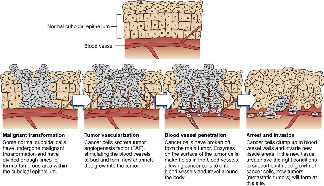
Metastasis occurs when cancer cells move from the primary location by breaking off from the original group and establishing remote colonies. These additional tumors are called metastatic or secondary tumors. Even though the tumor is now in another organ, it is still a cancer from the original altered tissue. For example, when breast cancer spreads to the lung and the bone, it is still breast cancer in the lung and bone—not lung cancer and not bone cancer. Metastasis occurs through many steps, as shown in Fig. 23-5.
Stay updated, free articles. Join our Telegram channel

Full access? Get Clinical Tree


