Brain Tumors


Brain Tumors


Jennifer E. Cahill

Terri S. Armstrong


OVERVIEW OF BRAIN TUMORS

The diagnosis of a brain tumor begins a journey of uncertainty, fear, and hope for the patient and family. Often the patient presents suddenly with an acute neurologic event such as seizure or after a more subtle onset of focal neurologic deficit or headache. Even when a definitive histopathological diagnosis is achieved, the particular clinical course for any given patient is impossible to predict, including the extent of neurologic and cognitive impairment; response to treatment; quality of life; and overall prognosis. Given these uncertainties, to care for the patient and family in a sensitive, compassionate, hopeful, and humanistic manner, the health care professional needs a broad knowledge base and specialized skill set. Within this framework of fundamental principles, concepts related to brain tumors and related management are explored in this chapter.

The Central Brain Tumor Registry of the United States (CBTRUS) estimates that in 2012, 66,290 new cases of primary malignant and nonmalignant brain and central nervous system tumors will be diagnosed.1 This number is higher than the incidence predicted by the American Cancer Society or the Surveillance, Epidemiology, and End Results (SEER) program, which collects primary malignant tumor data only. Primary brain and CNS tumors also occur more frequently in caucasians (85%). The median age of diagnosis of all primary brain tumors is 57 years.1 Although the lifetime risk of developing a brain tumor for both sexes and all ethnicities in the United States is less than 1%, brain tumors such as malignant gliomas can be highly fatal; representing 2% of cancer-related deaths in the United States.1 In children aging from 0 to 19 years, brain and central nervous system cancers are the second most common malignancy after leukemias.1 In 2012, an estimated 13,700 deaths were expected from primary malignant brain and central nervous system tumors in the United States.1

Most brain tumors are located in the frontal, temporal, parietal, and occipital lobes of the brain (supratentorial region). Nonmalignant meningiomas are the most common type of primary brain tumors (35%), followed by glioblastoma which accounts for another 16% of tumors.1 The incidence of pilocytic astrocytomas, germ cell tumors, and medulloblastomas is highest in children and decreases with age. For all other tumors, the incidence increases with age.1 Brain metastases or secondary brain tumors are found in 20% to 40% of cancer patients,2 with wide-ranging estimates of 98,000 to 170,000 new cases diagnosed in the United States each year. Brain metastases are not tracked by any of the current national registries so the true incidence remains unknown.3

While much work has attempted to discern the etiology of brain tumors, causality has not yet been identified for the majority of tumor types. Exposure to ionizing radiation is the only established risk factor.4, 5 Rare genetic syndromes, such as Li-Fraumeni and neurofibromatosis 1 and 2 confer a greater risk for brain tumors4, 5 and potential genetic etiologies have been reported; including polymorphisms at five loci encoding carcinogen metabolism, DNA repair, and cell cycle regulation.6 Recently, evidence has suggested a possible association between immunologic factors and gliomas; patients with selfreported allergies appear to have a reduced risk of glioma and patients with glioma were shown to have lower serum immunoglobulin E (IgE) levels.4 Of course, the results of these genetic and immunological associations cannot yet formally delineate the underlying mechanisms. Finally, evidence for an association with trauma, N-nitroso compounds, occupational risk factors, cell phone use, exposure to electromagnetic fields, and viral etiology remain indeterminate.4, 5, 7


CHART 20-1 World Health Organization Classification of Central Nervous System Tumors


  1. Tumors of neuroepithelial tissue


  2. Tumors of cranial and spinal nerves


  3. Tumors of the meninges


  4. Hematopoietic neoplasms


  5. Germ cell tumors


  6. Cysts and tumor-like lesions


  7. Tumors of the anterior pituitary


  8. Local extensions from regional tumors


  9. Metastatic tumors


Classification of Brain Tumors

Classification of brain tumors is based primarily on histopathologic characteristics, which are important considerations for determining treatment options. For clinical purposes, other conventional classifications are helpful in understanding the nature of brain tumors and will also be reviewed.


Histopathologic Basis of Classification

Although once there was a tumor-node-metastasis (TNM) staging system for brain tumors, this has been subsequently withdrawn. In brain tumors, size (T) is less relevant clinically than the particular grade and location of the tumor, nodal status (N) is incongruous with the fact that the brain and spinal cord lack lymph nodes and metastatic spread (M) rarely applies, given that most tumors do not spread outside of the central nervous system. Instead, primary brain tumors are broadly classified by the presumed tissue of origin, with the degree of malignancy based on the degree of anaplasia, among other features. In specific tumors, location within the brain may also be considered. Several histologic classification schemas exist, the most common of which is the World Health Organization (WHO) system.8


CHART 20-2 Classification of Major Brain Tumors


Gliomas


Astrocytic tumors


Pilocytic astrocytomas (grade I)


Astrocytoma (grade II)


Anaplastic astrocytoma (grade III)


Oligodendroglial tumors


Oligodendroglioma


Anaplastic (malignant) oligodendroglioma


Mixed oligoastrocytoma


Anaplastic oligoastrocytoma


Medulloblastoma


Unclassified (mostly gliomas)


Meningiomas


Menigiomas (multiple histologic types)


Atypical types


Anaplastic (malignant) meningioma


Pituitary adenoma


Neurinoma (schwannoma, acoustic neuroma)


Craniopharyngioma, dermoid, epidermoid, teratoma


Angiomas


Sarcomas


Miscellaneous (pinealoma, chordoma, granuloma, lymphoma)


Adapted from the WHO classification.


The WHO system was initially developed in the 1970s and is continually revised based on current pathological evidence. This system grades tumors on a continuum, utilizing the degree of anaplasia and survival into nine categories (Chart 20-1).8 Tumors originating from neuroepithelial tissue account for the most prevalent glial tumors. The neuroepithelial category is further subclassified in Chart 20-2, with tumors of astrocytic origin being the most common subclassification.5 It is increasingly recognized that these classification systems do not take into account additional differences in the molecular basis for tumors. For example, lower grade tumors (Grade II and III) can remain low grade or evolve into a more malignant Grade IV tumor called glioblastoma. Grade IV tumors will also occur spontaneously, without progression from a lower grade. These tumors are often referred to as de novo whereas those that undergo malignant transformation to a higher grade are considered secondary glioblastoma. Although this differentiation is not currently used to determine treatment approach, understanding the differences in the process involved in the growth of these tumor types is rapidly evolving and is a potential target for new therapies.


Conventional Classifications

Other conventional classification systems are based on several distinguishing clinical considerations including primary versus secondary (metastatic) formation, neuroembryonic origin, anatomic location, and malignant versus benign orientation.


  • Primary versus metastatic brain tumors: primary brain tumors originate from the various cells and structures that constitute the brain. Metastatic brain tumors (also called secondary brain tumors) originate from sites outside the brain, most often from primary tumors of the lung, breast, and melanomas. Primary brain tumors rarely metastasize outside of the central nervous system. Carcinomatosis is a condition in which carcinoma is widespread throughout the body; when lesions in the brain spread throughout the CNS to the spinal fluid and meninges, the term carcinomatous meningitis or more commonly, leptomeningeal disease is used.


  • Neuroembryonic origins: nervous system tumors originate from the ectodermal (outer) layer of the embryo. Chapter 5 briefly describes early embryonic development. At 16 days, the neural plate appears, changing to the neural groove and neural tube by the third week. Those neuroectodermal cells not incorporated into the neural tube form neural crests. The neural tube and neural crests contain two types of undifferentiated cells called neuroblasts and glioblasts (spongioblasts). The neuroblasts become the basic unit of structure in the nervous system and are then called neurons. The glioblasts form a variety of cells that support, insulate, and metabolically assist the neurons. They are collectively called glial cells. Glial cells are subclassified into astrocytes (star-shaped cells), oligodendrocytes (glial cells with few processes), and ependymal cells (line the ventricles). This is the basis for the broad category of brain tumors called gliomas. Gliomas are further subdivided into astrocytomas, oligodendrogliomas, and ependymomas.


  • Anatomic location: the anatomic location of the lesion affects signs and symptoms as well as presentation. Tumors can be extra-axial (located outside of the brain parenchyma) or intraaxial. Intra-axial tumors can be further defined by location by the specific site of the lesion such as the frontal or temporal lobe, pons, or cerebellum. Location may be noted by using the tentorium cerebelli as a reference point to differentiate between supratentorial, located above the tentorium (i.e., cerebral hemispheres), and infratentorial, located below the tentorium (i.e., brainstem or cerebellum). Knowing the location of the lesion helps to predict probable deficits based on an understanding of the normal function of that anatomic area. In addition, location is an important variable in selecting treatment options and prognosis.


  • Malignant versus benign: the benign versus malignant distinction is made on the basis of histologic properties. Benign tumors contain well-differentiated cells that retain much of the cellular characteristics of the progenitor cell. Malignant tumors contain undifferentiated, primitive-appearing cells. Generally, cells that are well differentiated are related to a better prognosis than cells that display greater degree of anaplasia. However, when applied to the classification of brain tumors, these terms can be somewhat misleading. A tumor that is considered to be histologically benign may be surgically inaccessible, as with a deep tumor requiring extensive dissection of tissue or one located in a vital area, such as the pons or medulla. A benign tumor that is partially or completely surgically inaccessible may continue to grow, and cause neurological deficits, if the tumor does not respond to other treatment options such as chemotherapy or radiation. These deficits can result in loss of significant neurological function and life, even though they are histologically “benign.”

Brain tumors are additionally classified with regard to rate of growth or mitotic activity. Benign tumors are typically characterized by low mitotic activity and slower rates of growth. Brain tumors with slow growth are referred to as low-grade tumors. Malignant tumors display considerable mitotic activity and increased cellular proliferation. Brain tumors with a high growth rate are referred to as high-grade tumors.


Pathophysiology of Brain Tumors

The pathophysiology of brain tumors can be viewed from the perspective of molecular considerations and the effects of the tumor, both directly on cerebral tissue and indirectly through the development of increased intracranial pressure (ICP).


Molecular Considerations

Transformation of glial or neuronal cells into brain tumors is a complex process that is still incompletely understood. Brain tumors arise in association with multiple, specific structural molecular genetic alternations (i.e., mutations) within cells. These mutations can cause the cell to proliferate inappropriately as well as result in other malignancy attributes, such as the loss of differentiated characteristics of the tissue of origin, acquisition of the ability to invade surrounding normal tissues and metastasize, and the ability to resist antineoplastic therapies.9 Two types of genetic molecular alterations trigger these changes in cellular behavior. The first change results in the complete cessation or partial decrease in cellular activities that physiologically restrain growth. The genes that regulate this change are known as tumor suppressor genes. The second change inappropriately activates genes that typically enhance cellular proliferation. Known as proto-oncogenes, these genes encode proteins that act as growth factors or growth factor receptors, mediators of signaling pathways, or regulators of gene expression. Mutations convert proto-oncogenes to oncogenes, which function in various ways to promote neoplastic changes, such as alterations in cell cycle progression, abnormalities in signal transduction pathways, glial cell invasion, and angiogenesis.9


The Effects of a Space-Occupying Lesion Within the Brain

Tumors directly affect the brain through compression of cerebral tissue, through invasion or infiltration of cerebral tissue, and sometimes through erosion of bone. A brain tumor usually grows as a spherical mass until it encounters a more rigid structure, such as bone or the falx cerebri. The encounter with an aplastic substance necessitates a change in the contour of the neoplasm. Neoplastic cells can also grow diffusely, with multiple cells infiltrating tissue spaces without forming a definite mass. The tumor enlarges because of cell proliferation, necrosis, fluid accumulation, hemorrhage, or the accumulation of degenerative by-products within the mass.

The clinical effects of a tumor within the brain and cranial vault depend on the location of the tumor, rate of growth, and consequences of increased ICP. A slow-growing tumor may become large
before clinical signs and symptoms of increased ICP are noted. This happens because the tumor’s volume is accommodated in the intracranial space over a long period of time, often years. A meningioma is a slow-growing tumor that can become large before signs and symptoms of increased ICP are noted. Conversely, depending on its location, evidence of a meningioma may be noted early because of focal deficits. With a fast-growing tumor, such as glioblastoma multiforme (GBM), there is little time for the intracranial compartment to accommodate the lesion, and signs of increased ICP may be noted in a shorter time frame. In summary, an understanding of the pathophysiologic effects of brain tumors requires an understanding of the Monro-Kellie hypothesis and increased ICP along with the specific tumor type, location, and related focal deficits. See Chapter 13 for a discussion of increased ICP and its management.

In most patients with brain tumors, vasogenic edema develops in the surrounding tissue as a result of compression. At the cellular level, an increase in permeability of capillary endothelial cells of the cerebral white matter results in seepage of plasma into the extracellular space and between the layers of the myelin sheath. This alters the electrical potential of cells, impairing cellular activity. Cerebral edema may also develop rapidly from alterations in the blood-brain barrier caused by substances released from tumor cells.10 As cerebral edema increases, a mass effect develops, and signs and symptoms of increased ICP become apparent. These signs and symptoms continue to develop as a tumor grows. The resulting increase in cerebral edema can result in cerebral herniation syndromes and death (see Chapter 13).


SIGNS AND SYMPTOMS OF BRAIN TUMOR

There are no definitive signs and symptoms of a brain tumor and patients present with widely variable symptom complaints or no symptoms at all. A patient’s particular clinical presentation depends on the size, location, compression or infiltration of specific cerebral tissue, related cerebral edema, and the presence of increased ICP. Brain tumors are sometimes discovered as asymptomatic masses, often as incidental findings on unrelated diagnostic imaging. When present, symptoms of a brain tumor are nonspecific and nondiagnostic, necessitating further evaluation with objective imaging techniques such as computed tomography (CT) scan or magnetic resonance imaging (MRI). Signs and symptoms associated with a brain tumor may be general or focal in nature.


  • General: headaches, nausea and vomiting, changes in the level of consciousness, and seizures


  • Focal: specific focal deficits, such as hemiparesis, or syndromes related to specific cerebral areas, such as expressive aphasia or acromegaly (Fig. 20-1)


Frontal Lobe

Multiple higher cognitive, executive, behavioral, speech, and motor functions are controlled by the frontal lobe. Focal deficits can be traced to either anterior frontal or posterior frontal findings.


Anterior Frontal Lobe. Patients with tumors in the anterior frontal lobe present with a wide range of higher-level cognitive function and personality changes such as short- and long-term memory deficits, difficulty in concentration and vigilance, slowing of mental processes and reaction time, abulia, and difficulty with calculations, problem solving, insight, abstraction, and synthesis of ideas. Collectively, anterior frontal lobe symptoms are called the frontal lobe syndrome. Personality and behavioral changes may include emotional lability, flat affect, lack of initiative and spontaneity, loss of self-restraint, and loss of social behavior.

Posterior Frontal Lobe. Broca’s area is located in the posterior-inferior frontal lobe. If a tumor is located in or around Broca’s area in the dominant hemisphere, fluent speech deficits such as word finding may be noted. The primary motor strip is located in the posterior frontal lobe. Tumors in this area can cause focal findings of motor weakness such as monoparesis or focal seizure activity.


Parietal Lobe

The parietal lobe contains the sensory discrimination and association areas for body orientation, vision, and language. Parietal lobe tumors cause deficits in sensation, inability to recognize common objects, and neglect syndromes such as the lack of awareness of the opposite side of the body. If the lesion is in the dominant hemisphere and is located in the left angular gyrus of the parietal lobe, Gerstmann’s syndrome may be present (i.e., finger agnosia, loss of right-left discrimination, acalculia, and agraphia). Seizure activity and homonymous hemianopsia are also possible with a parietal lobe tumor. A collection of symptoms associated with parietal lobe dysfunction is called the parietal lobe syndrome. Common symptoms include the following.


Sensory Changes


  • Hypo- or hyperesthesia (impaired sensation with increased or decreased tactile sensitivity)


  • Paresthesia (abnormal sensation involving tingling, crawling, or burning feeling on the skin)


  • Loss of two-point discrimination (unable to determine by feeling if the skin is touched by one or two points simultaneously)


Recognition Deficits


  • Inability to recognize letters or numbers


  • Astereognosis (inability to recognize an object by feeling its size and shape)


  • Autotopagnosia (inability to locate or recognize parts of the body)


  • Anosognosia (loss of awareness or denial of specific motor or sensory deficits)


  • Finger agnosia (inability to identify or select specific fingers of the hand such as thumb)


Orientation Deficits and Neglect Syndromes


  • Loss of right-left discrimination


  • Difficulty in going through doorways without knocking self on one side


  • Neglect syndrome (a tendency to ignore the part of the environment or body opposite to the tumor)


  • Construction apraxia (if asked to draw a face of a clock, ignoring the side of the clock opposite to the tumor)


Temporal Lobe

Neoplasms of the temporal lobe may cause psychomotor (complex partial) seizures, weakness, visual field deficits (often loss in the upper quadrant opposite the lesion), and memory deficits, most often for recent events. When the dominant side is involved, speech and language deficits are frequent because Wernicke’s area is located in the dominant temporal lobe. Psychomotor seizures with visual, auditory, or olfactory hallucinations; automatism; and amnesia for events of the attack can occur. They may begin with an aura of peculiar sensations of the abdomen, epigastrium, or thorax.


Occipital Lobe

Tumors of the occipital lobe are infrequent compared with lesions involving the other cerebral lobes. When neoplasms do occur, the symptoms tend to be associated with vision. Symptoms include homonymous quadrantanopia (loss of vision in one fourth of the visual field), visual hallucinations, and failure to recognize familiar objects.


Pituitary and Hypothalamus Region

The pituitary gland and hypothalamus are closely related by location and endocrine production. Common symptoms resulting from tumors in these areas are visual deficit caused by optic atrophy and paralysis of one or more of the extraocular muscles; headache; and endocrine dysfunction of the pituitary gland with the subsequent precipitation of various syndromes, such as Cushing’s syndrome, giantism, acromegaly, and hypopituitarism. In addition, tumors of the hypothalamus can affect fat and carbohydrate metabolism, water balance, sleep patterns, appetite, and sexual drive.


Lateral and Third Ventricles

If the tumor remains small, the patient may be asymptomatic. If the tumor grows into the cerebral hemispheres, deficits will depend on the particular function of the area involved. Tumors that grow within the ventricle may become of sufficient size to obstruct the flow of cerebrospinal fluid (CSF) resulting in noncommunicating hydrocephalus. If this occurs, headache, vomiting, and other symptoms of rapidly increased ICP will be noted. The patient may experience relief of symptoms by changing the position of the head. In this case, the position of the obstructing tumor is altered, thereby allowing the normal CSF flow pattern to be re-established.


Brainstem

Tumors of the brainstem may produce multiple symptoms such as lower cranial nerve deficits (swallowing, articulation, and gag reflex), motor and sensory deficits, vertigo, hiccups, ataxia, incoordination, nystagmus, dysphagia, nausea, and vomiting throughout the illness. Sudden death can occur from encroachment on vital centers (respiratory or cardiac arrest). Obstructive hydrocephalus may develop from encroachment on the ventricular system.


Midbrain

Neoplasms of the midbrain are rare. If present, they may result in occlusion of the cerebral aqueducts, cerebellar symptoms if the red nucleus is involved, Parinaud’s syndrome (i.e., conjugate paralysis of upward gaze) if the quadrigeminal plate is involved, abnormal posturing, and ptosis and diminished light reflex as the tumor enlarges.


Fourth Ventricle

Tumors of the fourth ventricle obstruct the flow of CSF (noncommunicating hydrocephalus) and infiltrate and compress the brainstem or cerebellum. See Chapter 13 for a discussion of hydrocephalus. Headache, vomiting, and nuchal rigidity are common symptoms. Sudden death caused by compression of the cardiorespiratory center is possible. The lower cranial nerves, which control the gag and swallowing reflexes, become impaired, making aspiration a constant concern.


Cerebellum

Growth of a tumor in the cerebellar area is accompanied by cerebellar signs (ataxia, incoordination, nystagmus, vertigo, nausea), obstruction of flow of CSF, and potential for brainstem compression. The usual signs of increased ICP (headache, vomiting, and classic changes in vital signs) are common, particularly with CSF obstruction.


ASSOCIATED INCREASED INTRACRANIAL PRESSURE SIGNS

Many tumors are associated with increased ICP and present with or without localizing signs. They include medulloblastoma, ependymoma of the fourth ventricle, hemangioblastoma of the cerebellum, pinealoma, colloid cyst of the third ventricle, and craniopharyngioma. These tumors are listed in Table 20-1.

The signs and symptoms associated with increased ICP are discussed in Chapter 13. Only papilledema and obstruction of CSF flow, as they relate to brain tumors, are discussed in this section.


Papilledema

Papilledema, seen in about 70% of patients with brain tumors, is associated with visual changes, such as decreased visual acuity, diplopia, and deficits in the visual fields. The visual pathways extend through the four lobes of the cerebral hemispheres. Therefore, it is reasonable to expect a high incidence of visual disturbances with supratentorial lesions. Dysfunction of the abducens nerve (cranial nerve VI), a symptom commonly seen in brain tumor lesions, results in an inability to move the eye outward on the horizontal plane. It is not uncommon for patients to be referred to a neurologist to rule out intracranial lesion after these subtle visual deficits or disturbances are first identified and evaluated through routine eye examinations by an optometrist or ophthalmologist.


Obstruction to Flow of Cerebrospinal Fluid

Tumor encroachment from within or outside the ventricles or subarachnoid space interferes with the normal flow of CSF. This obstruction of CSF results in obstructive hydrocephalus. If the


tumor encroachment occurs slowly, the development of hydrocephalus will be gradual. However, rapid tumor growth produces acute precipitation of signs and symptoms, such as a massive spike in ICP with rapid deterioration in neurological status.


TABLE 20-1 BRAIN TUMORS


TYPE OF TUMOR


DESCRIPTION


LOCATION OR DEMOGRAPHIC DATA


SIGNS AND SYMPTOMS


TREATMENT


PROGNOSIS


Common Brain Tumors


Astrocytoma (grades I and II)


Constitutes 25-30% of all cerebral gliomas


Grade I: well-defined cells


Grade II: cell differentiation less defined ↑ Cellularity


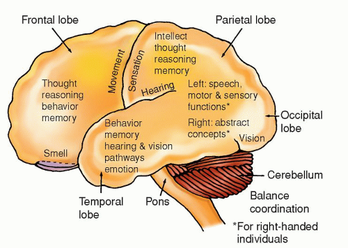
Usually found in cerebrum, cerebellum, hypothalamus, optic nerve and chiasma, and pons


Cerebral hemisphere tumors most often found in adults 20-40 yr


Neurological deficits depend on specific location of tumor and if it is supra- or infratentorial


Onset of a focal or generalized seizure in previously seizurefree person is most common first sign


Surgery: gross total removal is treatment of choice, but complete removal rarely possible; partial removal may prolong life; tumor recurrence often associated with malignant progression


Radiation and Chemotherapy: controversial; not done for grade I


5-6 yr survival on average


Range, 2-20 yr


Anaplastic astrocytoma (grade III)


Cellularity anaplastic: cellular atypia, ↑ mitosis


15-28 mos average survival


Glioblastoma multiforme (GBM) (also known as astrocytoma, grade IV)


Constitutes 20% of all intracranial tumors and 55% of all gliomas


Malignant, rapidly growing


Composed of heterogeneous cells


Necrotic and hemorrhagic areas within tumor common


Usually found in a frontal lobe


40-60 yrs most common and with increasing age


Male predilection


Memory loss, neurobehavioral changes, seizures, speech deficits, hearing/auditory (H/A), visual deficits


Diffuse cerebral symptoms


Surgery: resection and debulking to relieve compression and ICP


Radiation with concurrent temozolomide followed by adjuvant temozolomide


14-16 mos average survival


Astrocytoma of optic nerves and chiasma (spongioblastoma)


Most common in children; sometimes seen in young adults


As the tumor grows, it enlarges the optic foramen with little distortion of surrounding structures


Slow-growing tumor


Found along the optic nerves


Girls > boys, with 2:1 predilection


Early symptoms include Dim vision Hemianopsia Optic atrophy


Blindness


Proptosis


Hypothalamic imbalance


Surgery: removal possible but tumor often inaccessible


Radiation: usually poor response


10 yrs or more


Ependymoma (low grade and anaplastic)


Tumor of childhood and young adults


Arises from lining of ventricles


Slow-growing


In ventricles, particularly fourth; can attach itself to roof or floor of ventricle, or grow directly into cerebral hemisphere


Seen in children and adults up to 30 yrs, most often in men


Supratentorial more common in adults; infratentorial in children


Rapid elevation in ICP secondary to CSF obstruction


S&S vary by location


If fourth ventricle, ↓ level of consciousness, severe H/A, VS changes with ↑ ICP, N/V, pupillary changes, hemiplegia, hemiparesthesia, seizures


If in cerebellar area, ataxia


Surgery: removal if surgically accessible; depends on location


Radiation: for most


Chemotherapy: usually not helpful


Shunting procedure: prn to reduce ↑ ICP from obstructive hydrocephalus


About 5-10 yrs, depending on location


Oligodendroglioma (low grade and anaplastic)


Calcification noted on radiologic examination in about 50% of patients


Cerebral hemispheres, particularly frontal and temporal lobes


Found in patients 20-40 yrs


Depends on location


Seizures are first symptoms in 50% of patients


Surgery, chemotherapy for those with loss of heterozygosity of 1p and 19q chromosomes and radiation for those who are intact


5-10 yrs, depending on grade


Mixed gliomas


Named for predominant tumor cell present


Composed histologically of two or more cell types of astrocytoma/glioblastoma, oligodendroglioma, or ependymoma in any combination


Any place where various glioma types can be found


Depend on location of tumor


Depends on type of tumor


Surgery, radiation, chemotherapy


≥5 yrs or more


Meningioma


Extra-axial tumor arising from dural elements


Firm, encapsulated; can erode into bone


Have estrogen and progesterone receptors; grow rapidly during pregnancy


Slow-growing; can become large before symptoms appear


Recur if not completely removed; can become malignant with reoccurrence


Compresses brain


Predilection for areas proximal to venous sinuses


Most common in women; average age, 50 yrs


Parasagittal sinus


Lateral convexities


Sphenoid ridge


Suprasellar


Olfactory groove


Neurological deficits caused by compression and depending on area involved


Progressive H/A, memory loss, or cognitive changes; paraparesis; seizures; urinary incontinence


Gradual development of hemiparesis, speech abnormalities; other related to area of compression


Extraocular nerve palsy, proptosis, seizures


Bitemporal hemianopsia, optic atrophy, pituitary-related hormonal imbalance


Anosmia, visual deficits, dementia, pupillary abnormalities


Surgery: complete removal, if possible, or partial dissection


Radiation: after subtotal resection and at tumor recurrence


Immunotherapy for atypical meningiomas


“Cure” with total removal


Many years with partial excision with radiation


Metastatic brain tumors


20-40% of cancer patients have metastasis to brain from other parts of the body (lungs, breast, lower GI most common)


Spread to brain by blood


Usually well differentiated from other brain tissue; lesion may be single or multiple


Can occur anywhere


Seen as individual tumor or multiple tumors


Depend on location H/A, paresis, and cognitive deficits most common


Surgery: resection if possible, for singular lesion


Radiation: with multiple lesions


Gamma knife radiosurgery (for < three lesions)


Chemotherapy: similar as for primary tumor; methotrexate with oral leucovorin rescue common


Prognosis usually based on primary cancer


1-3+yrs average


Malignant melanomas


Rare


Cerebral hemispheres from a primary lesion in skin


Depend on location


Surgery, radiation, chemotherapy


Few months to few years


Primary cerebral lymphoma


Cellular tumor


Behaves much like a glioblastoma


Occurs in adults 40-50 yrs; more common in immunocompromised patients (immunosuppressive therapy for organ transplant or people with AIDS)


May arise in any part of brain


May be either monofocal or multifocal


Neurocognitive and personality changes


Focal signs or ↑ ICP signs


Biopsy followed by Decadron


Chemotherapy


Both radiation and chemotherapy are effective


After initial response, relapse common


Average survival, 1-4 yrs


Cerebellopontine Angle Tumors (includes several categories of tumors located in this anatomic area)


Miscellaneous astrocytomas and meningiomas


Can be confused with an acoustic neuroma without visualization


Definitive diagnosis made by surgical exposure, biopsy, and histologic examination


Cerebellopontine angle


Variation of those seen with acoustic neuroma (see below)


Surgery: if possible; difficult surgical access (near vital centers)


Radiation: may be selected over surgery


Depends on the type of tumor


Acoustic neuroma (schwannoma)


Arises from sheath of Schwann cells


Usual size: pea to walnut


Considered a benign tumor but located in an often inaccessible area


Slow-growing


Bilateral tumors are possible; when they occur, they result from a hereditary problem of chromosome 22; the tumors are part of central neurofibromatosis


Seen most often in patients 30-49 yrs


Involves vestibular branch of CN VIII


Small tumors are confined to internal auditory canal and involve CN VIII


Large tumors extend outside internal auditory meatus


Large tumors displace CN VII and compress CN V along with CN VIII; they may also encroach on CN IX and CN X, and possibly cerebellum


Depend on size; deficits noted on affected side


Small tumor (confined to internal auditory canal and involving CN VIII) and include: Tinnitus/vertigo


Hearing loss; most notable when using telephone or when source of sound is close to affected ear


Dizziness


Large tumor (outside auditory meatus):


S&S listed above and


Facial: loss of taste on anterior tongue, difficulty closing lower eyelid, facial weakness


Trigeminal: facial paresthesia/anesthesia, difficulty chewing


Glossopharyngeal and vagus (difficulty swallowing, hoarseness)


Cerebellar involvement (ataxia/incoordination, possibly hydrocephalus, ↑ ICP from obstruction of CSF flow secondary to displacement of pons and medulla)


Surgery: microsurgical complete removal or debulking of larger tumors (debulking to preserve CNs involved in the tumors)


Suboccipital retrosigmoid approach for smaller tumors


Translabyrinthine approach for larger tumors


With large tumors, the tumor may entwine other CNs that would cause severe deficits if tumor were completely excised


Radiation: focused radiation (proton beam, gamma knife) alternative in older patients; scar tissue a possible problem if later surgery needed. Also used in younger patients


Cure with small tumor and total resection; generally good outcome


Tumor regrowth possible if subtotal resection


Possible permanent hearing loss, loss of facial sensation on affected side, or facial droop


Decreased or absent corneal reflex


Cherdoma


Arises from embryonic remnants


May appear as a cerebellopontine angle tumor


Predilection M > F


Occurs in patients 30-49 yrs


Found in clivus (35%) dorsum of sellae to foramen magnum and (50% in sacrococcygeal area)


Loss of vision


Extraocular muscle paralysis


Paralyzed muscles of swallowing


Noted on MRI or CT scan


Surgery: excision (approach varies depending on tumor location)


Radiation: conventional or proton beam


Tumors tend to recur


Poor prognosis with aggressive and metastatic tumors


Pituitary Tumors


Pituitary adenomas*


Classified by type of: Hormones secreted


Effects (functioning or nonfunctioning)


Grade of sella turcica enlargement or erosion


Suprasellar extension


Hormone(s) secreted


Prolactin (most common)


Growth hormone


ACTH


Nonfunctioning: produce S&S from compression of adjacent structures (e.g., optic nerves, bitemporal hemianopsia)


Functioning (hormonesecreting): cause endocrine syndromes (e.g., acromegaly)


Enclosed adenomas:


I—sella normal; floor may be indented


II—sella enlarged, floor intact


III—invasive adenomas; localized erosion of the floor


IV—entire floor diffusely eroded


Classified A-D by suprasellar extension


A: No suprasellar extension


B: Suprasellar bulge does not reach floor of third ventricle


C: Tumor reaches third ventricle, distorting chiasmatic recess


D: Tumor fills third ventricle almost to foramen of Munro


Most pituitary tumors in anterior lobe


Both lobes can be damaged from compression of parasellar tumors


In general:


Visual disorders (diminished vision with a scotoma; bitemporal hemianopsia)


Paresis of extraocular muscles


H/A


Various endocrine disorders (see below)


Abnormal sella turcica region on CT scan


Endocrine disorders:


Prolactin-secreting adenoma


Galactorrhea


Amenorrhea


Infertility


Loss of pubic hair


Impotence


↑ Serum prolactin


ACTH-secreting adenoma


Adrenal hyperplasia


Cushing’s syndrome*


Growth hormone-secreting adenoma


Giantism before puberty or closure of epiphyses


Acromegaly after puberty or closure of epiphyses (enlarged jaw, nose, tongue, hands, feet)


Thickening of soft tissue of face


Enlarged heart and pulmonary disease


Diabetes mellitus


Serum growth hormone levels >10 ng/mL


Serious complications:


Pituitary apoplexy syndrome: acute onset of ophthalmoplegia, blindness, drowsiness, and coma; death possible


Depends on the size and type of the tumor, patient’s age, and endocrine and visual deficits; surgery, radiation, or drug therapy separately or in combination


Surgery: for smaller tumors, transsphenoidal microsurgery to remove total tumor and preserve or normalize pituitary


Radiation: conventional radiation therapy or proton beam, if available


Hormonal replacement: postsurgery, hormonal replacement possible


Other drug treatment: bromocriptine may be used to inhibit prolactin; for some patients, this is only treatment necessary for prolactinsecreting tumors


Curable with complete resection


In others, very good outcome


Developmental Tumors (seen sometimes in adults)


Craniopharyngioma


Thought to arise from Rathke’s pouch


Solid or cystic tumors


Can compress the pituitary and may even amputate the pituitary stalk


About 75% with calcified areas


Tumor growth is directed upward, resulting in invagination of the third ventricle and possible blockage of CSF flow


Optic chiasm elevated by tumor, resulting in traction on optic nerves


In or about the sella pituitary area


Usually affects children


Signs and symptoms of grossly ↑ ICP because of CSF flow block-age


Pituitary or hypothalamic dysfunction


Visual disturbance


Surgery: resection by intracranial or transsphenoidal approach


Radiation: after surgery; tumor radiosensitive


Excellent if tumor is excised with microsurgery, cure rate, 80%


Recurrence if only subtotal resection performed, even with radiation


Epidermoid and dermoid cysts


Cysts of congenital origin arising from the ectodermal layer; cysts lined with stratified squamous epithelium


Epidermoid cysts contain keratin, cellular debris, and cholesterol; dermoid cysts contain hair and sebaceous glands


On bones of skull or within brain


Depends on location


Surgery: complete removal is usually possible


Very good


Genetically Related Autosomal Dominant Diseases


Von Recklinghausen’s disease (neurofibromatosis)


Genetic origin because of autosomal dominant mendelian trait


Skin, nervous system, bones, endocrine glands, and other organs are sites of congenital anomalies, in addition to the multiple tumors of skin


Firm, encapsulated lesions attach to the nerve


Benign, multiple, circumscribed dermal and neural tumors with increased skin pigmentation (cosmetically offensive)


Tumors late in childhood or in early adolescence


Spots of hyperpigmentation (café au lait) and cutaneous and subcutaneous tumors


Surgery: possible, depending on the location of the tumor


Radiation: tumor is radioresistant


Depends on involved area


Hemangioblastoma (with Von Hippel-Lindau disease)


Vascular tumor


Slow-growing


Cerebellum (as a single or multiple lesion); less common in the medulla and cerebral hemispheres; tumor in adults


Dizziness


Unilateral ataxia


Signs and symptoms of ↑ ICP


Possible spinal cord involvement


Surgery: complete removal, if possible


Radiation: with recurrence


Usually curable


ACTH, adrenocorticotropic hormone; AIDS, acquired immunodeficiency syndrome; CN, cranial nerve; CSF, cerebrospinal fluid; CT, computed tomography; GI, gastrointestinal; H/A, headache; ICP, intracranial pressure; MRI, magnetic resonance imaging; N&V, nausea and vomiting; S&S, signs and symptoms; VS, vital signs.


* Cushing’s syndrome comprises moon faces, “buffalo hump,” abdominal striae, pendulous abdomen; ecchymosis, hypertension, muscle weakness, osteoporosis, and high cortisol levels.

Only gold members can continue reading. Log In or Register to continue

Stay updated, free articles. Join our Telegram channel

Jul 14, 2016 | Posted by in NURSING | Comments Off on Brain Tumors

Full access? Get Clinical Tree

Get Clinical Tree app for offline access