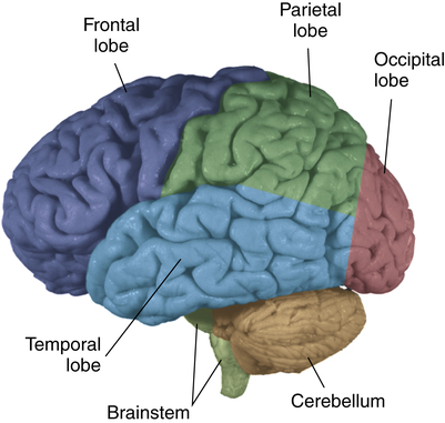
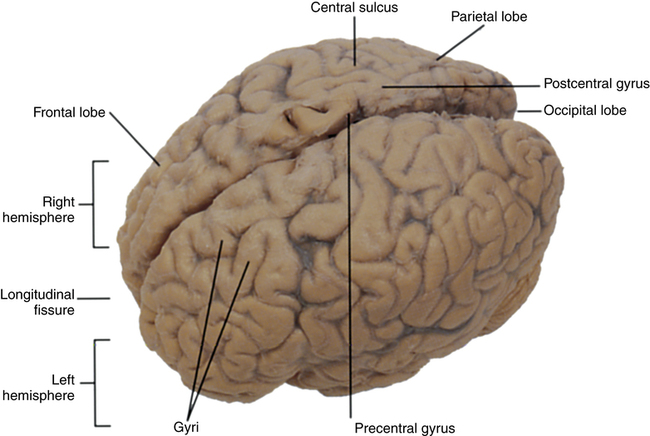
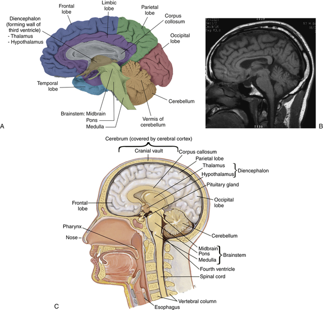
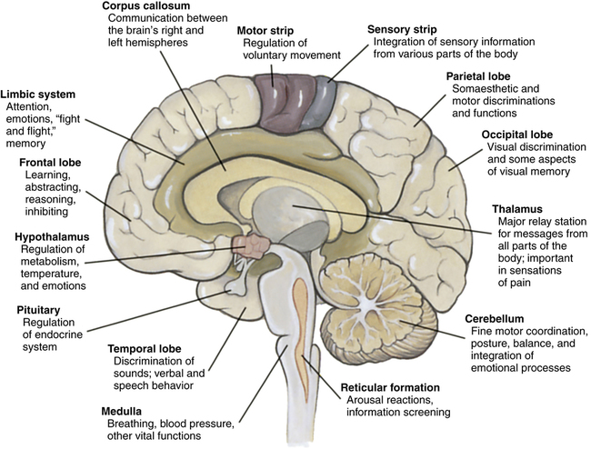
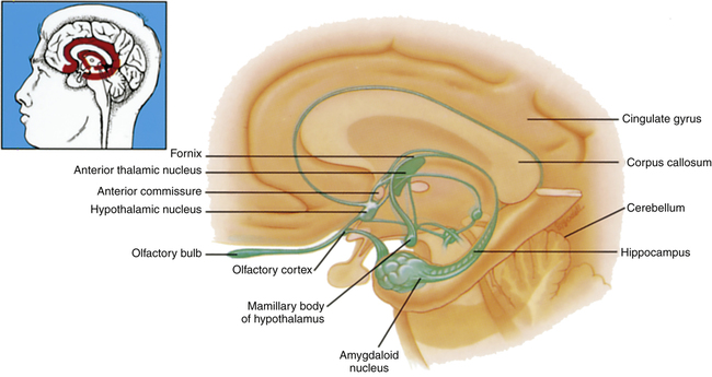
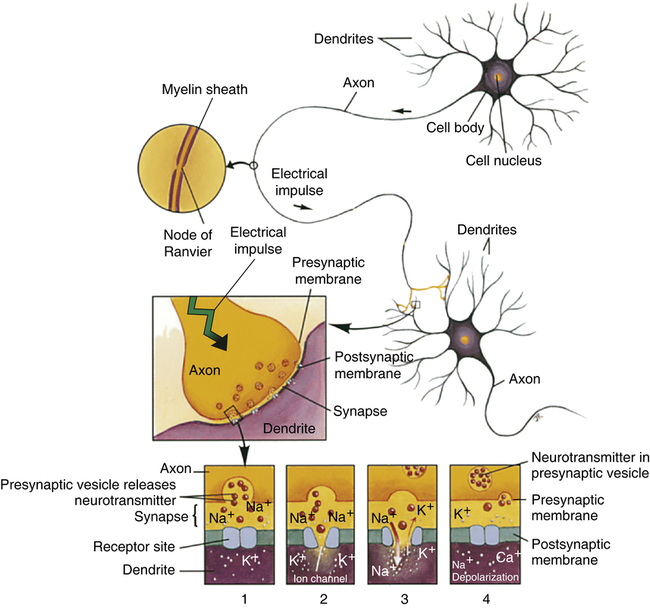
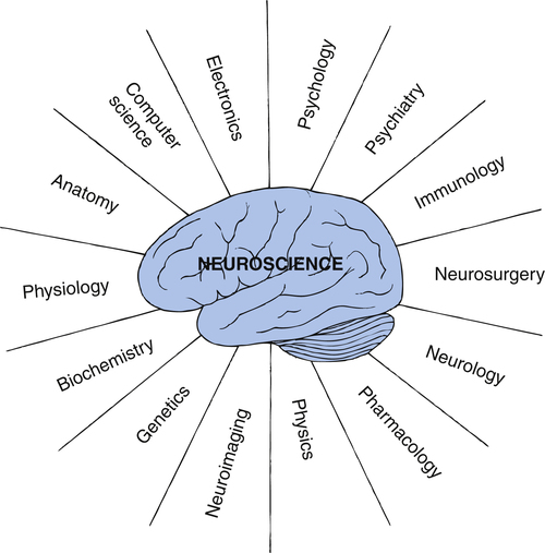
1. Apply knowledge about the structure and function of the brain to psychiatric nursing practice. 2. Describe how neuroimaging is used in psychiatry. 3. Examine the impact of biological rhythms and sleep on a person’s abilities and moods. 4. Describe how psychoneuroimmunology relates to mental health and illness. 5. Discuss genetics and the impact of the Human Genome Project on the understanding of mental illness. Interest in the brain and human behavior is as old as the human race. Records dating back to 3000 BC in Egypt describe brain anatomy and various brain injuries. Currently the field of neuroscience is rapidly expanding and includes many disciplines that combine for a more complete understanding of the human brain and how it is integrated with the body and the human environment (Figure 5-1). All nurses should have a working knowledge of the normal structure and function of the brain, just as all nurses should know about the structure and function of the heart. The brain weighs about 3 pounds. It is composed of trillions of groups of cells that have formed highly specific structures and sophisticated communication pathways that have changed over millions of years of evolution (Box 5-1). A brief review of key brain regions is presented in Figures 5-2 through 5-6 and in Box 5-2. Neurotransmitters are manufactured in the neuron and released from the axon, or presynaptic cell, into the synapse, which is the space between neurons. From there the neurotransmitters are received by the dendrite, or postsynaptic cell, of the next neuron. This neurotransmission process makes communication among brain cells possible (Figure 5-7). Neurons are very specialized, and neurotransmitters perform vital functions in the normal working brain. Their absence or excess can play a major role in brain disease and behavioral disorders. A single neurotransmitter can affect other brain chemicals as well as several different subtypes of receptor cells, each located along tracts connecting different regions of the brain. Thus the same neurotransmitter can have one effect in one part of the brain and different effects in another part of the brain. Nearly all known neurotransmitters fall into one of two categories: small amine molecules (monoamines, acetylcholine, amino acids) and peptides. These are described in Table 5-1. TABLE 5-1 NEUROTRANSMITTERS AND NEUROMODULATORS IN THE BRAIN
Biological Context of Psychiatric Nursing Care
Structure and Function of the Brain
SUBSTANCE
LOCATION
FUNCTION
Amines
Amines are neurotransmitters that are synthesized from amino acid molecules such as tyrosine, tryptophan, and histidine. Found in various regions of the brain, amines affect learning, emotions, motor control, and other activities.
Monoamines
Norepinephrine (NE)
Derived from tyrosine, a dietary amino acid; located in brainstem (particularly locus ceruleus)
Effect: can be excitatory or inhibitory
Levels fluctuate with sleep and wakefulness. Plays a role in changes in levels of attention and vigilance. Involved in attributing a rewarding value to a stimulus and in regulation of mood. Plays a role in affective and anxiety disorders. Antidepressants block reuptake of NE into presynaptic cell or inhibit monoamine oxidase from metabolizing it.
Dopamine (DA)
Derived from tyrosine, a dietary amino acid; located mostly in brainstem (particularly substantia nigra)
Effect: generally excitatory
Involved in control of complex movements, motivation, and cognition and in regulating emotional responses. Many drugs of abuse (e.g., cocaine, amphetamines) cause DA release, suggesting a role in sensation of pleasure. Involved in movement disorders seen in Parkinson disease and in many of the deficits seen in schizophrenia and other forms of psychosis. Antipsychotic drugs block DA receptors in postsynaptic cells.
Serotonin (5-HT)
Derived from tryptophan, a dietary amino acid; located only in brain (particularly in raphe nuclei of brainstem)
Effect: mostly inhibitory
Levels fluctuate with sleep and wakefulness, suggesting a role in arousal and modulation of general activity levels of CNS,∗ particularly onset of sleep. Plays a role in mood and probably in delusions, hallucinations, and withdrawal symptoms of schizophrenia. Involved in temperature regulation and pain-control system of body. The hallucinogenic drug LSD acts at 5-HT receptor sites. Plays a role in affective and anxiety disorders. Antidepressants block its reuptake into presynaptic cells.
Melatonin
Further synthesis of serotonin produced in pineal gland
Effect: mostly inhibitory
Induces pigment-lightening effects on skin cells and regulates reproductive and immune function.
Acetylcholine
Synthesized from choline; located in brain and spinal cord but is more widespread in peripheral nervous system, particularly neuromuscular junction of skeletal muscle
Effect: can have an excitatory or inhibitory effect
Plays a role in sleep-wakefulness cycle. Signals muscles to become active. Alzheimer disease is associated with degeneration in acetylcholine neurons. Myasthenia gravis (weakness of skeletal muscles) results from reduction in acetylcholine receptors.
Amino Acids
Glutamate
Found in all cells of body, where it is used to synthesize structural and functional proteins; also found in CNS, where it is stored in synaptic vesicles and used as a neurotransmitter
Effect: excitatory
Implicated in schizophrenia; glutamate receptors control the opening of ion channels that allow calcium (essential to neurotransmission) to pass into nerve cells, propagating neuronal electrical impulses. Its major receptor, NMDA, helps regulate brain development. This receptor is blocked by drugs (e.g., PCP) that cause schizophrenic-like symptoms. Overexposure to glutamate is toxic to neurons and may cause cell death in stroke and Huntington disease.
Gamma-aminobutyric acid (GABA)
A glutamate derivative; most neurons of CNS have receptors
Effect: major transmitter for postsynaptic inhibition on CNS
Drugs that increase GABA function, such as benzodiazepines, are used to treat anxiety and epilepsy and to induce sleep.
Histamine
Located in diencephalon, particularly hypothalamus (see Figure 5-4)
Effect: can be excitatory or inhibitory
May play a role in alertness and learning. Is being investigated as potential mechanism for side effects commonly associated with psychotropic medications (weight gain, hyperlipidemia). Same substance as involved with immunological/allergic responses.
Peptides
Peptides are chains of amino acids found throughout the body. New peptides are continually being identified, with 100 neuropeptides active in the brain, but their role as neurotransmitters is not well understood. Although they appear in very low concentrations in the CNS, they are very potent. They also appear to play a “second messenger” role in neurotransmission; that is, they modulate messages of nonpeptide neurotransmitters through G protein–linked receptors.
Endorphins, enkephalins, dynorphins, and endomorphins
Widely distributed in CNS
Effect: generally inhibitory
The opiates morphine and heroin bind to these endogenous opioid receptors on presynaptic neurons, blocking release of neurotransmitters and thus reducing pain.
Substance P
Spinal cord, brain, and sensory neurons associated with pain
Effect: generally excitatory
Found in pain transmission pathway. Blocking release of substance P by morphine reduces pain. ![]()
Stay updated, free articles. Join our Telegram channel

Full access? Get Clinical Tree


Biological Context of Psychiatric Nursing Care
Get Clinical Tree app for offline access

