Back Pain and Spinal Disorders


Back Pain and Spinal Disorders


Andrea L. Strayer

Joanne V. Hickey


This chapter focuses on selected spinal disorders common in neuroscience clinical practice, such as back pain and intervertebral disc disease. Other conditions such as osteoporosis and metastatic lesions are addressed only as risk factors rather than as primary conditions for a comprehensive discussion.

Back pain is a common and challenging health problem. Low back pain affects almost 90% of us at some time during our lifetime. It is second only to upper respiratory tract disease as a cause of temporary disability in all age groups. Because it ranks so high among the reasons for seeking health care, back pain has an enormous economic impact on providing health care. The economic toll to society is further increased by the lost productivity of sufferers and the income-associated disability costs. Likewise, neck pain is also a public health problem. Most patients with neck pain or low back pain suffer from musculoskeletal strain and will recover in 4 to 6 weeks. Others with more serious underlying causes require more aggressive treatment. Acute spine problems can become chronic conditions characterized by periods of exacerbation and temporary relief. Even after surgical intervention, some patients continue to experience symptoms of varying severity or have “failed back syndrome.” Management of acute back or neck pain is challenging and controversial. Currently, scientific evidence in regards to many aspects of neck or low back pain is inconclusive.


CONDITIONS RELATED TO BACK OR NECK PAIN

Several conditions such as normal degenerative changes of aging increase the risk of back or neck pain. Other conditions are “red flags” for serious underlying problems and must be addressed rapidly (Table 18-1). Degenerative changes of aging are discussed later in this chapter. Other conditions mentioned elsewhere are summarized in Table 18-2 (Figs. 18-1, 18-2, 18-3, 18-4).


Degenerative Changes Associated With Aging

The normal aging process decreases the fluid content in the nucleus pulposus, which at birth is about 90% and by age 70 is 70%.1 Degeneration of the annulus fibrosus leads to tears in the rings of the annulus. Together these changes alter the absorption and redistribution of forces placed on the spine. These altered biomechanics may lead to disc bulging, herniation, and abnormal movement between vertebrae. This is commonly referred to as the degenerative cascade.2

As the degenerative cascade continues, further disc degeneration will eventually lead to changes in the endplates of the vertebral bodies. With motion, bone of one vertebra contacts the bone of the next, creating osteophytes in the area of the vertebral body and facet joints. Alternatively, disc bulging may ossify to form osteophytes. Ligament laxity and facet hypertrophy also develop. Disc degeneration, bony overgrowth, ligament laxity and buckling, and facet hypertrophy contribute to the creation of central canal and/or foraminal narrowing.3, 4 Spinal canal narrowing can lead to spinal cord compression in the cervical spine and cauda equina compression in the lumbar spine. Foraminal narrowing can lead to radiculopathy in the cervical and lumbar spine.


Osteoporosis

Osteoporosis is characterized by decreased bone mineral density (see Figure 18-4). Bone is normally in a continuous process of bone reabsorption (osteoclast activity) and bone formation (osteoblast activity). When there is more bone reabsorption than formation, osteoporosis occurs.5 Peak bone mass occurs in the third decade of life, and decreases thereafter. Increasing age predisposes the person to net bone reabsorption, as does decreased estrogen (postmenopausal) and androgens, poor nutrition, alcohol and tobacco use, and corticosteroid use. According to the World Health Organization, when young healthy Caucasian women reach the age of 75 years, approximately 30% will have osteoporosis.6

Trabecular bone accounts for a large portion of the vertebral body and 90% of the load-bearing resistance. Because of the increased metabolic activity and more surface area of trabecular (also referred to as cancellous or spongiform) bone, it is especially affected by osteoporosis. Osteoporosis leads to a decreased density of horizontal trabeculae, which results in less tolerance to stress and biomechanical loads. This puts the patient at risk for fractures, which may lead to pain, alterations in daily activities, and disability.7, 8 Fractures often occur with minimal or no significant trauma. Low bone density also has implications for surgical or nonsurgical management options including surgical options and technique. The ultimate goal is for the patient to achieve an optimal outcome.


Low Back Pain

The guideline entitled Adult acute and subacute low back pain, published by the Institute for Clinical Systems Improvement, sets a national standard for patient management.9 The guideline’s recommendations, based on scientific evidence, are the consensus of
expert practitioners. Acute low back problems are defined as activity intolerance resulting from lower back or back-related leg symptoms of shorter than 6 weeks’ duration. Evidence and recommendations found in this guideline form the basis for the conservative care of the patient with acute low back pain outlined in this chapter.


TABLE 18-1 “RED FLAGS” TO RECOGNIZE IN THE HISTORY AND PHYSICAL EXAMINATION OF PATIENTS WITH NECK OR BACK PAIN


  • Possible tumor or infection


    • Tumor: pain in people under age 20 or age 50 or over; history of cancer


    • Infection: fever, chills, unexplained weight loss (note risk factors for spinal infection that include recent bacterial infection, e.g., urinary tract infection, IV drug abuse, immunosuppression)


    • Either tumor or infection: pain that worsens when lying down or the severity of which increases at nighttime


  • Possible fracture


    • Major trauma (e.g., motor vehicle accident, fall from height)


    • Minor trauma or even strenuous lifting in older patients or those who potentially have osteoporosis


  • Possible cauda equina syndrome


    • Lost or diminished sensation in the saddle area (e.g., perianal, perineal)


    • Recent onset of bladder dysfunction (e.g., retention, increased frequency, overflow)


    • Severe or progressive neurological deficit in the lower extremity


    • Poor rectal tone


    • Major motor weakness (quadriceps—weakness for knee extension; ankle plantar flexors, evertors, and dorsiflexors [foot drop])


  • Possible spinal cord compression (myelopathy)


    • Gait disturbance; tripping, falling


    • Hand clumsiness; trouble buttoning, zipping, writing; hand weakness


    • Lhermitte’s sign: shooting, electric shock sensations down back, usually noted with neck flexion


    • Hyper-reflexia, spasticity, and clonus


About 90% of patients recover spontaneously from low back pain within 4 to 6 weeks.10 A subsequent episode of low back pain is managed as a new acute episode. The focus of guidance has shifted from managing pain to helping patients improve activity tolerance and counseling patients to engage in lifestyle modifications to eliminate or reduce risk factors for future exacerbations of neck or back pain.11


Diagnosis of Back Problems

Because back pain is a symptom related to many conditions and disease processes, proper diagnosis is critical for treatment and a satisfactory outcome.12 During the initial encounter, usually at a clinic or primary care physician’s office, the history and physical examination are directed at the following.


  • Rule out signs or symptoms of potentially dangerous underlying conditions, that is, “red flags”; see Table 18-1.


  • Collect a detail-focused history emphasizing limits to normal lifestyle, functional loss (including bladder and bowel dysfunction), and degree of pain.


  • Perform a regional back examination (e.g., deformity, vertebral point tenderness, muscle spasm).


  • Perform a neurological screening examination with particular attention to muscle strength, sensory dermatomes (pinprick and light touch), reflexes, evidence of muscle atrophy, flexibility (forward bending), and observation of the patient walking (e.g., posture, on toes, on heels, squat).


  • Perform the straight-leg raising test for low back pain (sometimes called Lasègue’s sign); Figure 18-5 shows a way of testing for sciatic nerve root tension.

In the absence of signs of serious problems, there is no need for special diagnostic studies because 90% of patients recover spontaneously within 4 to 6 weeks. These are patients who have a lumbosacral sprain. However, if a serious underlying condition is found or if there is rapid progression of neurological deficits, urgent diagnostic studies and definitive care should be sought.13


Initial Management

Patients with back problems are almost always treated on an outpatient basis. The cornerstones of early management are education and reassurance, patient comfort, and activity modification.


Education and Reassurance

In the absence of “red flags,” patients need to be reassured that most people recover from back problems within 4 to 6 weeks. Education is a key element to recovery and prevention of future problems. The nurse often assumes the role of educator. Key elements of patient education during an acute episode of pain include the following.


  • Gradually reintroduce activities as symptoms improve. Walking; gentle, gradual stretching; sitting for only short periods of time; and changing positions frequently are recommended.


  • Ice alternating with heat, or whichever is preferred, may help decrease inflammation. Advise not to apply ice for more than 20 minutes of every hour and ensure heating pads are at a safe temperature.14


  • Instruct on core strengthening and low back stabilization exercises.


  • Stress will increase back pain. Take measures to decrease stress.


  • Constipation will increase back pain; constipation prevention should not be overlooked.

A frank discussion on lifestyle modification that is advisable for the patient to reduce or eliminate risk factors of future episodes is often a difficult but important topic. Prevention of further episodes is the goal. To be successful, the patient needs to actively participate in good neck and back health. Education for the prevention of neck or back pain includes stretching exercise, aerobic activity, smoking cessation, and attaining the appropriate weight.15


Patient Comfort

Pain and discomfort are the usual reasons for seeking health care. The safest effective medication for acute low back problems is acetaminophen (no more than 3000 mg/d). Nonsteroidal anti-inflammatory drugs (NSAIDs), such as ibuprofen, are also recommended, but caution must be exercised because they may cause gastric irritation as well as renal and allergic side effects. Muscle relaxants may also be ordered. About 30% of patients experience drowsiness, which interferes with daytime activities. Narcotics are avoided if at all possible because of possible dependency. Application of heat or cold to the concentrated area of pain may provide some relief. While it is often recommended, evidence does not support the use of skin traction, massage, spinal manipulation, or acupuncture as effective treatment modalities.16, 17, 18


TABLE 18-2 CONDITIONS RELATED TO SPINAL PAIN


CONDITIONS


DESCRIPTION, SIGNS, AND SYMPTOMS


MANAGEMENT OR TREATMENT


Deformity


Scoliosis


  • Abnormal sagittal, coronal, and axial curvature of the spine.


  • May be idiopathic or degenerative


  • Predisposes the patient to disc and vertebral disease


Osteoporotic compression fractures


Cause anatomic alterations; may result in the following:


  • Malalignment of one vertebra with the next


  • Disproportionate stress to selected areas of the vertebral column


  • A narrowed space within the spinal canal


  • Decreased bone density leads to decreased tolerance to stress and increased risk of compression fractures.


  • One third of women over age 65 will develop a spinal fracture. Significant pain can accompany the fracture. Some are afflicted with multiple fractures.


Possible management approaches depend on exact etiology, degree of disability, and age of the patient.


  • Physical therapy


  • Braces


  • Fusion with extensive instrumentation


  • Treatment includes bracing, activity restrictions, and pain management.


  • Surgery may be indicated if there is uncontrolled pain, progressive deformity, neurological deficit, or spinal instability.


Neoplasms


  • Metastatic lesions involving the vertebrae or spinal cord


  • Primary tumors of the dura or spinal cord


  • Metastasis is most often from prostate, lungs, breast, or gastrointestinal tract; deficits depend on the level of the lesion.


    – They cause pain and neurological deficits (e.g., bowel or bladder dysfunction, paresis, paresthesia).


  • With primary tumors, deficits depend on the spinal level involved; signs and symptoms are the same as for metastatic lesions.


Surgical decompression may be necessary; alternatively, irradiation with or without surgery (see Chapter 21) may be advised.


Infections


  • Abscess and/or osteomyelitis secondary to infections elsewhere in the body


  • Infections related to surgical procedures


  • Possible organisms include Staphylococcus, tubercle bacillus, Aspergillus, and Streptococcus


  • Pain is the chief complaint; other deficits relate to the dermatomal level. If cervical spine epidural abscess is present, spinal cord compression can result.


Management may include the following.


  • Immobilization


  • IV antibiotics for 4-6 wk


  • Surgical drainage


  • Surgical decompression and possible reconstruction and spinal fusion, if necessary


Degenerative Diseases of the Vertebral Column


  • Spondylosis: degeneration of the intervertebral discs leads to disc collapse. There may be bulging of the annulus and buckling of the ligamentum flavum as well as decrease in neural foramen height. This leads to alterations in load transmission, which lead to osteophyte formation at the vertebral bodies and posterior facet joints. The degenerative process can lead to either stiffness between levels or instability from hypermobility. In the cervical spine, spondylosis can progress to cause central canal stenosis and spinal cord compression. If the patient has myelopathy on physical examination, it is called cervical spondylotic myelopathy.


  • Pain can result from nerve root compression, fatigue, and additional stress on the vertebral column.


  • Disc protrusion can cause collapse of the disc space, resulting in narrowing of the intervertebral foramen and compression of the nerve roots.


Conservative management (effective in the majority of patients)


  • Activity modification


  • Stretching, strengthening, and aerobic exercise


  • Attaining appropriate weight Smoking cessation Psychosocial stressor management


  • Drug therapy (non-narcotic analgesics; nonsteroidal anti-inflammatory agents [ibuprofen]; other types of anti-inflammatory drugs) Surgical approach


  • If conservative treatment is ineffective, operative intervention may be warranted.


  • Spondylolisthesis: slipping of one vertebra on an adjacent vertebra


  • Most spondylolisthesis are degenerative.


  • The most frequently affected area is L-5, followed by L-4.


  • Symptoms are mild early in the course of the disease, then progress (lower back pain radiating to the thighs and legs; tenderness over L-4 and L-5; sensory and motor weaknesses).


  • Narrowing of the spinal canal and thecal sac are possible as a result of disc displacement.


  • There is also narrowing of the neural foramen, which can cause nerve root compression and radiculopathy.


  • Management includes conservative pain control and physical therapy


  • Surgery for thecal sac or nerve root decompression is generally a decompressive laminectomy. A fusion may be indicated.


Inflammatory Diseases


  • Rheumatoid arthritis: a generalized disease process affecting the connective tissue of the spine, hips, and hands


    – Cervical atlantoaxial area commonly affected


    – Cervical spine involvement is observed in 15-70% of patients


    – Women ages 25-45 yrs affected three times more often than men


  • As the disease progresses, occipitocervical instability can result from upper cervical spine ligament laxity, articular cartilage destruction, and bone abnormalities.


  • Cord compression can result from basilar invagination and instability.


  • Patients often present with neck pain.


Treatment of choice for upper cervical spine instability is surgical stabilization.


  • Ankylosing spondylitis


    – Predominantly affects young men


    – Sacroiliac joints primary sites


    – Involves destruction of the joints and ankylosis


    – Slowly progressive disease; can result in complete calcification of the anterior longitudinal ligament with resulting immobilization of the spine


    – The spine is brittle and vulnerable to fracture because of the immobility of the spine.


  • The chief symptom is pain in the center lower back.


  • Morning stiffness is common, and decreased hip mobility may also occur.


  • Slow, progressive course lasts several years.


  • In early disease, symptoms precede roentgenographic changes; as the disease advances, the spine looks like a “bamboo spine” on x-ray studies.


  • Back pain, stiffness, and limitation of movement are the most common symptoms.


Management is symptomatic.


  • Pain control


  • Physical therapy


  • Other approaches, depending on the age of the patient and the degree of disability


Trauma


  • Sprains and strains, including whiplash


  • Spondylolysis: stress fracture of the pedicle


  • Can be unilateral or bilateral


  • Often is a repetitive injury in young athletes, such as gymnasts. Spondylolysis can lead to a spondylolisthesis.


  • Treatment is rest from the sport and physical therapy.


  • Surgery is only indicated if conservative treatment is not beneficial.


Referred Pain from Viscera


  • A patient may have back pain secondary to referral from viscera, such as the gallbladder and kidneys.


  • A thorough physical examination will reveal underlying cause.


  • Treatment depends on underlying cause.


Activity Modification

Up to 6 Weeks. Activity is altered to avoid undue back irritation and debilitation from inactivity. Bed rest is not recommended. Prolonged bed rest (i.e., more than 4 days) has potentially debilitating effects, and its efficacy in the treatment of acute back problems is unproved.19 For people with severe limitations resulting from leg pain, 2 days of bed rest may be necessary. It is important to continue routine activity, temporarily avoiding specific activities that increase stress on the spine and aggravate symptoms. Consideration should be given to the patient’s specific circumstances.

To avoid undue stress to the back, patients need education on proper body mechanics (how to lift, sit, walk, and bend). Sitting, although generally a safe activity, may aggravate symptoms for some patients. Avoiding debilitation is accomplished by low-stress aerobic conditioning, such as walking, stationary biking, and swimming, with the time of exercise gradually increasing. Temporary activity restrictions for the workplace may be necessary, depending on the type of work an individual performs. Depending on gender and severity of symptoms, restrictions may need to be placed on the number of pounds that should be lifted without assistance. These recommendations range from 20 lb (9.07 kg) for patients with severe symptoms regardless of gender, to 60 lb (27.2 kg) and 35 lb (15.8 kg), respectively, for men and women with mild symptoms.

After 6 Weeks. If after 6 weeks the patient has not recovered, and there is a question of an underlying problem or physiologic evidence of tissue insult or nerve impairment, imaging and possibly nerve conduction studies are ordered.


  • Magnetic resonance imaging (MRI) is the “gold standard” for diagnosis of neural and soft-tissue problems (Fig. 18-6).


  • Nerve conduction studies are ordered to evaluate for nerve compromise (radiculopathy) if the radiographic study, history, and clinical examination do not correlate and further diagnostic study is warranted.


  • Other laboratory tests (e.g., erythrocyte sedimentation rate, complete blood count, and urinalysis) are helpful to screen for nonspecific medical problems; a bone scan is helpful if a spinal cord tumor, infection, or occult fracture is suspected. Generally, this would be completed with the initial evaluation if “red flags” are present.

More specific information is found in the Adult acute and subacute low back pain.9


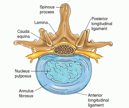
Figure 18-1 ▪ Axial view of the lumbar spine, showing an intervertebral disc, the contents of the spinal canal, and the elements of the posterior bony arch.


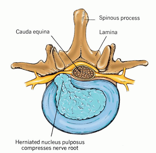
Figure 18-2 ▪ Ruptured vertebral disc. (From: Chaffee, E. E., & Lytle, I. M. [1980]. Basic physiology and anatomy. Philadelphia: J. B. Lippincott.)


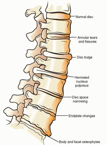
Figure 18-3 ▪ The degenerative cascade. (From: Jeong, G. K., & Bendo, J. A. [2004]. Spinal disorders in the elderly. Clinical orthopaedics and related research, 425, 110-125.)


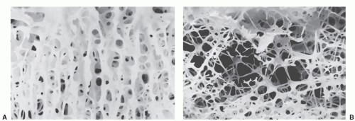
Figure 18-4 ▪ Normal bone (A). Osteoporotic bone (B). (From: Gill, S. S., & Einhorn, T. A. [2004]. Metabolic bone disease of the adult and pediatric spine. In: Frymoyer, J. W. & Wiesel, S. W. (Eds.), The adult and pediatric spine. (3rd ed., pp. 121-140). Philadelphia: Lippincott Williams & Wilkins.)


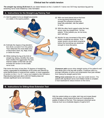
Figure 18-5 ▪ Straight-leg raising (SLR) test. (A) Instructions for SLR when the patient is lying down. (B) Instructions for SLR when the patient is sitting. (From: Bigos, S., Bowyer, O., Braen, G., Brown, K. C., Deyo, R. A., Haldeman, S., … Weinstein, J [1994]. Acute low back problems in adults. Clinical Practice Guideline, Quick Reference Guide Number. 14. Rockville, MD: U.S. Department of Health and Human Services, Public Health Service, Agency for Health Care Policy and Research, AHCPR Pub. No. 95-0643.)


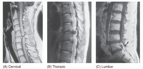
Figure 18-6 ▪ MRI views of herniated discs. (A) cervical. Midsaggital T1-weighted MRI shows herniated disc (arrow) at C5-C6 producing compression of the spinal cord. (B) thoracic. Midsagittal T2-weighted MRI shows hypointense herniated disc (arrow) in midthoracic region resulting in compression of the spinal cord. (C) lumbar. Midsagittal postcontrast T1-weighted MRI of the same patient shows that herniated disc (arrow) is not attached to any parent disc. There is peripheral disc enhancement. (From: Castillo, M. [1999]. Neuroradiology companion [2nd ed.]. Philadelphia: Lippincott-Raven.)


Neck Pain

Neck pain is also a symptom in many conditions and disease processes; proper diagnosis is critical for treatment and a satisfactory outcome. Neck pain diagnosis and initial management generally follow the same algorithm as low back pain. Initially, the patient is seen and evaluated by the primary care physician. As with low back pain, “red flags” are ruled out. A detailed history is obtained, focusing on the neck and upper extremities. Neurological screening examination will focus on the strength, sensation, and reflexes of the upper extremities. Finger fine motor and dexterity as well as the Hoffmann’s reflex and Babinski’s reflex are also evaluated for any signs of myelopathy.20, 21

As with low back pain, in the absence of signs of serious problems, there is no need for special diagnostic studies. The majority of patients has musculoskeletal strain and recovers spontaneously within 4 to 6 weeks. However, if a serious underlying condition is found or if there is rapid progression of neurological deficits, urgent diagnostic studies and definitive care should be sought.

Initial management, education and reassurance, patient comfort, and activity alterations follow the same general guidelines as low back pain.


HERNIATED INTERVERTEBRAL DISCS

Herniation of cervical and lumbar intervertebral discs can cause significant pain and alterations in functional activity. The lumbar spine is most frequently affected by a herniated disc followed by the cervical spine. Thoracic herniation is uncommon. Patients may complain of arm or leg pain and be diagnosed with radiculopathy (pain and/or paresthesias in the distribution of a nerve root).

Men suffer from intervertebral disc herniation much more frequently than women. Most patients with disc disease are between 30 and 50 years old. About 90% to 95% of lumbar herniation occurs at the L-4 to L-5 or L-5 to S-1 levels.22 When the cervical region is involved, the most common levels are C-6 to C-7 (C-7 radiculopathy) and then C-5 to C-6 (C-6 radiculopathy).23 Multiple herniation sites occur in 10% of patients (Fig. 18-6).


Etiology

Trauma accounts for approximately 50% of all disc herniation. Examples of traumatic incidents include lifting heavy objects while in a flexed position (most common), slipping, falling on the buttocks or back, and suppressing a sneeze in the lumbar area. In the cervical area, flexion/extension injury of the neck can cause traumatic herniation. In some patients, no history of trauma can be identified. The cumulative result of repeated minor injuries is a chronic condition that places the patient at high risk for herniation.24 The herniation syndrome also occurs in association with other degenerative processes, such as osteoarthritis or ankylosing spondylitis (Marie-Strümpell’s spondylitis) and spinal stenosis. Patients with congenital anomalies, such as scoliosis, appear to be predisposed to disc injury because of malalignment of the vertebral column.


Signs and Symptoms by Location


Lumbar Area

More than 90% of all clinically significant lower extremity radiculopathy is due to disc herniation at the L-4 to L-5 or L-5 to S-1 level. The signs and symptoms of herniated lumbar discs are grouped in the following general categories: pain, postural deformity, motor changes, sensory changes, alterations of reflexes, and nerve tension signs.25


Pain. Pain is the first and most characteristic symptom of a herniated disc. Pain varies in terms of quality, radiation, severity, and timing. Most describe the leg pain as sharp, shooting, burning, stabbing, or aching. The term sciatica is sometimes used to describe a syndrome of lumbar back pain that spreads down one leg to the ankle and is intensified by coughing and sneezing. The nerve roots L-4, L-5, S-1, S-2, and S-3 give rise to the sciatic nerve. The pain in the buttock is described as deep, aching, or gnawing. The intensity of pain is influenced by leg position.

Pain from a herniated disc is aggravated and intensified by coughing, sneezing, straining, stooping, standing, sitting, blowing the nose, spasms of the paravertebral muscles, and any jarring movement while walking or riding. The character of pain ranges from mild discomfort to excruciating agony. Prolonged sitting is particularly painful. Pain may be alleviated by changing positions frequently, lying on the back with knees flexed and a small pillow at the head. Other patients prefer the lateral recumbent position, lying on the unaffected side with the knee flexed on the affected side.

Postural Deformity. The patient walks cautiously, bearing as little weight as possible on the affected side. The gait may be described as stiff or antalgic, and movement is deliberate to prevent jarring. Climbing stairs is particularly painful.

Motor Deficits. Slight motor weakness may be experienced, although major weakness does occur. Motor weakness can be difficult to evaluate because of the defensive reaction precipitated by pain. Weakness may be evident on plantarflexion or dorsiflexion of the foot (L-4) and dorsiflexion of the great toe (L-5, extensor hallucis longus [EHL]) and occasionally of the hamstring and quadriceps muscles. Weakness with plantarflexion correlates to the S-1 level. Foot drop occurs with significant dorsiflexion weakness. Atrophy of the affected muscles may develop, although it is not a common finding and can be minimal.

Cauda equina syndrome (CES) is caused by compression of the spinal nerves in the spinal canal at the level of the cauda equina. It can also be caused by any space-occupying lesion at the level from approximately L-2 to S-1. Symptoms include often bilateral progressive leg or foot weakness, urinary retention or incontinence, bowel incontinence, and saddle anesthesia. CES is rare; however, severe neurological compromise from a large herniated disc represents a surgical emergency.

Sensory Deficits. The most common sensory impairments from root compression are paresthesias and numbness, particularly of the leg and foot. Note the specific areas of decreased sensation in the foot and leg using the pinprick method of sensory testing. Assess if the sensory deficit is dermatomal, that is, if it follows the distribution of a single nerve. Tenderness may be noted over the L-5 and S-1 vertebral spinous processes and along the tracking of the sciatic nerve.

Alterations of Reflexes. Depending on the level of the disc herniation, knee or ankle reflexes may be absent or diminished and there may be a negative Babinski’s reflex because of the lower motor neuron component.

Nerve Root Tension Signs. Other signs found on the clinical examination commonly associated with evaluating patients with possible lumbar herniated disc disease include the straight-leg raising test and crossed straight leg test. The straight-leg raising (SLR) test (also called Lasègue’s sign) is helpful in determining limitations of lower limb range of motion due to pain and the location of the pain during testing (Fig. 18-5). Normally, it is possible when lying on the back to move the straightened leg about 90 degrees with only some slight discomfort in the hamstring muscles. The sciatic nerve becomes stretched with movement and creates traction on the proximal nerve roots. Traction and stretching of the nerve roots begin when the leg is at a 30- to 40-degree angle. In the patient with a herniated low back disc, the stretching of the sciatic nerve during passive, straight-leg raising creates traction on the irritated nerve roots, thereby producing severe pain. Note the angle degree at which the patient experiences pain. This may be as little as 20 to 30 degrees. Repeating the Lasègue’s maneuver on the unaffected leg produces pain of decreased severity on the contralateral side, also referred to as the crossed straight-leg test.


Specific Lumbar Levels

The most common sites for lumbar disc herniation are the L-4 to L-5 and L-5 to S-1 levels, and in that order. Lesions at the L-3 to L-4 level are rare. Each level presents a characteristic syndrome of symptoms that is distinct from that of other levels (Figs. 18-7, 18-8, 18-9, 18-10; Table 18-3). Note that multiple levels of disc herniation can exist.


L-3 to L-4 Level. Pain (L-4 radiculopathy) is located in the lower back, hip, posterolateral thigh, and anterior leg. Paresthesias are experienced in the middle section of the anterior thigh. Weakness may be noted in the quadriceps muscles, which may also demonstrate atrophic changes. The knee-jerk reflex is diminished.

L-4 to L-5 Level. Pain (L-5 radiculopathy) is perceived in the hip, groin, posterolateral thigh, lateral calf, dorsal surface of the foot, and first or second and third toes. Paresthesias may be experienced over the lateral leg and web of the great toe. There is tenderness at the femoral head and lateral gluteal region. There may be weakness with dorsiflexion of the great toe and foot. Foot drop can occur. Functionally, the foot cannot be picked up at the ankle, making walking difficult and putting the patient at risk for falls. The patient has difficulty walking on the heels. Atrophy, if present, is minor. Reflexes are usually not diminished.

L-5 to S-1 Level. Pain (S-1 radiculopathy) is perceived in the midgluteal, posterior thigh, and calf regions down to the heel and the outer surface of the foot on the side of the fourth and fifth toes. Paresthesias are found in the posterior calf and in the lateral heel, foot, and toe. Tenderness is especially apparent in the area about the sacroiliac joint. Weakness in plantarflexion
of the foot may be evident. The patient has difficulty walking on the toes. The hamstring muscles may also show signs of weakness. If atrophy is present, the gastrocnemius and the soleus muscles are affected. The ankle-jerk reflex is often diminished or absent.


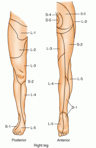
Figure 18-7 ▪ Dermatomes of the leg.


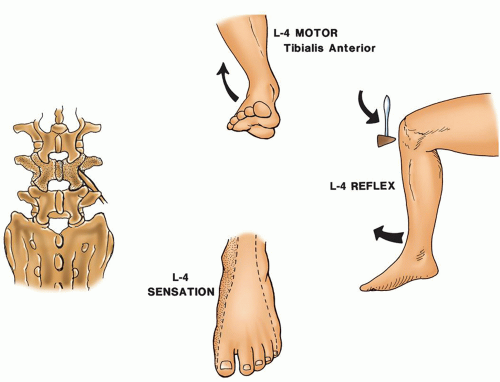
Figure 18-8 ▪ L-4 neurologic level. (From: Scherping, S.C. [2004]. History and physical examination. In: Frymoyer, J. W. & Wiesel, S. W. [Eds.]. The adult and pediatric spine [3rd ed., pp. 49-68]. Philadelphia: Lippincott Williams & Wilkins.)


TABLE 18-3 NEUROLOGICAL LEVELS OF THE LOWER EXTREMITIES25


ROOT


REFLEX


MUSCLES


SENSATION


L-2


Iliopsoas


Anterior thigh, groin


L-3


Patellar


Quadriceps


Anterior and lateral thigh


L-4


Patellar


Anterior tibialis


Medial leg and medial foot; medial malleolus


L-5


Extensor hallucis longus, hip abductors


Lateral leg and dorsum of foot; first web space


S-1


Achilles


Peroneus longus and brevis, gastroc soleus


Lateral foot; little toe


Remission of Pain. Pain associated with herniated disc disease can be recurrent. Patients may also present with a history of one or more similar episodes. Between acute exacerbations, pain may be completely absent or substantially diminished. The time between acute exacerbations is highly variable, with some having only weeks between episodes and some having complete relief with no recurrence.


Cervical Area

The cervical region of the spine is also prone to disc degeneration and spondylosis, as well as trauma, which predisposes the affected person to a wider range of pathologic conditions.


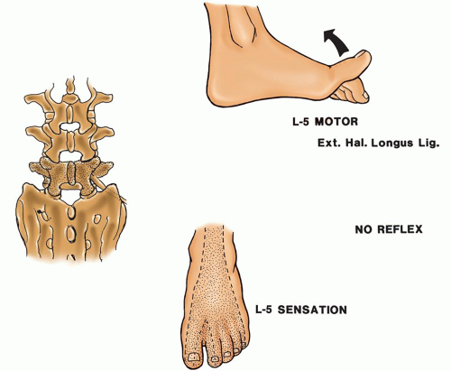
Figure 18-9 ▪ L-5 neurologic level. (From: Scherping, S.C. [2004]. History and physical examination. In: Frymoyer, J. W. & Wiesel, S. W. [Eds.]. The adult and pediatric spine [3rd ed., pp. 49-68]. Philadelphia: Lippincott Williams & Wilkins.)

The most common cause of radiculopathy is decreased disc height and the associated degenerative changes (spondylosis) of the lower cervical region. Unlike the lumbar spine, disc herniation is only responsible for 20% to 25% of cases of radiculopathy, while degenerative changes account for 70% to 75% of cases.23 Symptoms usually develop without any apparent precipitating event. However, symptoms may follow trauma, such as whiplash or hyperextension injuries. On radiographic investigation, there is disc degeneration and spondylosis, exhibited as degenerative changes in the intervertebral and facet joints. These degenerative changes lead to neuroforaminal narrowing and subsequent nerve compression. The most common sites of cervical herniation are at the C-5 to C-6 and C-6
to C-7 levels, with pain along the affected sensory dermatomes. Anatomically, there is little free room within the spinal canal to accommodate any extraneous material. The cervical spinal cord is firmly positioned by the ligamenta denticulata. Disc protrusion in the cervical area can result not only in root compression but also in cord compression because of the lack of free space. The particular presenting symptoms depend on the anatomic point and degree of disc protrusion.


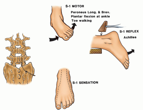
Figure 18-10 ▪ S-1 neurologic level. (From: Scherping, S.C. [2004]. History and physical examination. In: Frymoyer, J.W. & Wiesel, S.W. [Eds.]. The adult and pediatric spine [3rd ed., pp. 49-68]. Philadelphia: Lippincott Williams & Wilkins.)

Jul 14, 2016 | Posted by in NURSING | Comments Off on Back Pain and Spinal Disorders

Full access? Get Clinical Tree

Get Clinical Tree app for offline access