7 Anatomy and Physiology
General Terminology
Important terms of direction to review include superior (above), inferior (below), anterior (facing forward), posterior (toward the back), medial (toward the midline), and lateral (away from the midline or toward the sides). Proximal and distal are terms of direction usually used in reference to limbs. Proximal means closer to the point of attachment, and distal refers to further away from the point of attachment. Figure 7-1 depicts the directional terms.
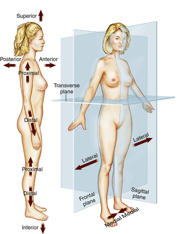
Figure 7-1 Planes and directions of the body.
(From Patton KT, Thibodeau GA: Anatomy and physiology, ed 7, St Louis, 2010, Mosby.)
Additional useful terminology is defined later in this chapter.
Histology
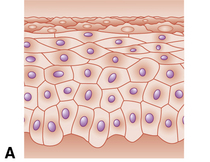
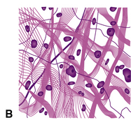
Histology is the study of tissues. A tissue is a group of cells that act together to perform specific functions. The four fundamental tissues are epithelial, connective, muscle, and nerve tissues (Figure 7-2). Epithelial cells cover, line, and protect the body and its internal organs. Connective tissue is the framework of the body, providing support and structure for the organs. Nerve tissue is composed of neurons and connective tissue cells that are referred to as neuroglia. Muscle tissues have the ability to contract or shorten. Muscle tissue is classified as voluntary muscle (skeletal muscles) or involuntary muscle (smooth muscle and cardiac muscle tissue).

Skin
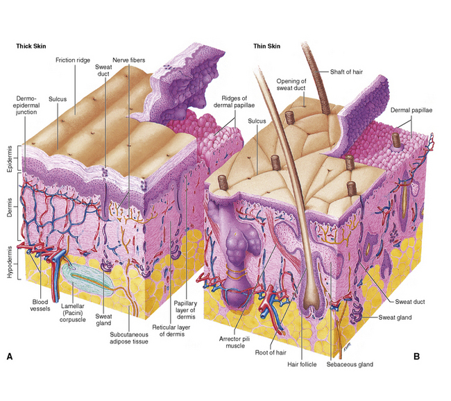
The appendages of the skin include hair and nails. Both are composed of a strong protein called keratin. Skin structure is illustrated in Figure 7-3. Hair, nails, and skin may show changes in disease that may be used in the diagnosis of clinical conditions. For example, skin cancer is a clinical condition that is associated with the skin.
Skeletal System
The axial skeleton (Figure 7-4) consists of the 28 bones of the skull. These are separated into the 14 facial bones and the 14 bones of the cranium. The facial bones are two nasal bones, two maxillary bones, two zygomatic bones, one mandible (the only moveable bone of the skull), two palatine bones, one vomer, two lacrimal bones, and two inferior nasal conchae. The bones of the cranium are the single occipital, frontal, ethmoid, and sphenoid and the paired parietal, temporal, and ossicles of the ear (malleus, incus, and stapes).
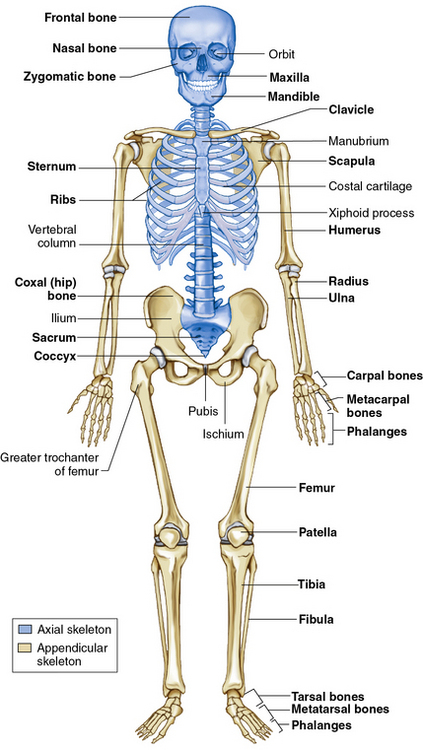
Figure 7-4 Anterior view of the skeleton.
(From Patton KT, Thibodeau GA: Anatomy and physiology, ed 7, St Louis, 2010, Mosby.)
The axial skeleton also has 33 bones of the vertebral column, as depicted in Figure 7-5. There are seven cervical vertebrae, 12 thoracic vertebrae, five lumbar vertebrae, five sacral vertebrae (fused to form the sacrum), and the coccygeal vertebrae (known as the tailbone). The final portion of the axial skeleton consists of the bones of the thorax, the sternum, and the 12 pairs of ribs.
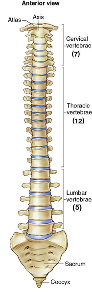
Figure 7-5 Anterior view of the vertebral column.
(From Patton KT, Thibodeau GA: Anatomy and physiology, ed 7, St Louis, 2010, Mosby.)
The appendicular skeleton (see Figure 7-4) includes the girdles and the limbs. The upper portion consists of the pectoral or shoulder girdle, the clavicle and scapula, and the upper extremity. The bones of the arm are the humerus, the radius and ulna, the carpals (wrist bones), the metacarpals (bones of the hand), and the phalanges (bones of the fingers). The lower portion of the appendicular skeleton is made up of the pelvic girdle or os coxae. Each of the os coxae consists of a fused ilium, ischium, and pubis. Bones of the lower extremity include the femur (thighbone), the tibia and fibula, the tarsals (ankle bones), the metatarsals (bones of the foot), and the phalanges.
Stay updated, free articles. Join our Telegram channel

Full access? Get Clinical Tree


