The Endocrine System
Objectives
1. Identify the location of each endocrine gland.
3. Summarize the effects of the thyroid hormones.
4. Describe common diagnostic tests for the endocrine system.
1. Assess for specific age-related changes of the endocrine system in an elderly patient.
3. Perform a focused assessment on a patient who may have an endocrine disorder.
Key Terms
adenohypophysis (ă-DĔN-ŏ-hī-PŎF-ă-sĭs, p. 823)
adrenocorticotropic hormone (ă-DRĔN-ŏ-KŎR-tĭ-kō-TRŌ-pĭk, p. 823)
endocrine (ĔN-dŏ-krĭn, p. 821)
exocrine (Ĕk-sŏ-krĭn, p. 821)
fructosamine assay (p. 827)
glucocorticoids (glū-kō-KŎR-tĭ-kŏydz, p. 821)
glucose tolerance test (p. 827)
hemoglobin A1c (A1C) (HĒ-mō-glō-bĭn, p. 827)
hormones (HŎR-mōnz, p. 822)
hypersecretion (hī-pĕr-SĔ-KRĒ-shŭn, p. 823)
hyposecretion (hī-pō-SĔ-KRĒ-shŭn, p. 823)
insulin (ĬN-sū-lĭn, p. 822)
mineralocorticoids (mĭn-ĕr-ăl-ō-KŎR-tĭ-kŏydz, p. 821)
negative feedback (p. 823)
parathormone (păr-ă-THŎR-mōn, p. 821)
pressor (p. 821)
target cells (p. 822)
target tissues (p. 822)
thyrocalcitonin (thī-rō-KĂL-sĭ-TŌ-nĭn, p. 819)
thyroid panel (THĪ-rŏyd, p. 827)
thyroxine (THĪ-rŏk-sĭn, p. 819)
triiodothyronine (trī-ī-ō-dō-THĪ-rō-nĕn, p. 819)
 http://evolve.elsevier.com/deWit/medsurg
Overview of anatomy and physiology of the endocrine system
What are the organs and structures of the endocrine system?
What are the functions of the endocrine system?
• Alter chemical reactions and control the rate at which chemical activities take place within cells.
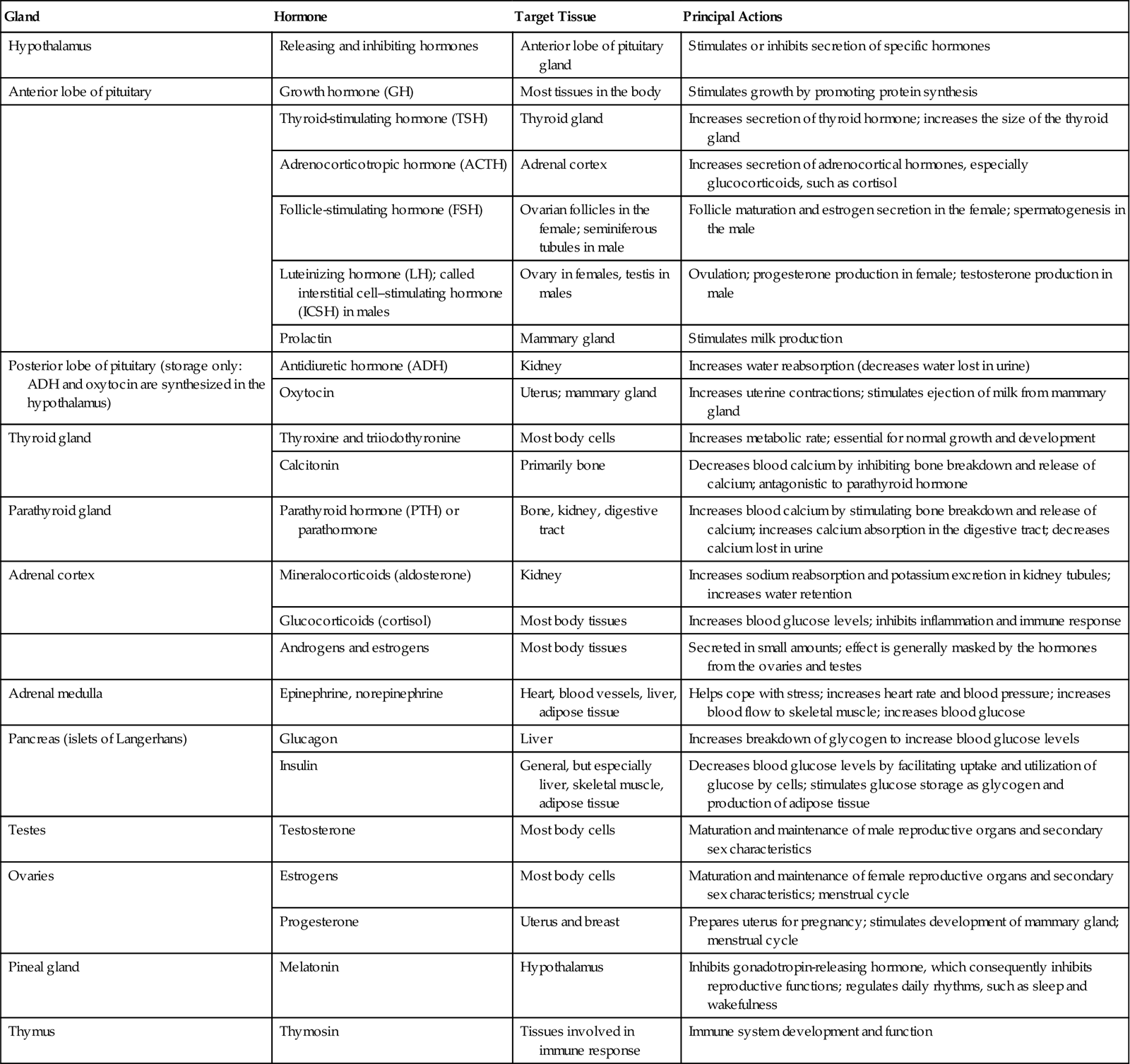
• Activate a particular mechanism in the cell, such as the system that controls cellular growth and reproduction. The hormones produced by the endocrine system, the target organs on which they act, and the principal actions of each hormone are presented in Table 36-1.
Table 36-1
The Principal Endocrine Glands and Their Hormones
| Gland | Hormone | Target Tissue | Principal Actions |
| Hypothalamus | Releasing and inhibiting hormones | Anterior lobe of pituitary gland | Stimulates or inhibits secretion of specific hormones |
| Anterior lobe of pituitary | Growth hormone (GH) | Most tissues in the body | Stimulates growth by promoting protein synthesis |
| Thyroid-stimulating hormone (TSH) | Thyroid gland | Increases secretion of thyroid hormone; increases the size of the thyroid gland | |
| Adrenocorticotropic hormone (ACTH) | Adrenal cortex | Increases secretion of adrenocortical hormones, especially glucocorticoids, such as cortisol | |
| Follicle-stimulating hormone (FSH) | Ovarian follicles in the female; seminiferous tubules in male | Follicle maturation and estrogen secretion in the female; spermatogenesis in the male | |
| Luteinizing hormone (LH); called interstitial cell–stimulating hormone (ICSH) in males | Ovary in females, testis in males | Ovulation; progesterone production in female; testosterone production in male | |
| Prolactin | Mammary gland | Stimulates milk production | |
| Posterior lobe of pituitary (storage only: ADH and oxytocin are synthesized in the hypothalamus) | Antidiuretic hormone (ADH) | Kidney | Increases water reabsorption (decreases water lost in urine) |
| Oxytocin | Uterus; mammary gland | Increases uterine contractions; stimulates ejection of milk from mammary gland | |
| Thyroid gland | Thyroxine and triiodothyronine | Most body cells | Increases metabolic rate; essential for normal growth and development |
| Calcitonin | Primarily bone | Decreases blood calcium by inhibiting bone breakdown and release of calcium; antagonistic to parathyroid hormone | |
| Parathyroid gland | Parathyroid hormone (PTH) or parathormone | Bone, kidney, digestive tract | Increases blood calcium by stimulating bone breakdown and release of calcium; increases calcium absorption in the digestive tract; decreases calcium lost in urine |
| Adrenal cortex | Mineralocorticoids (aldosterone) | Kidney | Increases sodium reabsorption and potassium excretion in kidney tubules; increases water retention |
| Glucocorticoids (cortisol) | Most body tissues | Increases blood glucose levels; inhibits inflammation and immune response | |
| Androgens and estrogens | Most body tissues | Secreted in small amounts; effect is generally masked by the hormones from the ovaries and testes | |
| Adrenal medulla | Epinephrine, norepinephrine | Heart, blood vessels, liver, adipose tissue | Helps cope with stress; increases heart rate and blood pressure; increases blood flow to skeletal muscle; increases blood glucose |
| Pancreas (islets of Langerhans) | Glucagon | Liver | Increases breakdown of glycogen to increase blood glucose levels |
| Insulin | General, but especially liver, skeletal muscle, adipose tissue | Decreases blood glucose levels by facilitating uptake and utilization of glucose by cells; stimulates glucose storage as glycogen and production of adipose tissue | |
| Testes | Testosterone | Most body cells | Maturation and maintenance of male reproductive organs and secondary sex characteristics |
| Ovaries | Estrogens | Most body cells | Maturation and maintenance of female reproductive organs and secondary sex characteristics; menstrual cycle |
| Progesterone | Uterus and breast | Prepares uterus for pregnancy; stimulates development of mammary gland; menstrual cycle | |
| Pineal gland | Melatonin | Hypothalamus | Inhibits gonadotropin-releasing hormone, which consequently inhibits reproductive functions; regulates daily rhythms, such as sleep and wakefulness |
| Thymus | Thymosin | Tissues involved in immune response | Immune system development and function |
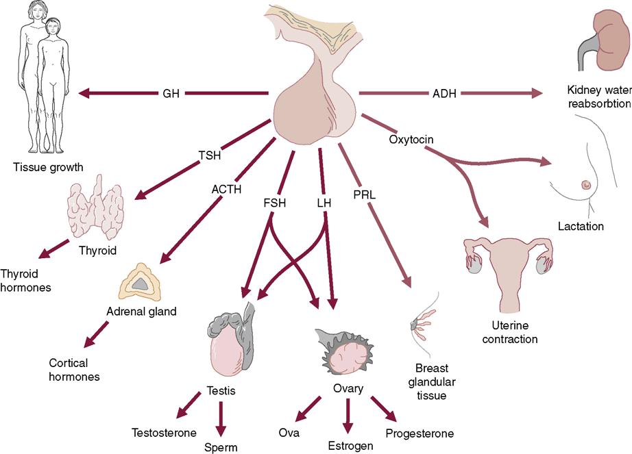
What are the effects of the pituitary hormones?
What are the effects of the thyroid hormones?
What are the functions of the parathyroid glands?
What are the functions of the hormones secreted by the adrenal glands?
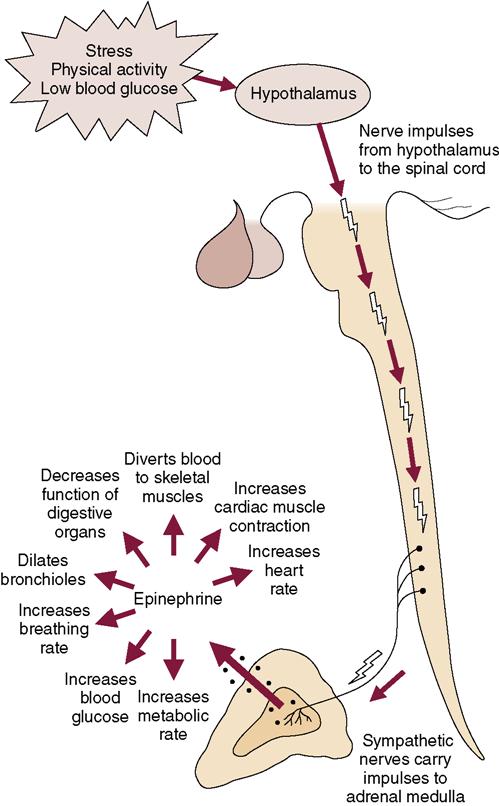
• Epinephrine prepares the body to meet stress or emergency situations and prevents hypoglycemia (Figure 36-2). Norepinephrine functions as a pressor (causing blood vessel constriction) hormone to maintain blood pressure.
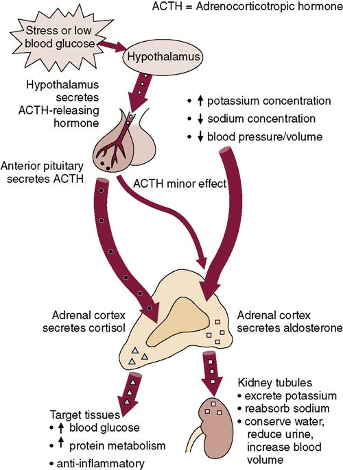
• The two major types of hormones secreted by the adrenal cortex are the mineralocorticoids and the glucocorticoids (Figure 36-3).
• Both aldosterone and cortisol are controlled by adrenocorticotropic hormone (ACTH)–releasing hormone from the hypothalamus and ACTH secreted by the anterior pituitary (see Figure 36-3).
What is the hormonal function of the pancreas?
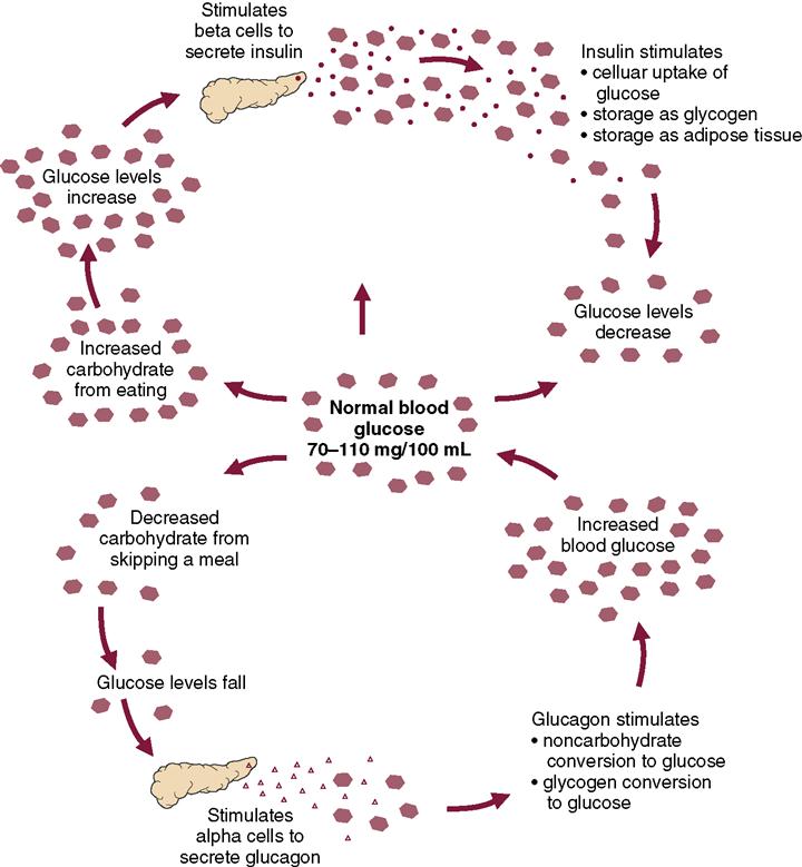
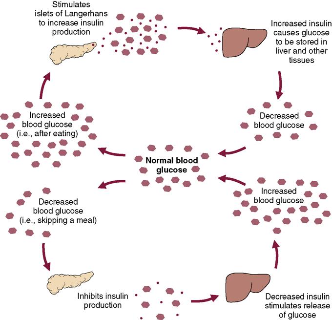
• The beta cells are responsible for producing and secreting insulin. Insulin is needed for the cells of the body to be able to use glucose as fuel (Figures 36-4 and 36-5).
What are the effects of aging on the endocrine system?
The endocrine system
The endocrine system regulates metabolism, growth and development, and sexual function and reproductive processes. A primary function of the endocrine system is to synthesize and release hormones directly into the bloodstream and the body fluids. The cells and tissues that are affected by a specific hormone are called its target cells or target tissues.
Some of the endocrine hormones, such as the thyroid hormones, affect practically every cell in the body. Others, such as the sex hormones, exert their special effects on only one kind of organ. Moreover, hormones from one endocrine gland can affect another endocrine gland. The pituitary, for example, secretes several different kinds of hormones that affect other endocrine glands. For this reason, the pituitary gland is often referred to as the “master gland” of the body.
The endocrine system and the nervous system are the two major control systems of the body, and their regulatory functions are interrelated. However, the endocrine system typically controls body processes that occur slowly, such as cell growth, whereas the nervous system controls body processes that occur more rapidly, such as breathing and body movement.
The secretion of a particular hormone normally depends on the need. If an endocrine gland receives a message that its particular hormone is in short supply, it will synthesize and release more of that hormone. If, on the other hand, the hormonal need of a target tissue is being satisfied, production or secretion of the hormone will be inhibited—a concept known as negative feedback.
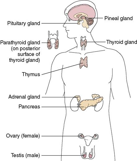
Some glands, such as the adrenal medulla and posterior pituitary, receive their information about hormone levels in the body directly, and respond only to stimulation of nerve endings within the glands themselves. However, the posterior pituitary gland indirectly receives notice to either release or inhibit hormones: stimulation comes by way of the hypothalamus and the anterior lobe of the pituitary (the adenohypophysis). The hypothalamus contains special nerve endings that produce releasing and inhibiting hormones; these hormones are then absorbed into capillaries of a portal system that transports the hormones to the adenohypophysis (the anterior lobe of the pituitary). Thus the hypothalamus controls the secretion of hormones from the pituitary. The pituitary, in turn, controls the release or inhibition of hormones from other glands. Many of the hormones of the anterior pituitary are “tropic” hormones; that is, they tend to cause a change in the endocrine gland that is the target of the specific pituitary hormone. An example is adrenocorticotropic hormone (ACTH), which acts on the adrenal cortex. (If you break down this term, you can easily see that the components of adrenal–cortex–tropic tell you exactly where or what type of hormone this is and where it comes from.) The major endocrine glands can be found in Figure 36-6; see Table 36-1 for the various tropic hormones and target tissues.
Stay updated, free articles. Join our Telegram channel

Full access? Get Clinical Tree


