Fetal Development
Objectives
1. Define each key term listed.
2. Describe the process of gametogenesis in human reproduction.
3. Explain human fertilization and implantation.
4. Describe embryonic development.
5. Describe fetal development and the maturation of body systems.
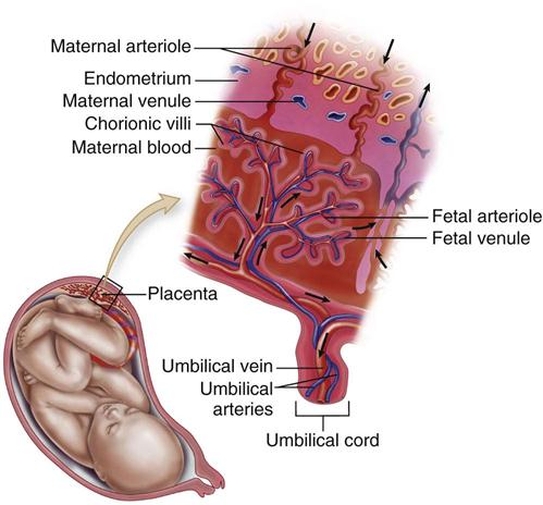
6. Describe the development and functions of the placenta, the umbilical cord, and the amniotic fluid.
7. Compare fetal circulation to circulation after birth.
8. Explain the similarities and differences in the two types of twins.
Key Terms
age of viability (p. 37)
amniotic sac (p. 35)
autosomes (p. 31)
chorion (KŌ-rē-ŏn, p. 34)
decidua (dĕ-SĬD-yū-ă, p. 34)
diploid (DĬP-loid, p. 31)
dizygotic (DZ) (dī-zī-GŎT-ĭk, p. 41)
fertilization (p. 32)
gametogenesis (găm-ĕ-tō-JĔN-ĕ-ĭs, p. 32)
germ layers (p. 34)
haploid (HĂP-loid, p. 31)
monozygotic (MZ) (mŏn-ō-zī-GŎT-ĭk, p. 41)
oogenesis (ō-ō-JĔN-ĕ-sĭs, p. 31)
placenta (plă-SĔN-tă, p. 38)
spermatogenesis (spŭr-mă-tō-JĔN-ĕ-sĭs, p. 31)
teratogens (TĔR-ă-tō-jĕnz, p. 31)
Wharton’s jelly (p. 39)
 http://evolve.elsevier.com/Leifer
The human body contains many millions of cells at birth, but life begins with a single cell created by the fusion of a sperm with an ovum. Deoxyribonucleic acid (DNA) programs a genetic code into the nucleus of the cell; the nucleus controls the development and function of the cell. Defects in the DNA code can result in inherited disorders. The genes and chromosomes contained within the DNA determine the uniqueness of the traits and features of the developing person.
Normal human chromosomes begin in pairs, one supplied by the mother and the other by the father. Each body cell contains 46 chromosomes, made up of 22 pairs of autosomes (body chromosomes) and 1 pair of sex chromosomes that determine the sex of the fetus. Each chromosome contains genes that involve heredity. Cell division then occurs, which is the basis of human growth and regeneration.
Biological development is not isolated. It is influenced by the external environment, such as maternal drug use (teratogens that cause damage to growing cells include some prescribed medications), maternal undernutrition, or smoking by the mother, and it is now known that sounds such as music are heard by the fetus and are recognized by the newborn. All these factors influence prenatal growth and development. Early prenatal care is essential to the optimum outcome of the pregnancy (see Chapter 4).
Cell Division and Gametogenesis
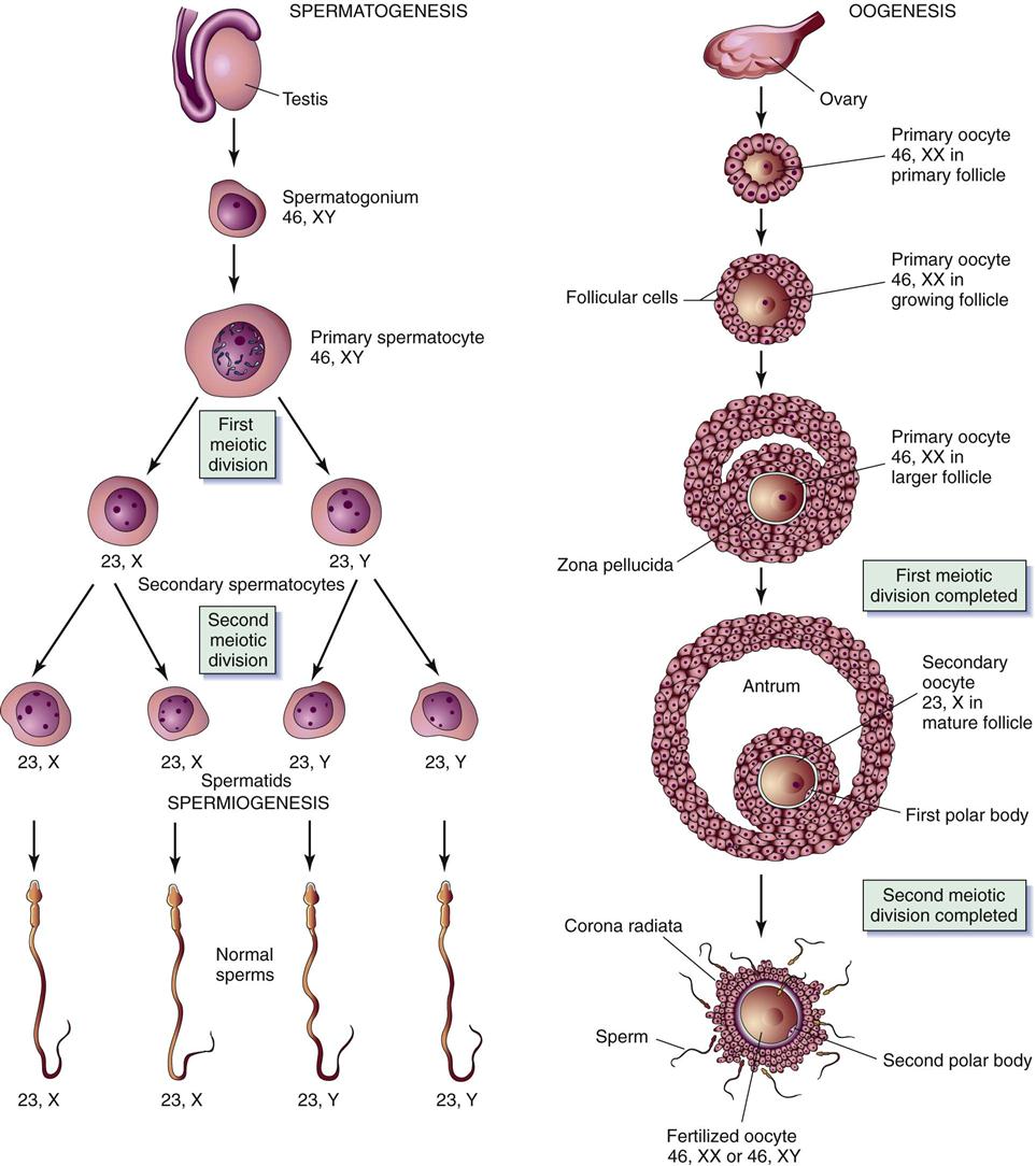
The division of a cell begins in its nucleus, which contains the gene-bearing chromosomes. The two types of cell division are mitosis and meiosis. Mitosis is a continuous process by which the body grows and develops and dead body cells are replaced. In this type of cell division, each daughter cell contains the same number of chromosomes as the parent cell. The 46 chromosomes in a body cell are called the diploid number of chromosomes. The process of mitosis in the sperm is called spermatogenesis, and in the ovum it is called oogenesis.
Meiosis is a different type of cell division in which the reproductive cells undergo two sequential divisions. During meiosis the number of chromosomes in each cell is reduced by half to 23 chromosomes per cell, each including only one sex chromosome. This is called the haploid number of chromosomes. This process is completed in the sperm before it travels toward the fallopian tube and in the ovum if it is fertilized after ovulation. At the moment of fertilization (when the sperm and the ovum unite), the new cell contains 23 chromosomes from the sperm and 23 chromosomes from the ovum, thus returning to the diploid number of chromosomes (46); traits are therefore inherited from both the mother and the father. The formation of gametes by this type of cell division is called gametogenesis (Figure 3-1).
Fertilization
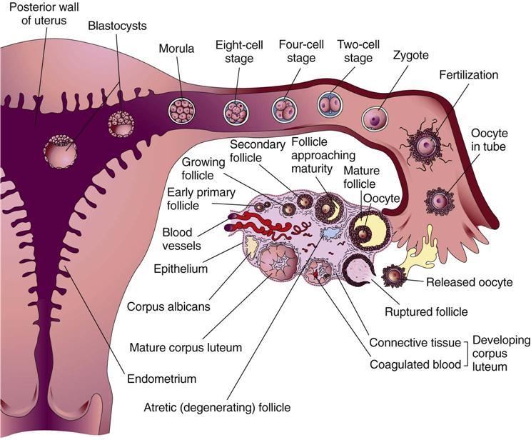
Fertilization occurs when a sperm penetrates an ovum and unites with it, restoring the total number of chromosomes to 46. It normally occurs in the outer third of the fallopian tube, near the ovary (Figure 3-2). The sperm pass through the cervix and the uterus and into the fallopian tubes by means of the flagellar (whiplike) activity of their tails and can reach the fallopian tubes within 5 minutes after coitus. As soon as fertilization occurs, a chemical change in the membrane around the fertilized ovum prevents penetration by another sperm.
The time during which fertilization can occur is brief because of the short life span of mature gametes. The ovum is estimated to survive for up to 24 hours after ovulation. The sperm remains capable of fertilizing the ovum for up to 5 days after being ejaculated into the area of the cervix.
 Nursing Tip
During sexual counseling the nurse should emphasize that the survival time of sperm ejaculated into the area of the cervix may be up to 5 days and that pregnancy can occur with intercourse as long as 5 days before ovulation.
Sex Determination
The sex of human offspring is determined at fertilization. The ovum always contributes an X chromosome (gamete), whereas the sperm can carry an X or a Y chromosome (gamete). When a sperm carrying the X chromosome fertilizes the X-bearing ovum, a female child (XX) results. When a Y-bearing sperm fertilizes the ovum, a male child (XY) is produced (Figure 3-3).
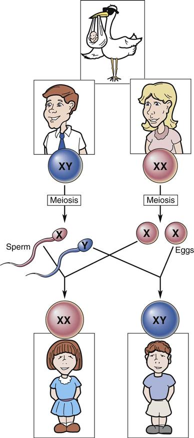
Because sperm can carry either an X or a Y chromosome, the male partner determines the sex of the child. However, the pH of the female reproductive tract and the estrogen levels of the woman’s body affect the survival rate of the X- and Y-bearing sperm as well as the speed of their movement through the cervix and the fallopian tubes. Thus the female has some influence on which sperm fertilizes the mature ovum.
 Nursing Tip
Both mother and father influence the sex of their offspring, although the father contributes the actual sex chromosome. This fact may reduce blame in some cultures when the child is not of the desired sex.
Inheritance
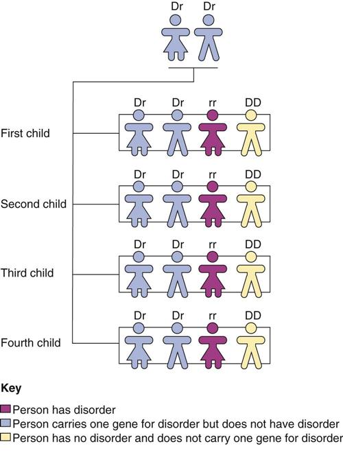
Each gene (a segment of the DNA chain) is coded for inheritance. The coded information carried by the DNA in the gene is responsible for individual traits, such as eye and hair color, facial features, and body shape. Genes carry instructions for dominant and recessive traits. Dominant traits usually overpower recessive traits and are passed on to the offspring. If only one parent carries a dominant trait, an average of 50% of the offspring will have (and thus display) that dominant trait. If each parent carries a recessive trait, there is a higher chance that one of the offspring will display that trait (Figure 3-4).
Tubal Transport of the Zygote
The zygote is the cell formed by the union of the sperm and the ovum, and it is transported through the fallopian tube and into the uterus. Fertilization normally occurs in the outer third of the fallopian tube. During transport through the fallopian tube, the zygote undergoes rapid mitotic division, or cleavage. Cleavage begins with two cells, which subdivide into four and then eight cells to form the blastomere. The size of the zygote does not increase; rather, the individual cells become smaller as they divide and eventually form a solid ball called the morula (see Figure 3-2).
The morula enters the uterus on the third day and floats there for another 2 to 4 days. The cells form a cavity, and two distinct layers evolve. The inner layer is a solid mass of cells called the blastocyst (see Figure 3-2), which develops into the embryo and the embryonic membranes. The outer layer of cells, called the trophoblast, develops into an embryonic membrane, the chorion. Occasionally the zygote does not move through the fallopian tube and instead becomes implanted in the lining of the tube, resulting in a tubal ectopic pregnancy (see Chapter 5).
Implantation of the Zygote
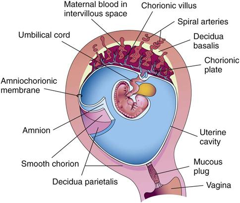
The zygote usually implants in the upper section of the posterior uterine wall. The cells burrow into the prepared lining of the uterus, called the endometrium. The endometrium is now called the decidua; the area under the blastocyst is called the decidua basalis and gives rise to the maternal part of the placenta (Figure 3-5).
Development
Cell Differentiation
During the week between fertilization and implantation, the cells within a zygote are identical to one another. After implantation the cells begin to differentiate and develop special functions. The chorion, the amnion, the yolk sac, and the primary germ layers appear.
Chorion
The chorion develops from the trophoblast (outer layer of embryonic cells) and envelops the amnion, embryo, and yolk sac. It is a thick membrane with fingerlike projections called villi on its outermost surface. The villi immediately below the embryo extend into the decidua basalis on the uterine wall and form the embryonic or fetal portion of the placenta (Figure 3-6).
Amnion
The amnion is the second membrane; it is a thin structure that envelops and protects the embryo. It forms the boundaries of the amniotic cavity, and its outer aspect meets the inner aspect of the chorion.
The chorion and the amnion together form an amniotic sac filled with fluid (bag of waters) that permits the embryo to float freely. Amniotic fluid is clear, has a mild odor, and often contains bits of vernix (fetal skin covering) or lanugo (fetal hair on the skin). The volume of amniotic fluid steadily increases from about 30 mL at 10 weeks of pregnancy to 350 mL at 20 weeks. The volume of fluid is about 1000 mL at 37 weeks. In the latter part of pregnancy the fetus may swallow up to 400 mL of amniotic fluid per day and normally excretes urine into the fluid. The following are functions of amniotic fluid:
Yolk Sac
On the ninth day after fertilization a cavity called the yolk sac forms in the blastocyst. It functions only during embryonic life and initiates the production of red blood cells. This function continues for about 6 weeks, until the embryonic liver takes over. The umbilical cord then encompasses the yolk sac, and the yolk sac degenerates.
Germ Layers
After implantation the zygote in the blastocyst stage transforms its embryonic disc into three primary germ layers known as ectoderm, mesoderm, and endoderm. Each germ layer develops into a different part of the growing embryo. The specific body parts that develop from each layer are listed in Box 3-1.


