CHAPTER 29. Infusion Therapy in Children
Anne Marie Frey, BSN, CRNI® and Janet Pettit, MSN, NNP-BC, CNS
Anatomical and Physiological Differences in Children, 550
Pediatric Developmental and Assessment Considerations, 551
Fluid Therapy, 554
Fluid-Volume Deficit, 555
Other Infusion Therapies, 555
Peripheral Access, 558
Central Access, 563
Other Intravascular Routes, 566
Administration Equipment, 567
Alternative-Site Infusion Therapy, 568
Summary, 568
Administering intravenous infusions to neonates and children poses unique challenges to the clinicians responsible for their care. Not only are children very different from adults, but they also display variations among their different age groups, including physical, physiological, developmental, cognitive, and emotional variables. The nurse performing infusion techniques in children should be knowledgeable of the child’s developmental stage and highly skilled in the basic principles of safe administration of infusion solutions and medications. This skill set includes the ability to calculate small doses and low infusion rates, select appropriate venipuncture sites and equipment, and develop creative measures to distract curious little minds and hands. This chapter focuses on the needs of children as they relate to infusion therapy and on the unique aspects of caring for children and their families.
ANATOMICAL AND PHYSIOLOGICAL DIFFERENCES IN CHILDREN
PHYSIOLOGY
Thermoregulation
The large surface area in relation to volume, the thin layer of subcutaneous fat, and unique methods for producing heat predispose the neonate to excessive heat loss. Premature and sick infants are predisposed to cold, stress, and hypoglycemia, demonstrating an increased metabolism, which results in higher oxygen and caloric requirements and ultimately acidosis (Hockenberry and Wilson, 2007).
Measures must be taken to protect the neonate from hypothermia during all aspects of care, including obtaining vascular access and performing site care. A neutral thermal environment (one that permits the infant to maintain a normal core temperature with minimum oxygen consumption and calorie expenditure) must be provided during all caregiving functions, including venipuncture. Use of radiant warming beds, incubators, warming lamps, warm blankets, and head coverings are all measures that may be employed to protect the infant from heat loss.
Vessel size
Venous and arterial vessels in the infant and child are smaller than those in the adult. Although the vessels are anatomically positioned in the same locations throughout life, the small size and presence of subcutaneous fat may contribute to difficulty locating suitable veins. Vasodilation, caused by application of heat to the extremity, before performing venipuncture may facilitate venous identification and catheter insertion. Other vein location measures include transillumination (Goren et al, 2001) and use of infrared and near-infrared technologies.
Renal function
The newborn has an anatomically complete renal system; however, in the infant younger than 6 weeks of age, glomerular filtration and the ability to concentrate urine function less precisely than in the mature child. Mature kidney function is complete by approximately 2½ to 3 years of age. The young infant’s renal system has difficulty coping with changes in fluid and electrolyte status. Dehydration, conditions of hyperosmolality, and sometimes overhydration are problems for the infant. Not only are infants more prone to develop these conditions, but also the effects progress more rapidly. Infants have a greater urine volume per kilogram of body weight. Expected urine output with adequate intake should be 0.5 to 1.0 mL/kg/hour for the newborn and 1.0 to 2.0 mL/kg/hour for the infant (Hockenberry and Wilson, 2007).
Hepatic function
Throughout the first year of life, the liver remains immature and this resultant decrease in hepatic function affects the ability to metabolize drugs, form plasma proteins and ketones, store glycogen and vitamins, and break down amino acids. Digestive and metabolic processes are usually completely developed by the beginning of toddlerhood.
Endocrine function
The endocrine system in the newborn is intact anatomically; however, it functions at an immature level. The entire endocrine system interrelates and affects body homeostasis. Decreased functioning of this system affects the ability of the infant to cope with stress. For example, endocrine-secreted hormones that affect fluid and electrolyte equilibrium and metabolism include adrenocorticotropic hormone, antidiuretic hormone, and vasopressin. The immature endocrine system, in concert with other immature body systems, predisposes the infant to such conditions as dehydration and unstable blood glucose levels.
BODY COMPOSITION
Subcutaneous fat
The percentage of body fat gradually increases over the first 6 months of life with the highest percentages of body fat seen in the toddler and prepubescent years. Additional adipose tissue may add to the difficulty of locating veins for IV access. In addition, the percentage of body fat can affect therapeutic requirements of lipid-soluble IV medications such as the benzodiazepines (e.g., diazepam [Valium], lorazepam [Ativan], and midazolam [Versed]).
Circulating blood volume
In children, the circulating blood volume is much greater per unit of body weight than in the adult, but the absolute blood volume is small (Table 29-1). In a small infant, hypovolemia can occur from only a small amount of blood loss. The following equations compare the relative impact of an absolute-volume loss on a 7-kg infant and a 70-kg adult:
| Age | mL/kg of body weight |
|---|---|
| Neonate | 85-90 |
| Infant | 75-80 |
| Child | 70-75 |
| Adolescent | 65-70 |


Fluid and electrolyte metabolism
The ratio of fluid to body mass is greater in the newborn than at any other time of life; this ratio decreases steadily with age. The newborn has the largest proportion of free water in the extracellular spaces. This results in higher levels of total body sodium and chloride and lower levels of potassium, magnesium, and phosphate than in the adult. At a rate of exchange seven times greater than that of the adult, the infant exchanges approximately half of its total extracellular fluid daily. The basal metabolism rate is also twice as great in relation to body weight in the infant than it is in the adult. Insensible water losses are greater in infants because of their larger body surface/body weight ratio. These factors, in addition to the inability of the kidneys to concentrate urine in infants, can cause dehydration, acidosis, and overhydration (Fann, 1998).
The daily fluid requirement (per kilogram of body weight) for an infant is three times greater than that of an adult and increases under stress. The normal daily caloric requirements are also greater than those of the adult because of the child’s increased basal metabolism rate and rapid growth rate. Children who are ill, especially those with fever or who have had major surgery, require more calories than are required for regular maintenance (see Table 17-7).
PEDIATRIC DEVELOPMENTAL AND ASSESSMENT CONSIDERATIONS
Successful pediatric infusion therapy relies on more than technical skill and knowledge of the physiological differences between children and adults. An understanding of the psychological aspects of the child’s growth and development and the application of appropriate interventions is essential when interacting with children. Table 29-2 outlines the various developmental stages of childhood and suggests nursing actions for IV placement for each stage.
| Age group | Developmental stage and characteristics | Nursing implications |
|---|---|---|
| Infant (birth to 1 yr) | Basic Trust vs. Mistrust Develops trust as basic needs are met. Separation anxiety and fear of strangers with infant >6 months. Communicates by crying. | Keep infant warm. Use a pacifier. Avoid feeding immediately before procedure (risk of vomiting and aspiration). Use assistants other than family to restrain infant. Encourage parental tactile contact and soothing verbal stimuli immediately after procedure and throughout duration of therapy. Protect site from infant’s reach. |
| Toddler (1-3 yr) | Autonomy vs. Shame Has little understanding of cause and effect. Communicates by crying, pointing, using basic words. May calm with security items. May regress in developmental milestones. | Prepare immediately before procedure. Use simple and honest explanations; tell child of impending IV insertion right before procedure. Use positioning for comfort techniques to restrain. Transitional objects provide comfort (blanket, toy). Likes rewards (e.g., stickers). N ote: Secure anchoring of IV site is essential for this age group. |
| Preschool (4-6 yr) | Initiative vs. Guilt Follows directions but has short attention span. Needs support with invasive procedures and may perceive pain as punishment. Involve in decisions when possible. | Prepare just before procedure. Use equipment to teach (with dolls and stuffed animals). Use short simple words (e.g., “small straw” for “catheter”). Explain that holding still is a big help and that it is OK to cry. Curious about IV but able to keep from touching with frequent reminders. Never bribe or threaten. (“If you don’t drink, you’ll get an IV.”) Praise cooperation. |
| School age (6-12 yr) | Sense of Industry Understands directions and likes to see cause and effect. Magical thinking exists. May try to delay procedure, likes sense of control, participation. Can conceptualize the element of time. | Prepare several hours in advance. Provide privacy. Provide distraction during procedure. Parents present at child’s choice. Allow child to help and give tasks, such as ripping tape, holding still, slow breathing. Explain each step. Offer choices as much as possible. Reassure that crying is OK. |
| Adolescence (13-19 yr) | Sense of Identity Vacillates between dependence and independence. Questions authority figures. Exaggerated response to pain; minor illness magnified. Fears altered body image. | Preparation several hours to days in advance is vital. Show equipment, and explain function and allow time for questions. Offer choices such as site location, if peers or family can be present. Explain therapy as to an adult patient. Provide privacy. Teach adolescent to report observations about IV. |
HISTORY
Although in most cases the assigned nurse or nurse practitioner will obtain a nursing history, the infusion nurse can contribute to this ongoing database by assessing criteria related to infusion therapy. This includes information such as IV history, current plan of treatment, expectations of the child and family, and special considerations that might affect IV access, such as hand dominance or thumb sucking if peripheral IV access in the upper extremities is considered. Reviewing maternal prenatal and delivery records, as well as an assessment of maternal health in the immediate postpartum period, contributes to the history of the newborn. Additionally, events in the immediate newborn transition period may be critical.
PHYSICAL ASSESSMENT
Before and during the course of infusion therapy, the clinical assessment of the child should include weight; height; vital signs; condition of skin, mucous membranes, and fontanels; urine volume; and neurological status. Some elements of the clinical assessment may be performed simultaneously with the nursing history interview. When possible, the young child should be allowed to sit in the parent’s lap or the parent should be allowed to be in proximity to the child. This helps the child feel more secure and enhances the child’s ability to cooperate. The physician or nurse practitioner, along with the assigned staff nurse, obtain and document physical assessment data.
Height and weight
Height and weight measures are essential for accurate calculations to be made in fluid and medication administration. The comparison of prior weight with current weight is an accurate indicator of fluid-volume deficit or fluid-volume excess. Children younger than 2 years of age are more susceptible to weight changes resulting from fluid balance than from a change in body mass. In this age group, the proportion of body fluid to total body weight is greater and most of this fluid is in the extracellular space.
Changes in weight must be monitored closely. Approximately 1 g of body weight is equal to 1 mL of body fluid. Thus weight loss or gain of 1 kg (2.2 pounds) within 24 hours represents 1 L of fluid loss or gain. Weight changes in a 24-hour period of plus or minus 50 g in an infant, 200 g in a child, or 500 g in an adolescent may be significant, and the physician should be notified. For consistency, infants should be weighed without clothing and on the same scale each time. With small infants, the infusion equipment should be weighed or the weight approximated before venipuncture, and that amount should be subtracted from the total weight obtained. Calculations for fluid replacement are based on the percentage of weight lost; the amount of fluid restriction is based on the amount of weight gained (Mott et al., 1990 and Cahill-Alsip and McDermott, 2001).
Vital signs
A listing of normal pediatric vital signs is provided in Table 29-3.
| ∗The normal range of the child’s temperature will depend on the measuring method used. Temperatures exhibit circadian rhythms at all ages. | |||||
| †From the National Heart, Lung, and Blood Institute: The fourth report on the diagnosis, evaluation, and treatment of high blood pressure in children and adolescents (pp 10-11), Bethesda, Md, 2005, Author. Retrieved 6/11/08 from www. nhlbi.nih.gov. | |||||
| ‡After age 12 years, a boy’s pulse rate is 5 beats/min slower than that of a girl in this age group. | |||||
| From McKinney ES et al: Maternal-child nursing, ed 3, St Louis, 2009, Saunders. | |||||
| Temperature∗ | |||||
|---|---|---|---|---|---|
| Age | Fahrenheit | Celsius | Pulse rate (Beats/min) | Respiratory rate (Breaths/min) | Blood pressure (mmHg) † |
| Newborn | 96.8-99 (axillary) | 36-37.2 (axillary) | 120-160 | 30-60 | Systolic: 60-99 (Doppler) Diastolic: 30-62 |
| 4 years | 97.5-98.6 (axillary) | 36.4-37 (axillary) | 80-125 | 20-30 | Girls Systolic: 91-104 Diastolic: 52-66 Boys Systolic: 93-107 Diastolic: 50-65 |
| 10 years | 97.5-98.6 (oral) | 36.4-37 (oral) | 70-110‡ | 16-22 | Girls Systolic: 102-115 Diastolic: 60-74 Boys Systolic: 102-115 Diastolic: 61-75 |
| 16 years | 97.5-98.6 (oral) | 36.4-37 (oral) | 55-90 | 15-20 | Girls Systolic: 111-124 Diastolic: 66-80 Boys Systolic: 116-130 Diastolic: 65-80 |
Body temperature may initially be elevated during dehydration but becomes subnormal as dehydration progresses. With each degree of rise or fall in body temperature, the basal metabolic rate increases. This increase in basal metabolic rate results in additional fluid and caloric maintenance requirements of 10% to 12% above maintenance requirements. In many health care settings, the traditional method of using the rectal route has been replaced by safer methods of body temperature recording, such as axillary, which is the primary route in infants, children up to 4 to 6 years of age, or any child who is uncooperative, unconscious, seizure-prone, or has had recent oral surgery. The oral route can be used in children older than 2 years of age who are cognitively able. Rectal temperature assessment has the risk of perforating the wall of the rectum, and is contraindicated in conditions such as thrombocytopenia, neutropenia, and imperforate anus, or after rectal surgery (Wilshaw et al, 1999). More recently, devices that measure tympanic temperature have been used; these devices provide quick measurements and are comfortable, but are accurate only if the probe fits well in the ear canal (usually in children older than 1 year of age).
The apical pulse is the best site for auscultation of the heart rate in an infant and child. As the child grows older, the heart rate decreases to adult values. An apical rate in a resting infant of 80 to 100 beats per minute is considered bradycardia, and an apical rate greater than 160 to 180 beats per minute is considered tachycardia. Changes in rate and rhythm may indicate changes in circulating blood volume or electrolyte imbalances.
The normal range of respiration in the infant is 30 to 60 breaths per minute, and diminishes slowly during the toddler stage to near adult levels. An infant with a respiratory rate greater than 60 breaths per minute is generally considered tachypneic. Apnea is defined as a 20-second or longer period without respiration. The infant’s respiratory rate may be increased in either fluid depletion or fluid overload. Alterations in respiratory rate may represent inadequate oxygenation or an attempt to compensate for metabolic acid-base imbalances (i.e., respiratory rate is increased in acidosis and is decreased in alkalosis). The child who is hyperventilating either from anxiety or from a disease process may develop respiratory alkalosis. Breath sounds and rate should be noted when respirations are assessed. Moist breath sounds (rales) may be an indication of fluid overload.
Blood pressure (BP) readings in children normally are much lower than adult levels and vary upward with age. To obtain an accurate blood pressure, the appropriate sized cuff should be used. The width of the BP cuff should be sufficient to cover 75% of the upper arm between the top of the shoulder and the olecranon. The length of the cuff must completely surround the circumference of the limb with or without overlapping. Sites other than the brachial artery that can be used to obtain a BP measurement include the radial, popliteal, dorsalis pedis, and posterior tibial arteries (Mott et al, 1990). If an electronic BP monitor is used, the manufacturer’s instructions and guidelines for correct cuff size should be followed. Even with an oscillometric device, movement interferes with the accuracy of measurement. The child should be quiet and the extremity stabilized during the procedure. Changes in BP may indicate a change in circulating blood volume. In fluid-volume deficit, the BP is usually decreased. When a fluid-volume overload exists, the BP is generally elevated. In infants and children, however, the BP should not initially be relied on as an accurate measurement of shock. Normal BP may be maintained as the circulatory system compensates with vasoconstriction, tachycardia, and increased cardiac contractility. The other assessment signs discussed, including skin color, temperature, fluid volume, quality of peripheral pulses, mental status, heart rate, and urinary output, are the best status indicators to determine shock (International Consensus on Science, 2000).
Skin
Skin color, turgor (elasticity), temperature, moisture, and texture all relate to the child’s state of hydration and nutrition. With fluid-volume deficit, the skin tends to be cool and dry and exhibits poor color return when pressure is gently applied to skin. Cyanosis and mottling usually occur in more advanced stages of fluid deficit. If fever is also present, the skin may be warm and moist. Skin turgor is generally a good indicator of fluid-volume status. Skin turgor is assessed by gently grasping the skin on the abdomen or inner thigh between the thumb and index finger and then quickly releasing. Good hydration is exhibited by an immediate return of the skin to its normal position. Fluid deficit may be present when the skin remains suspended (called tenting) for a brief period after being released (Barkin, 1990).
Edema is a sign of fluid-volume overload or fluid shift from the intravascular space to the interstitial space. In infants, edema is most noticeable in the periorbital and scrotal/perineal areas, especially after the infant has been lying flat for a period. The presence and severity of edema can be assessed by indenting the skin with a finger and then releasing to determine if pitting occurs.
Circulation, or end-organ perfusion, is assessed via the skin, brain, and kidneys. Decreased perfusion in the skin is an early sign of shock. As cardiac output decreases, the extremities become cool and blanched and capillary refill is delayed (more than 2 to 3 seconds in neonates and greater than 3 seconds with infants). Mottling, pallor, cyanosis, and peripheral cyanosis, except in newborns who normally have acrocyanosis, are additional signs of poor perfusion (International Consensus on Science, 2000).
Mucous membranes
Moistness of the mucous membranes provides information regarding the hydration status of a child. Dryness of the mucous membranes occurs early in dehydration. The area of the mouth where the gums and cheek meet is often the best place to assess moistness because it remains moist, even during mouth breathing. The lack of tears and the presence of dark circles around sunken eyes are signs of dehydration that occur later in the progression of the condition (Barkin, 1990 and Mott et al., 1990).
Fontanels
The anterior fontanel remains open until the child is approximately 2 years of age. This characteristic provides an additional tool to assess hydration status. The anterior fontanel is either completely flat or slightly sunken in a normal state, depressed in states of fluid deficit, and bulging with increased pressure caused by cerebral edema, hemorrhage, or fluid-volume excess.
Urine
An accurate record of urinary output is an important element in the management of fluid balance. A specific gravity value between 1.002 and 1.030 is usually an indication of fluid balance. Fluid restriction or deficit is reflected by a high specific gravity, whereas a low measurement reflects fluid retention or overload or the poor concentrating ability of the premature kidney. In young infants, however, the immature renal system is less able to concentrate urine, causing the results of the measurement to be less accurate (Mott et al, 1990).
When hourly output records are necessary, the child in intensive care may require a urinary catheter for assessment of accurate output; otherwise, the usual practice is to weigh the diapers. The weight of the dry diaper is subtracted from that of the wet diaper, allowing the nurse to determine urinary output. The weight in grams equals the volume voided in milliliters.
Neurological status
In young children, neurological status is more difficult to assess than in adults. Developmentally, young children are unable to respond verbally to questions regarding orientation to surroundings. Changes in behavior or mood, such as hyperirritability when touched or refusal to drink fluids, may indicate a change in the child’s neurological status. A child older than 2 months of age should be able to normally focus on his or her parent’s face and be attracted by bright objects. Failure to recognize parents may be an early sign of hypoperfusion to the brain. The family is usually the first to detect this but may only be able to interpret this change as “something wrong.” A usual sign of neurological involvement in the infant is a high-pitched cry. Changes in the level of consciousness may be an indication of fluid-volume or electrolyte imbalance. Infants are more sensitive to hypo- and hypernatremia and abruptly demonstrate signs of lethargy, somnolence, and hypersensitivity to noise and touch. Twitching, tremors, or convulsions may be noted in more advanced cases. Cerebral edema caused by fluid retention can be displayed by vomiting, restlessness, irritability, and even convulsions, as well as complaints of headache in older children (Barkin, 1990 and Mott et al., 1990).
FLUID THERAPY
MAINTENANCE FLUID REQUIREMENTS
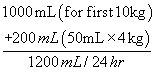
Maintenance fluid requirements for a 24-hour period are based on the child’s age and change in weight. The standard regimen for normal maintenance fluids is based on water and electrolyte output through insensible fluid losses (particularly transcutaneous and respiratory in the neonate), gastrointestinal fluids, and urine. Because the metabolic rate in infants and children is higher than that in adults, fluid losses are also greater than in adults, and maintenance requirements are increased. The amount of fluid lost before treatment is determined by comparison of preillness and current weights and by clinical signs of deficit. Maintenance fluid requirements for children are usually calculated according to the guidelines in Table 17-7 (Barkin, 1990, Mott et al., 1990 and Fann, 1998).
Example: Fluid maintenance for a 14-kg infant would be 1200 mL/24 hours:
REPLACEMENT (DEFICIT) THERAPY
Replacement therapy is divided into phases. The first phase is initial management or rapid delivery of fluid therapy; the second phase, repletion and maintenance therapy; and the third phase, early recovery.
Phase I: initial management
In children with fluid-volume deficit, replacement of vascular volume is essential. These children require immediate infusion of fluids (20 mL/kg 0.9% sodium chloride or lactated Ringer’s) infused rapidly over 20 to 60 minutes, especially if there is evidence of poor tissue perfusion or changes in vital signs and neurological status. If the child has a documented normal blood glucose level or is known to have diabetes, dextrose solutions should be omitted to prevent hyperglycemia, which may induce an osmotic diuresis. The child’s condition is then reassessed. If the response to the therapy is poor, an additional bolus of the initial solution is given. The child continues to be evaluated for the need of additional boluses, invasive monitoring, or the implementation of repletion (maintenance) therapy (Barkin, 1990 and Fann, 1998).
Phase II: repletion and maintenance therapy
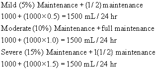
During this second phase (2 to 24 hours after onset of the deficit), replacement is combined with maintenance requirements. Acid-base and electrolyte disturbances are partially corrected. The simple formulas in Table 29-4 incorporate replacement and maintenance requirements for mild, moderate, and severe volume deficit.
| Amount of deficit | Formula for calculating amount of replacement fluid |
|---|---|
| Mild (5%) | Maintenance + (maintenance × 0.5) |
| Moderate (10%) | Maintenance + (maintenance × 1.0) |
| Severe (15%) | Maintenance + (maintenance × 1.5) |
Example: A 10-kg infant has a maintenance fluid requirement of 1000 mL/24 hours. The replacement amounts for 24 hours are shown in the following equations:
Phase III: early recovery
Early recovery, which can last from 24 to 96 hours, is aimed at correcting the remaining deficits occurring in hypertonic dehydration. These electrolyte deficits need to be corrected slowly so as not to impair neurological status. By this time, the child usually is well enough to ingest some fluids orally.
FLUID-VOLUME DEFICIT
Any abnormal fluid loss or reduction in fluid intake can lead to depletion of fluid in the extracellular and intracellular compartments. The most common cause of fluid loss in children is gastroenteritis with diarrhea accompanied by nausea or vomiting. Oral fluid intake is reduced in response to the symptoms, and therefore the amount of fluid lost through the stools or vomiting cannot be balanced. A fluid-volume deficit results, and physiological changes occur that progress as the condition worsens. The infusion of IV solutions into infants and children with fluid-volume deficit requires an understanding of the various types of deficit and the specific therapies required. In addition to being characterized by degree of fluid loss, fluid-volume deficit (dehydration) is also categorized by type—isotonic, hypotonic, or hypertonic, depending on the changes in the child’s serum sodium level (Table 29-5).
| ECF, extracellular fluid; GI, gastrointestinal; ICF, intracellular fluid. | |||
| Description | Isotonic | Hypotonic | Hypertonic |
|---|---|---|---|
| Type of Loss | |||
| Solute and water loss proportional | Greater solute loss than water | Greater water loss than solute | |
| ICF and ECF fluid shift | None | From ECF to ICF | From ICF to ECF |
| Plasma volume | Decreased | Decreased | Maintained |
| Serum sodium level (mEq/L) | 125-150 | <125 | >150 |
| Cause | GI fluid loss | GI fluid loss with hypotonic oral intake (glucose in water, ginger ale) | GI fluid loss with hypertonic oral intake (boiled skim milk) |
| Urine loss | Diabetes insipidus | ||
| Decreased oral intake | Fever | ||
| Hyperventilation | |||
| Clinical Signs | |||
| Skin | Poor turgor, cold, dry, dusky | Very poor turgor, cold, clammy, dusky | Fair turgor; cold, thick, and “doughy” skin |
| Eyes | Sunken | Sunken | Sunken |
| Mucous membranes | Dry | Slightly dry | Parched |
| Fontanels | Depressed | Depressed | Depressed |
| Pulse rate | Rapid | Rapid | Moderately rapid |
| Blood pressure | Low | Very low | Moderately low |
| Neurological status | Irritable or lethargic | Lethargic, coma, seizure | Hyperirritable, high-pitched cry, seizure |
| Infusion | Half of deficit replaced in first 8 hr, remaining over next 16 hr | Half of deficit replaced in first 8 hr, remaining over next 16 hr | Slow and gradual over 48 hr |
| Sodium level | 2-3 mEq/kg/24 hr | 2-3 mEq/kg/24 hr plus replacement; sodium should only be increased ≤2 mEq/L/hr up to serum level of 120 mEq/L | Sodium should not be reduced >2 mEq/L/hr |
| Potassium level | 2-3 mEq/kg/24 hr | 2-3 mEq/kg/24 hr | 2-3 mEq/kg/24 hr |
Hypovolemic shock is a common problem in children who are in need of emergency care. Trauma and burns are obvious causes, but hypovolemic shock can also occur with gastroenteritis and diabetic ketoacidosis. Sepsis in a child can develop into septic shock, which is not classified as hypovolemic but equally requires fluid resuscitation. The initial therapies for hypovolemic and septic shock are similar, and both require immediate vascular access. In traumatic shock, large volumes of blood and fluid are required; therefore IV access sites should be adequate to meet these needs. In emergency situations in which large volumes of fluids are needed, two peripheral catheters, in 24-, 22-, or 20-gauge size, or a short-term emergency central access device, such as a nontunneled femoral line or an intraosseous needle, can provide adequate vascular access in a child (International Consensus on Science, 2000, Hockenberry and Wilson, 2007 and Lavelle and Costarino, 2008). The clinical assessment and laboratory data determine the urgency and the type of therapy required. The degree of fluid loss is categorized according to the percentage of total body weight lost: mild (less than 5%), moderate (5% to 10%), and severe (more than 10%).
OTHER INFUSION THERAPIES
In addition to the administration of solutions, venous access devices in children are also used for therapies such as medication administration, parenteral nutrition (PN), and transfusion of blood products. An overview of these therapies as they relate to children is provided in this section. See Chapter 14 and Chapter 17 for additional information on blood component therapy and parenteral nutrition, respectively.
MEDICATION ADMINISTRATION
The most commonly used calculations for medication administration in pediatrics are those based on body weight and body surface area (calculated by using a nomogram). Dosing of pediatric medications is usually ordered as milligrams (mg) per kilogram (mg/kg), or in the case of chemotherapeutic agents, milligrams per meter squared (mg/m 2).
Techniques
All methods of IV medication administration, such as IV bolus or push and intermittent and continuous infusions, are used in the pediatric patient. In children, however, the dose and volume can be different from those used in adults, depending on the age and the size of the child. In neonates and infants, medications are commonly calculated to the tenths of a milligram or milliliter. Continuous infusions are used primarily to administer drugs that require maintenance of a constant blood level, and potent drugs that must be precisely titrated to individual needs.
Infusion of intermittent medications can be administered by several different methods. The in-line calibrated chamber is commonly used in general pediatric settings for continuous and intermittent infusions in small children. The medication is injected into the in-line calibrated chamber, diluted to a recommended concentration with a compatible IV solution, and infused at a prescribed rate. At the completion of the infusion, the usual practice is to flush the chamber with a volume of a compatible IV solution. The advantages of this method include eliminating the risk of fluid overload and providing accuracy in fluid delivery. A disadvantage of this method is that it is not practical for small infants who are fluid and volume restricted. Also, the drug must be compatible with the primary solution or a second IV administration set-up is required. Recent evidence shows that at least two times the flush volume of the administration set is necessary to clear medication from the line and ensure complete dosage delivery to the patient (Ford et al, 2003).
Administering medications via a syringe pump is commonly used for neonates and children and provides an accurate method of IV medication administration. The syringe pump can be connected via an extension set directly onto an intermittent venous access device or in a piggyback fashion into the primary administration set closest to the insertion site to ensure timely administration. A syringe containing the properly diluted medication is attached to a microbore tubing and pump set to infuse at a prescribed rate.
The retrograde method of medication infusion is less commonly used in the neonatal intensive care unit. A specific low-volume (less than 1 mL) retrograde administration set, with an access port at each end, is attached and primed along with the primary administration set. To administer medications via the system, a medication-filled syringe is attached to the port most proximal to the patient, an empty syringe is connected to the port most distal from the patient, the clamp between the port and the child is closed, and the medication is injected distally up the tubing (away from the child). The solution in the retrograde tubing is displaced upward in the tubing into the empty syringe. Both syringes are removed, the lower clamp is opened, and the medication is then infused into the patient at the prescribed rate. The medication volume is then automatically incorporated into the regulated amount of fluid to be infused. This method is often used in infants who cannot tolerate a rapid infusion rate or additional fluid volume; in this method, the medication infuses at the same rate set for the IV infusion.
PARENTERAL NUTRITION
Parenteral nutrition (PN) is administered when patients cannot or will not fully use their gastrointestinal tract for nutrition or when they require supplemental calories for growth or healing. The goal of parenteral nutrition is to meet anabolic needs and allow normal growth and development. Conditions requiring PN include prematurity, children with congenital or acquired anomalies of the digestive tract (e.g., inflammatory bowel disease, Hirschsprung’s disease, short-bowel syndrome, pancreatitis, necrotizing enterocolitis, omphalocele, gastroschisis), surgical conditions, or disease states that may increase the risk of starvation (e.g., cancer, cystic fibrosis, acquired immunodeficiency syndrome [AIDS]). Candidates for parenteral nutrition therapy are identified by medical diagnosis, physical examination, and nutritional evaluation, including such measurements as weight loss, below normal percentiles for height and weight on the growth chart, and abnormal laboratory values such as albumin level (Bilodeau, Poon, and Mascarenhas, 1998).
The parenteral nutrition solution is made up of several components, depending on the status of the child; these solutions can be individualized to fit calorie and nutrient needs (Heird, 1993 and ASPEN Board of Directors and the Clinical Guidelines Task Force). The basic components of PN are protein, carbohydrates, fat, electrolytes, vitamins, trace elements, and minerals. Protein, for growth, is administered in the form of crystalline amino acids; infants younger than 6 months require a special amino acid mixture that mimics that of breast milk (ASPEN 2002). Dextrose is used as the carbohydrate source, providing energy so that the body does not break down protein or fat to meet metabolic needs. Fat emulsions or lipids provide a high calorie content per volume, making them ideal sources of calories for children (ASPEN 2002). Fat emulsions also buffer the irritating effects that glucose tends to exert on the vein. Lipids should be infused cautiously in patients with infections, compromised renal function, or hyperbilirubinemia. Compromised liver function and an increased risk of fungal infections have been associated with lipid infusions (Weiss et al, 1991). Fat emulsions may be infused over a 24-hour period, or given over several hours with a break in therapy to allow clearance before proceeding to the next daily infusion. In VLBW preterm and small gestational age infants, lipids are cleared more slowly and patients are at a greater risk of developing hyperlipidemia.
When treating children, it is often difficult to balance fluid needs with calorie needs. A large volume may be necessary to infuse through a peripheral vein to meet a child’s caloric needs. Therefore PN is often concentrated in a smaller volume and administered via a central vein. Dextrose 10% may be administered peripherally; central access should be considered when the dextrose percentage exceeds 10%. Fluids containing greater than 10% dextrose concentration can be very irritating to the vein intima and cause local injury.
For children receiving long-term PN, continued growth can change the nutritional requirements, so careful monitoring is essential. PN can be administered in the home or hospital. Home PN is often administered via the central route and cycled over 12 to 18 hours, usually at night, so that the child may function normally during the day. Usually, home PN is initiated in the hospital. When the child is stable and the infusion process for home PN has been mastered by the caregiver(s) with appropriate participation of the child (depending on his or her level of development), home PN may be instituted.
Continuous monitoring is required to ensure safe, efficacious infusion of PN that meets the changing needs of the child. This includes monitoring the following on a scheduled basis:
• Parenteral nutrition base solution or formula
• Electrolytes, vitamins, and trace elements
• Fat emulsion
• Infusion rate
• Laboratory results
• Nutritional measurements
A multidisciplinary team approach can result in the successful clinical application and monitoring of PN while preventing complications.
< div class='tao-gold-member'>
Only gold members can continue reading. Log In or Register to continue
Stay updated, free articles. Join our Telegram channel

Full access? Get Clinical Tree


