Chapter 15
Male Genitalia and Hernias
If a man’s urine is like the urine of an ass, like beer yeast, like wine yeast or varnish, that man is sick … and through a bronze tube in the penis pour oil and beer and licorice.
From the Sushruta Samhita (ca. 3000 BCE)
General Considerations
Since the beginning of recorded history, the external genitalia and the urologic system have been of special interest to people. Kidney stones and urologic surgery were well described in antiquity. One of the earliest reported kidney stones was found in a young boy who lived about 7000 BCE.
Circumcision is one of the oldest known surgical procedures in medicine. Male circumcision has been widely practiced as a religious rite since ancient times. An initiatory rite of Judaism, circumcision is also practiced by Muslims, for whom it signifies spiritual purification. Although the origin is unknown, circumcision is often depicted on the walls of temples dating from 3000 BCE. In the Egyptian Book of the Dead, it is written, “The blood falls from the phallus of the Sun God as he starts to incise himself.” By the time of the Roman takeover of Egypt in 30 BCE, the practice of circumcision had a ritual significance, and only circumcised priests could perform certain religious rites. The Hindus regarded the penis and testicles as a symbol of the center of life and sacrificed the prepuce as a special offering to the gods.
The Bible has many urologic references. In Genesis 17 : 7, Abraham makes a covenant with God for the Jews. He is told in Genesis 17 : 14, “And the uncircumcised male who is not circumcised in the flesh of his foreskin, that soul shall be cut off from his people; he hath broken My covenant.” In Leviticus 12 : 3, the Jews were told, “And in the eighth day the flesh of his foreskin shall be circumcised.” Leviticus 15 : 2–17 deals with discharges that render a man unclean. Today, it is estimated that 1 in every 6 men worldwide is circumcised. There are more than 15 million postinfancy circumcisions a year, and thus it is one of the most common surgical procedures.
The Bible, Hindu literature, and Egyptian papyri described a disease now presumed to be gonorrhea. The Mesopotamian tablets described a variety of cures, such as this: “If a man’s penis on occasions of his pleasure hurts him, boil beer and milk and anoint him from the pubis.” Avicenna’s Canon of Medicine (1000 CE) was considered the authoritative medical text for centuries and described placing a louse in the penis to counteract a penile discharge.
Gonorrhea was probably first named by Galen in the second century CE. Gonorrhea is the Greek translation of “a flow of offspring.” Galen apparently thought that the purulent discharge was a leakage of semen. Many terms have been used to describe gonorrhea throughout the years. Perhaps the most common is clap, a name used for the past 400 years. It is thought that the term clap was derived from a specific area in Paris known for prostitution called Le Clapier.
Benign prostatic hyperplasia (BPH) is the most common benign neoplasm in aging men. It has been estimated that by 60 years of age, the prevalence is greater than 50%, and by 85 years of age, the prevalence approaches 90%. In addition, by 80 years of age, 1 in every 4 men requires some form of treatment for relief of symptomatic BPH. More than 300,000 surgical procedures are performed in the United States annually for BPH, most commonly a transurethral resection of the prostate.
Cancer of the genitourinary system is common. In the United States, in 2011, prostate cancer accounted for 29% of all cancer cases in men. It accounted for 11% of all cancer deaths and was the second most common cause of cancer deaths after lung and bronchus cancer (28%). In 2011, there were 240,890 new cases of prostate cancer and 33,720 deaths from the disease in the United States.
For reasons that remain unclear, the highest incidence rate for prostate cancer is in African American men and Jamaican men of African descent (54.8 per 100,000); for white persons, it is 23.7 per 100,000. The lowest incidence rate is in Asians and Pacific Islanders (10.7 per 100,000). For a 50-year-old man with a 75-year life expectancy, the lifetime risk for development of microscopic prostate cancer is 42%; the risk for development of clinically evident prostate cancer is 10%; and the risk for development of fatal prostate cancer is 3%. Genetic studies suggest that strong familial predisposition may be responsible for 5% to 10% of prostate cancers. Some recent studies have shown that a diet high in processed meat or dairy foods may be a risk factor, and obesity appears to increase the risk of aggressive prostate cancer. Approximately 95% of all prostate cancers arise from an area of the gland where it can be readily detected by rectal examination. More than 90% of all prostate cancers are discovered in the local or regional stages, for which the 5-year survival approaches 100%. During the past 25 years, the 5-year relative survival rate for all stages combined has increased from 69% to 99.6%. According to the most recent data, 10-year survival is 95% and the 15-year survival is 82%. Obesity and smoking are associated with an increased risk of dying from prostate cancer.
Cancer of the urinary bladder accounted for an additional 6% of cancer cases but only 4% of all cancer deaths in men and less than 1% in women. In 2011, there were 69,250 new cases (52,020 in men, 17,230 in women) of cancer of the urinary bladder in the United States and 14,990 deaths from the disease. Cancer of the urinary bladder is the fourth most common malignancy among men and the eighth most frequent among women. Approximately 260,000 new cases of urinary bladder cancer are diagnosed worldwide every year. The highest incidence rates for bladder cancer are found in industrialized countries such as the United States, Canada, France, Denmark, Italy, and Spain. The lowest rates are in Asia and South America, where the incidence is only approximately 30% as high as in the United States. Cigarette smoking is an established risk factor for cancer of the urinary bladder. It is estimated that approximately 50% of these cancers in men and 30% in women are linked to smoking. Occupational exposures may account for up to 25% of all urinary bladder cancers. Most of the occupationally accrued risk is attributable to exposure to a group of chemicals known as arylamines. Occupations with high exposure to arylamines include dye workers, rubber workers, leather workers, truck drivers, painters, and aluminum workers. People who live in communities with high arsenic levels in the drinking water also have an increased risk of cancer of the urinary bladder.
Although testicular cancer accounts for only 1% of all cancers in men, testicular carcinoma is the most common cancer in men in the 15- to 35-year-old age group. There were 8290 new testicular cancer cases in 2011 and 350 related deaths. Testicular cancer is four times less common in African-American men than in white men. The risk for development of testicular cancer in a man’s lifetime is approximately 1 per 500. Approximately 90% of all testicular tumors manifest as an asymptomatic testicular mass. Once these tumors are detected and treatment is begun, the cure rate can approach 90%, even when the tumor has spread beyond the testicle. Many patients have oligospermia or sperm abnormalities before therapy. Virtually all become oligospermic during chemotherapy with platinum-based agents. Many recover sperm production, however, and can father children, often without the use of cryopreserved semen. In a population-based study, 70% of patients actually fathered children. Men in whom testicular cancer has been cured have approximately a 2% to 5% cumulative risk of developing a cancer in the opposite testicle during the 25 years after initial diagnosis. The most important prognostic factor has been shown to be early detection by routine physical examination and self-examination. All men should be instructed in testicular self-examination.
There were 60,920 new cases of renal cancer in 2011. These included 92% renal cell carcinoma, 7% renal pelvis carcinoma, and 1% Wilms tumor, a childhood cancer that develops before the age of 5 years. The American Cancer Society reported 13,120 deaths from all types of renal cancer. Tobacco use is a strong risk factor for renal cancer with the largest increased risk for cancer of the renal pelvis, particularly in heavy smokers.
Erectile dysfunction (ED) is an extremely common problem. It has been estimated that more than 30 million American men have some degree of ED and that nearly a million new cases can be expected to develop annually. Studies have shown that ED affects not only a man’s physical and sexual satisfaction but also his general quality of life, with especially strong links to depression. In the Massachusetts Male Aging Study, 52% of men from 40 to 70 years of age had some degree of ED. Seventeen percent reported minimal dysfunction, 25% reported moderate dysfunction, and 10% reported complete dysfunction. This study also revealed the progressive nature of ED with increasing age. At 40 years of age, 5% of the American male population has complete ED, and at 70 years of age, 15% of the population has complete ED. Sixty-seven percent of men 70 years of age have some degree of ED. As the population continues to age, clinicians will treat more and more male patients for ED in the future.
Structure and Physiologic Characteristics
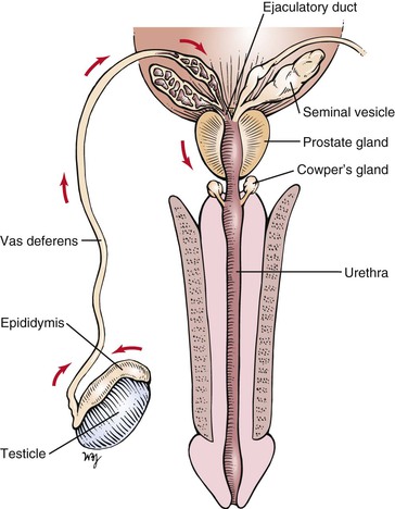
Cross-sectional and frontal views of the male genitalia are shown in Figure 15-1.
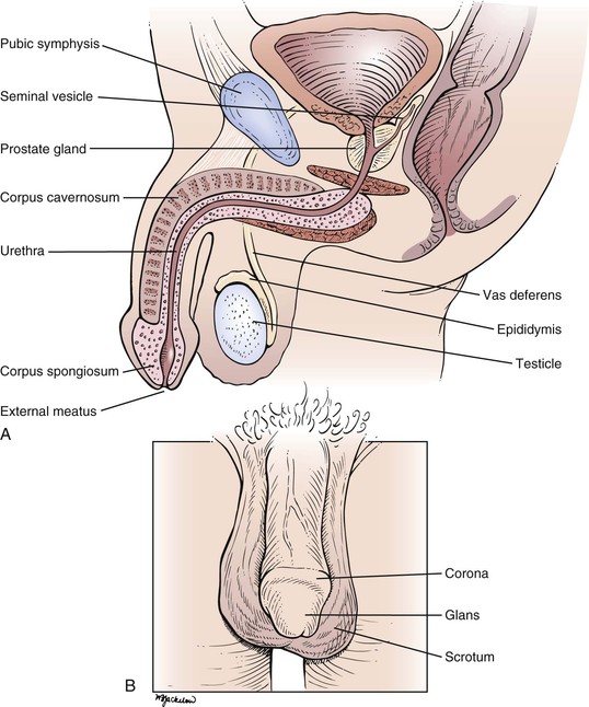
The penis is composed of three elongated, distensible structures: two paired corpora cavernosa and a single corpus spongiosum. The urethra runs through the corpus spongiosum. The penis has two surfaces, dorsal and ventral (urethral), and consists of the root, the shaft, and the head. The shaft is composed of erectile tissue that, when engorged with blood, produces a firm erection necessary for sexual intercourse. The corpora cavernosa also contain smooth muscle that contracts rhythmically during ejaculation.
On the dorsal aspect of the penis in the midline runs the dorsal vein, with an artery and a nerve on either side. The distal end of the corpus spongiosum expands to form the head, or glans penis. The glans penis covers the end of the corpora cavernosa. The glans has a prominent margin on its dorsal aspect, the corona. A slitlike opening on the tip of the glans is the external meatus of the urethra.
The skin of the penis is smooth, thin, and hairless. At the distal end of the penis, a free fold of skin called the prepuce (foreskin) covers the glans. Secreted mucus and sloughed epithelial cells called smegma collect between the prepuce and the glans, providing a lubricant during sexual intercourse. The prepuce can be retracted to expose the glans as far as the corona. During circumcision, the prepuce is removed.
The root of the penis lies deep to the scrotum, in the perineum. At the root, the corpora cavernosa diverge. Each corpus cavernosum is enveloped in a dense, fibroelastic covering called the tunica albuginea, and these tunicae fuse to form the median septum of the penis. A cross section through the penis is shown in Figure 15-2.
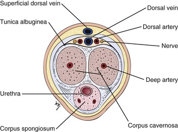
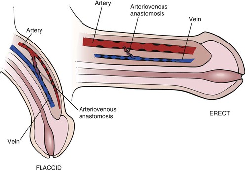
The blood supply to the penis is from the internal pudendal artery, from which the dorsal and deep arteries of the corpora cavernosa are derived. The veins drain into the dorsal vein of the penis. In the flaccid state, the venous channels and arteriovenous anastomoses are widely patent, whereas the arteries are partially constricted.
Erection is a complex hemodynamic and neurophysiologic event. In the flaccid state, the smooth muscles of the penile arteries and sinusoid spaces are contracted. The erectile state begins in the brain and requires relaxation of the smooth muscles of the penis. From the brain center, neural signals are sent to the corpora cavernosa, where synthesis and release of the neurotransmitter nitric oxide occur. Nitric oxide is the primary mediator responsible for endothelial and cavernous smooth muscle relaxation. Nitric oxide activates guanylate cyclase to produce cyclic guanosine monophosphate (GMP), which decreases intracellular calcium levels, allowing smooth muscle relaxation and an increase in arterial inflow and corporal veno-occlusion in the penis. Venous outflow is decreased because distention of the blood-filled sinusoidal spaces compresses the veins against the inner layer of the rigid tunica albuginea. In the erect state, the arteriovenous channels are closed, and the arteries are widely opened. Muscular pillars are present in the walls of the arteries, veins, and arteriovenous anastomoses, which aid in occluding the lumina. Phosphodiesterase, predominantly type V in penile tissue, catalyzes the conversion of cyclic GMP to GMP and results in detumescence. There are some new medications that selectively inhibit phosphodiesterase V. These agents enhance the effect of the nitric oxide–mediated increase in cyclic GMP levels and significantly improve erectile function and sexual function in men. The anatomy of erection is illustrated in Figure 15-3.
The urethra extends from the internal urinary meatus of the bladder to the external meatus of the penis. The urethra can be divided into three portions: the prostatic (posterior) portion, the membranous portion, and the cavernous (anterior) portion. The short posterior portion passes through the prostate gland. The common ejaculatory duct and several prostatic ducts enter at the distal end of this portion. The external urethral sphincter surrounds the membranous urethra, and on either side lie Cowper’s bulbourethral glands. The anterior urethra is the longest portion and passes through the corpus spongiosum. The ducts of Cowper’s glands enter the anterior urethra near its proximal end.
The scrotum is the pouch containing the testes; it is suspended externally from the perineum. The scrotum is divided into halves by the intrascrotal septum, one testis lying on each side. The wall of the scrotum contains involuntary smooth muscle and voluntary striated muscle. A major role of the scrotum is temperature regulation of the testes. The testes are maintained at approximately 2° C lower than the temperature of the peritoneal cavity, a condition necessary for spermatogenesis. The size of the scrotum is variable according to the individual and his response to ambient temperature. During exposure to cold temperatures, the scrotum is contracted and very rugate. In a warm environment, the scrotum becomes pendulous and smoother.
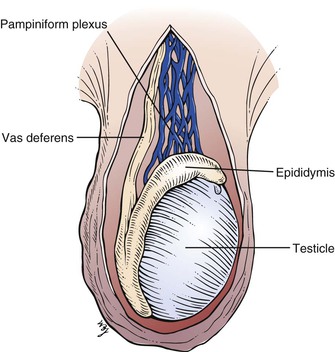
The testes, or testicles, are ovoid, smooth, and approximately 1.5 to 2 inches (3.5 to 5 cm) in length. The left testicle commonly lies lower than the right. The testes are covered with a tough fibrous coat called the tunica albuginea testis. Each testicle has a long axis directed slightly anteriorly and upward and contains long, microscopic, convoluted seminiferous tubules that produce sperm. The tubules end in the epididymis, which is comma-shaped and located on the posterior border of the testis. It consists of a head that is swollen and overhangs the upper pole of the testicle. The inferior portion, or tail, of the epididymis continues into the vas deferens. The testicular artery enters the testicle in its posterior midportion. The veins draining the testicle form a dense network called the pampiniform plexus, which drains into the testicular vein. The right testicular vein drains directly into the inferior vena cava, whereas the left drains into the left renal vein. The lymphatic drainage of the testes is to the preaortic and precaval nodes, not to the inguinal nodes. This is important to recognize because the testes are embryologically intra-abdominal organs, and neoplasms and inflammations of the testis produce adenopathy of these nodal chains. In general, inguinal adenopathy is rare.
The relationship of the testicle and epididymis is illustrated in Figure 15-4.
The vas deferens is a cordlike structure, easily felt in the scrotum. The vas deferens, testicular arteries, and veins form the spermatic cord, which enters the inguinal canal. The vas deferens passes through the internal ring and, after a convoluted course, reaches the fundus of the bladder. It passes between the rectum and the bladder and approaches the vas deferens of the opposite side near the seminal vesicles. Near the base of the prostate, the vas deferens joins with the duct of the corresponding seminal vesicle to form the ejaculatory duct, which passes through the prostate gland to enter the posterior urethra.
The prostate gland is approximately the size of two almonds, or approximately 1.5 inches (3.5 cm) long by 1.2 inches (3 cm) wide. Traversing the gland in the midline is the posterior urethra. On either side is an ejaculatory duct. The prostate is commonly divided into five lobes. The posterior lobe is clinically important because prostate carcinoma frequently affects this lobe. In the presence of cancer, the midline groove between the two lateral lobes may be obliterated. The middle and lateral lobes are above the ejaculatory ducts and are typically involved in benign hypertrophy. The anterior lobe is of little clinical importance.
The sources and direction of seminal fluid flow in the male genitalia are illustrated in Figure 15-5.
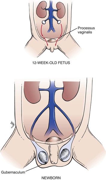
The descent of the testes is important to review at this time. In the normal full-term male newborn, both testes are in the scrotum at birth. The testes descend to this position just before birth. At approximately the twelfth week of gestation, the gubernaculum develops in the inguinal fold and grows through the body wall to an area that will ultimately lie in the scrotum. This tract marks the location of the future inguinal canal. A dimple called the processus vaginalis forms in the peritoneum and follows the course of the gubernaculum. By the seventh month of gestation, the processus vaginalis has reached the aponeurosis of the external oblique muscle. Each testis then begins its descent from the abdominal cavity through the internal ring to lie in the abdominal wall. During the eighth month, the testes descend along the inguinal canal; at birth, they are in the scrotum. At birth, the gubernaculum is barely distinguishable, and the processus vaginalis becomes obliterated within the spermatic cord. In approximately 5% of male infants, there is imperfect descent of the testis (cryptorchidism). The descent of the testes is illustrated in Figure 15-6.
The genital development stages for boys are illustrated in Figure 21-42 (and discussed in Chapter 21, The Pediatric Patient).
Review of Specific Symptoms
The most common symptoms of male genitourinary disease are as follows:
Pain
Sudden distention of the ureter, renal pelvis, or bladder may cause flank pain. Any patient with flank pain should be asked the following questions:
“Where did the pain begin? Can you point to the area?”
“Do you feel the pain in any other area of your body?”
“Did the pain start suddenly?”
“Have you ever had this type of pain before?”
“What seems to make the pain worse? Less?”
“Has the color of your urine changed?”
Gradual enlargement of an organ is usually painless. An aching pain in the costovertebral angle may be related to sudden distention of the renal capsule, which results from acute pyelonephritis or obstructive hydronephrosis. The spasmodic, colicky pain from upper ureteral dilatation may cause referred pain to the testis on the same side. Lower ureteral dilatation may cause pain referred to the scrotum. The pain of ureteral distention is severe, and the patient is restless and uncomfortable in any position. Bladder distention causes lower abdominal fullness and suprapubic pain, with an intense desire to urinate. Pain in the groin may result from pathologic processes in the spermatic cord, testicle, or prostate gland; from lymphadenitis of any cause; from hernia; from herpes zoster; or from a disorder that is neurologic in origin.
Testicular pain can result from nearly any disease of the testis or epididymis. Such diseases include epididymitis, orchitis, hydrocele, spermatic cord torsion, and tumor. Referred pain from the ipsilateral ureter must always be considered. Priapism is a painful, persistent erection of the penis that is not a result of sexual excitation. The sustained erection results from thrombosis of veins in the corpora cavernosa. This occurs in patients with sickle cell anemia or leukemia. The exact mechanism is unknown, but it appears to result from a blockage of venous drainage from the penis while the arteries remain patent. Chronic priapism often results in organic ED.
Dysuria
Pain on urination, called dysuria, is frequently described as “burning.” Dysuria is evidence of inflammation of the lower urinary tract. The patient may describe discomfort in the penis or in the suprapubic area. Dysuria also implies difficulty in urination. This may result from external meatal stenosis or from a urethral stricture. Painful urination is usually associated with urinary frequency and urgency. When the patient describes pain or difficulty in urination, ask the following questions:
“How long have you noticed a burning sensation on urination?”
“How often do you urinate each day?”
“How does your urination feel different?”
“Do you have a discharge from your penis?”
“Does the urine seem to have gas bubbles in it?”
“Have you noticed any solid particles in your urine?”
Fecaluria is the presence of fecal material in the urine and is rare. The passage of feculent-smelling material results from either an intestinovesicular fistula or an urethrorectal fistula. These fistulas occur as a consequence of ulceration from the bowel to the urinary tract. Diverticulitis, carcinoma, and Crohn’s disease are frequent causes.
Pus in the urine, or pyuria, is the body’s response to inflammation of the urinary tract. Bacteria are the most common cause of inflammation resulting in pyuria, although pyuria is also seen in patients with neoplasms and kidney stones. Cystitis and prostatitis are common causes of pyuria.
Changes in Urine Flow
Changes in urine flow include frequency and incontinence. Urinary frequency is the most common symptom of the genitourologic system. Frequency is defined as passing urine more often than normal. Nocturia is urinary frequency at night. There are several causes of frequency: decreased bladder size, bladder wall irritation, and increased urine volume. If an obstructed bladder cannot be completely emptied at each voiding, its effective capacity is diminished. The following questions, in addition to the ones pertaining to dysuria, should be asked to help define the problem.
“Do you find that you must wake up at night to urinate?”
“Can you estimate the amount of urine passed each time you urinate?”
“Do you have sudden urges to urinate?”
“Have you found that despite an urge to urinate, you cannot start the stream?”
“Has there been a change in the caliber of the stream?”
“Have you found that you must wait longer for the stream to start?”
“Do you have the sensation that after urination has stopped, you still have to urinate?”
“Do you have to strain at the end of urination?”
Prostatic hyperplasia is the most common cause of reduced usable bladder capacity in men. Symptoms include frequency of urination, nocturia, urgency, weak stream, intermittent stream, and a sensation of incomplete emptying. Long-standing prostatic hypertrophy can lead to a complete inability to urinate, necessitating catheterization (a condition known as urinary retention), or can lead to urinary tract infections or to bladder stones. Most bladder diseases, such as cystitis, cause frequency as a result of irritation of the bladder mucosa. Polyuria, or voiding large amounts of urine, is usually accompanied by excessive thirst, or polydipsia. Diabetes mellitus and diabetes insipidus are common causes of polydipsia.
Urinary incontinence is the inability to retain urine voluntarily. The urge to urinate may be so intense that incontinence may result. In addition to the questions regarding dysuria and frequency, ask the following:
In patients with chronically distended bladders, as in those with prostatic hypertrophy, there is always a large amount of residual urine. The pressure within the bladder is constantly elevated. A slight increase in intra-abdominal pressure raises the intravesicular pressure sufficiently to overcome bladder neck resistance, and urine escapes. Leakage may be steady or intermittent. This type of incontinence is overflow incontinence. Stress incontinence is leakage that occurs only when the patient strains. The primary defect is a loss of muscular support in the urethrovesicular region. Residual urine is insignificant. Any increase in intra-abdominal pressure causes leakage. This type of incontinence is more common in women and is discussed in Chapter 16, Female Genitalia.
Polyuria is the production of increased amounts of urine, frequently greater than 2 to 3 L/day. The normal daily urine output varies from 1 to 2 L/day. The most important diseases to differentiate are diabetes mellitus, diabetes insipidus, and psychologic diabetes insipidus. Ask the following questions:
“How long have you been passing large amounts of urine?”
“How often do you have to urinate at night?”
“Is there any variability in the urine flow from day to day?”
“Do you have excessive thirst?”
“Do you prefer water or other fluids?”
“What happens if you don’t drink? Will you still have to urinate?”
“Do you have any visual problems? Headaches?”
Patients with diabetes mellitus have a high osmotic load and have polyuria. Increased appetite is also common. Diabetes insipidus is caused by a vasopressin deficiency related to a lesion in the hypothalamus or pituitary gland. In these patients, the urine cannot become concentrated despite a rise in plasma osmolality. Patients with psychogenic diabetes insipidus, which is more common, have polyuria related to compulsive drinking of water. It is seen in patients with psychologic problems. The onset of polyuria is abrupt in patients with psychogenic diabetes insipidus, and they have no preference for the type of fluid they drink. In contrast, patients with true diabetes insipidus prefer water. Because true diabetes insipidus is related to intracranial lesions, it is not surprising that affected individuals suffer from headaches and visual disturbances, especially visual field abnormalities.
Red Urine
Red urine is often indicative of hematuria, or blood in the urine. However, there are many causes of red urine, and it should not automatically be assumed that red urine indicates bleeding. Vegetable dyes, drugs such as phenazopyridine (Pyridium®), and excessive ingestion of beets can cause red urine. When it is determined that the urine is red as a result of the presence of blood, the hematuria is termed gross hematuria. Hematuria may be the first symptom of serious disease of the urinary tract. Ask the following questions of any patient with the symptom of red urine:
“How long have you noticed red urine?”
“Have you had red urine previously?”
“Have you noticed clots of blood in the urine?”
“Did you have an upper respiratory infection or a sore throat a few weeks ago?”
“Are you aware of any bleeding problems?”
“Are you taking any medications?”
Individuals who participate in strenuous activities may traumatize blood cells as these cells travel through the small vessels in the feet. A condition called march hemoglobinuria may result, causing intravascular hemolysis and hemoglobinuria. The temporal relationship of blood in the urine is an important factor. Blood only at the beginning, or initial hematuria, usually has a source in the urethra. Blood only at the end of urination, or terminal hematuria, indicates a disorder at the bladder neck or the posterior urethra. Blood evenly distributed throughout urination is total hematuria and implies disease above the prostate gland or a massive hemorrhage at any level. Blood staining of undergarments without blood in the urine indicates pathologic processes in the external urethral meatus. Weight loss and hematuria are seen in renal cell carcinoma. Red urine that occurs 10 to 14 days after an upper respiratory infection may indicate acute glomerulonephritis.
Penile Discharge
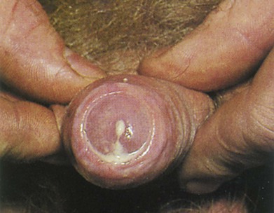
Discharge from the penis is a continuous or intermittent flow of fluid from the urethra. Ask the patient whether he has ever had a discharge and, if he has, whether it was bloody or purulent. Bloody penile discharges are associated with ulcerations, neoplasms, or urethritis. Purulent discharges are thick and yellowish-green and may be associated with gonococcal urethritis or chronic prostatitis. Determine when the discharge was first noted. Figure 15-7 shows a purulent penile discharge in a man with gonococcal urethritis. Gonorrhea is caused by Neisseria gonorrhoeae. After exposure, approximately 25% of men and more than 50% of women contract the disease. In men, the acute symptoms of dysuria and a purulent urethral discharge begin 2 to 10 days after exposure. In women, a vaginal discharge and dysuria develop days to weeks after exposure; however, in up to 50% of women, the infection may be asymptomatic.
Stay updated, free articles. Join our Telegram channel

Full access? Get Clinical Tree


