X
X-rays, conventional
Purpose of the test
An x-ray film provides a radiographic image of the organs or tissues to detect abnormalities such as tumors, perforations, abscesses, infections, foreign bodies, or fractures of the bone.
Basics the nurse needs to know
Conventional radiographs are low-cost procedures that may be used to provide a preliminary image, with low exposure to radiation for the patient. Computed tomography, magnetic resonance imaging, nuclear scan, and ultrasound have replaced many of the previous uses of radiographs, but the conventional radiograph is still a mainstay of imaging of musculoskeletal tissue, particularly in trauma cases (Renner, 2009).
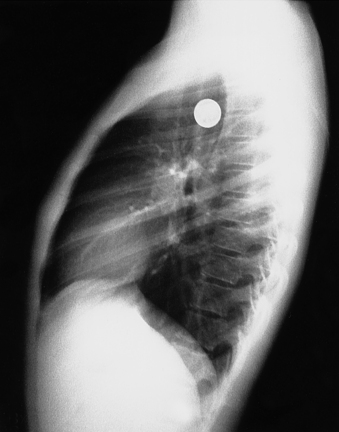
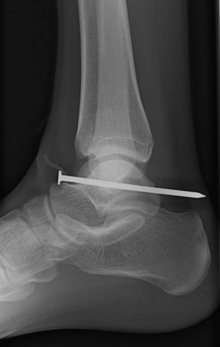
Metal objects absorb or block x-ray emissions and will appear white on the film. Before the radiograph is taken, patients must remove all metal objects and jewelry so that the underlying tissue can be visualized. Radiographs are very useful to image a metallic foreign body that is swallowed, aspirated, or has penetrated the body (Figures 105 and 106).
Chest radiograph
This film is obtained routinely for preoperative patients to screen for tuberculosis and other serious pulmonary or cardiac diseases. It also provides a preoperative comparison film for the postoperative patient in whom a pulmonary or cardiac complication develops. The chest radiograph is a basic radiologic procedure to identify broken ribs, a pneumothorax, or suspected pulmonary disorder (Figure 107). The chest radiograph also provides data about the heart, including its size and shape. In congenital and acquired cardiac disease, the enlargement of the heart and its atria or ventricles provides information about the improper function of the cardiac valves, pulmonary or aortic arterial hypertension, and venous pulmonary conditions that affect heart size.
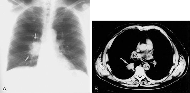
Figure 107. Lung cancer. A, The posteroanterior radiographic view of the lung shows an ill-defined mass (arrows). Its shaggy appearance is very suggestive of carcinoma. B, Follow-up with a computed tomography scan on the same patient provides a clear view of the tumor (arrow), its location and relationship to the mediastinal structures, including the pulmonary artery (PA) and aorta (AO).
(From Mettler FA: Essentials of radiology, ed 2, Philadelphia, 2005, Saunders.)
Stay updated, free articles. Join our Telegram channel

Full access? Get Clinical Tree


