Vascular Surgery


Vascular Surgery


2 How is carotid stenosis managed?


In symptomatic patients, if the stenosis is 70% to 99%, patients usually are advised to undergo carotid endarterectomy (CEA) for the best long-term prognosis—if their state of health allows them to tolerate the surgery. If stenosis is 50% to 69%, the data are less clear, and patient factors affect the decision. CEA is generally recommended for men, patients aged 75 or older, patients with recent stroke (not transient ischemic attack), and patients with hemispheric symptoms other than transient monocular blindness (amaurosis fugax). Female patients, patients younger than 75 years, and those with mild symptoms generally do better with medical management if stenosis is 50% to 69%. If stenosis is less than 50%, medical management is indicated.


Patients should not undergo carotid endarterectomy after a stroke that leaves them severely disabled, but small, nondisabling strokes are not contraindications to surgery. Carotid endarterectomy should not be performed during a transient ischemic attack or stroke in evolution. Surgery is always done electively, not on an emergent basis.


In asymptomatic patients, if the stenosis is 60% to 99%, CEA is indicated. If stenosis is less than 60%, medical management is indicated. Medical management includes antihypertensive agents, statins, and antiplatelet therapy.


The role of carotid angioplasty and carotid stenting in carotid stenosis is not yet clearly defined. Carotid endarterectomy remains the treatment of choice for suitable carotid stenosis.


Because medical therapy has improved since the initial studies comparing CEA with medical management were performed, medical management of lower grade carotid stenosis and asymptomatic carotid stenosis is gaining favor. This is an area that is still being clarified in the medical literature and likely will not be tested on the USMLE.


4 What are the classic findings in a patient with an abdominal aortic aneurysm? How is it evaluated?
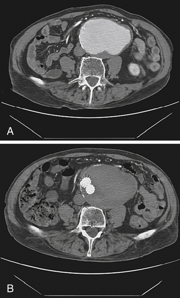

Abdominal aortic aneurysm (Fig. 39-1) classically presents as a pulsatile abdominal mass that may cause abdominal pain. If pain is present, rupture/leak of the aneurysm should be suspected, although an unruptured aneurysm may cause some degree of pain. Ultrasound or computed tomography (CT) scan is used for initial evaluation and diagnostic confirmation in stable patients, as well as for serial monitoring.


5 How is an abdominal aortic aneurysm managed? What clues indicate that the aneurysm has ruptured?


If the aneurysm is smaller than 5 cm, you can follow it with serial ultrasound examinations to ensure that it is not enlarging. These smaller aneurysms should be managed with risk factor reduction (smoking cessation and treatment of hypertension and dyslipidemia). If the aneurysm is larger than 5 cm (or if you are told that it is enlarging rapidly), surgical correction should be advised if the patient can tolerate the surgery.


A pulsatile abdominal mass plus hypotension requires emergent laparotomy for a presumed ruptured aneurysm, which carries a mortality rate of roughly 90%. The management of an abdominal aortic aneurysm dissection depends on the location of the dissection. Patients who survive the initial tear typically complain of a severe sharp or tearing sensation in the back or chest. Acute dissections involving the ascending aorta are considered surgical emergencies. Dissections confined to the descending aorta are treated medically unless the dissection progresses or continues to bleed.

Stay updated, free articles. Join our Telegram channel

Apr 8, 2017 | Posted by in NURSING | Comments Off on Vascular Surgery

Full access? Get Clinical Tree

Get Clinical Tree app for offline access