3 Tissues, organs, systems and homeostasis
Tissues
A tissue consists of cells and the products of cells specially developed for carrying out a special function. In the body there are four main types of tissue (Table 3.1):
Table 3.1 Classification of tissues
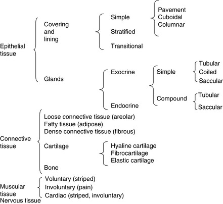 |
Epithelial tissue
Covering and lining epithelia
Covering epithelia may be classified according to the arrangement and shape of their cells.
Simple epithelium is composed of a single layer of cells attached to a basement membrane; it is very delicate and is found where there is little wear and tear (Fig. 3.1).
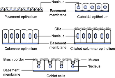
Goblet cells are cells that secrete mucim, which dissolves in water to become mucus.
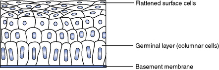
Stratified epithelium is made up of many layers of cells (Fig. 3.2). The deepest cells, called the germinal layer, lie on the basement membrane and are columnar. As they divide, and this occurs frequently, the parent cells are pushed nearer the surface and become flattened. The cells on the surface are rubbed off and are continually replaced from below. If the surface of the epithelium is dry, as on the skin, the surface cells die because the blood supply is below the basement membrane, and the scaly surface which develops, keratin, constitutes a waterproof layer. If the surface is moist, as in the mouth, the cells survive until they are rubbed off, and so keratin is not formed.
Glands
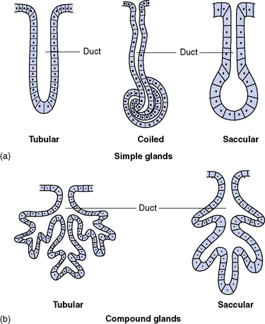
Exocrine glands
Exocrine glands (Fig. 3.3) pour their external secretions out through a duct. The secretions of many of these glands contain enzymes, which are chemical substances produced by the cells of the gland. These enzymes produce chemical changes when they come into contact with specific substances but do not themselves enter into the reaction.
Connective tissue
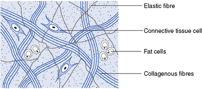
Elastic fibres are fine branching fibres that are highly elastic.
Loose connective tissue
This type of tissue, also called areolar tissue, consists of a loose network of both collagenous and elastic fibres with small scattered groups of fat cells and some fibroblasts (Fig. 3.4). Some blood vessels and nerves are found in the tissue but these are not very numerous. Areolar tissue forms a transparent skin, as thin as tissue paper, but very tough, and it is found between and around the organs of the body.
< div class='tao-gold-member'>
Stay updated, free articles. Join our Telegram channel

Full access? Get Clinical Tree


