The respiratory system

17 The respiratory system


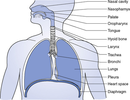
All living cells require a constant supply of oxygen to carry on their metabolism. Oxygen is in the air, and the respiratory system is constructed in such a way that air can be taken into the lungs, where some of the oxygen is extracted for use by the body and at the same time carbon dioxide and water vapour are given up. The organs of the respiratory system (Fig. 17.1) are:


image


The nose


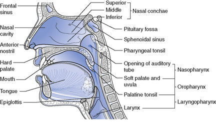
The external nose is the visible part of the nose, formed by the two nasal bones and by cartilage. It is both covered and lined by skin and inside there are hairs that help to prevent foreign material from entering. The nasal cavity (Fig. 17.2) is a large cavity divided by a septum. The anterior nares are the openings that lead in from the outside world and the posterior nares are similar openings at the back, leading into the pharynx. The roof is formed by the ethmoid bone at the base of the skull and the floor by the hard and soft palates at the roof of the mouth. The lateral walls of the cavity are formed by the maxilla, the superior and middle nasal conchae of the ethmoid bone, and the inferior nasal concha. The posterior part of the dividing septum is formed by the perpendicular plate of the ethmoid bone and by the vomer, while the anterior part is made of cartilage.


The three nasal conchae project into the nasal cavity on each side and greatly increase the surface area of the inside of the nose. The cavity of the nose is lined throughout with ciliated mucous membrane, which is extremely vascular; atmospheric air is warmed as it passes over the epithelium, which contains many capillaries. The mucus moistens the air and entraps some of the dust, and the cilia move the mucus back into the pharynx for swallowing or expectoration. The nerve endings of the sense of smell are situated in the highest part of the nasal cavity round the cribriform plate of the ethmoid bone.


Some of the bones surrounding the nasal cavity are hollow. The hollows in the bones are called the paranasal sinuses, which both lighten the bones and act as sounding chambers for the voice, making it resonant. The maxillary sinus lies below the orbit and opens through the lateral wall of the nose. The frontal sinus lies above the orbit towards the midline of the frontal bone. The ethmoidal sinuses are numerous and are contained within the part of the ethmoid bone separating the orbit from the nose, and the sphenoidal sinus is in the body of the sphenoid bone. All the paranasal sinuses are lined with mucous membrane and all open into the nasal cavity, from which they may become infected.


The pharynx


The roof of the pharynx is formed by the body of the sphenoid bone and, inferiorly, it is continuous with the oesophagus (see Fig. 17.2). At the back it is separated from the cervical vertebrae by loose connective tissue, while the front wall is incomplete and communicates with the nose, mouth and larynx. The pharynx is divided into three sections: the nasopharynx, which lies behind the nose; the oropharynx, which lies behind the mouth; and the laryngopharynx, which lies behind the larynx.


The nasopharynx is that part of the pharynx that lies behind the nose above the level of the soft palate. On the posterior wall, there are patches of lymphoid tissue called the pharyngeal tonsils, commonly referred to as the adenoids. This tissue sometimes enlarges and blocks the pharynx, causing mouth breathing in children. The auditory (or acoustic) tubes open from the lateral walls of the nasopharynx and through them air is carried to the middle ear. The nasopharynx is lined with ciliated mucous membrane, which is continuous with the lining of the nose.


The oropharynx lies behind the mouth below the level of the soft palate, with which its lateral walls are continuous. Between the folds of these walls, which are called the palatoglossal arches, are collections of lymphoid tissue called the palatine tonsils. The oropharynx is part of the respiratory tract and alimentary tract but it cannot be used for swallowing and breathing simultaneously. During swallowing, breathing stops momentarily and the oropharynx is completely blocked off from the nasopharynx by the raising of the soft palate. The oropharynx is lined with stratified epithelium.


The larynx


The larynx is continuous with the oropharynx above and with the trachea below (see Fig. 17.2). Above it lies the hyoid bone and the root of the tongue. The muscles of the neck lie in front of the larynx, and behind the larynx lies the laryngopharynx and the cervical vertebrae. On either side are the lobes of the thyroid gland. The larynx is composed of several irregular cartilages joined together by ligaments and membranes (Fig. 17.3).


The thyroid cartilage is formed of two flat pieces of cartilage fused together in the front to form the laryngeal prominence or Adam’s apple. Above the prominence is a notch called the thyroid notch. The thyroid cartilage is larger in the male than in the female. The upper part is lined with stratified epithelium, the lower part with ciliated epithelium.


The cricoid cartilage lies below the thyroid cartilage and is shaped like a signet ring with the broad portion at the back. It forms the lateral and posterior walls of the larynx and is lined with ciliated epithelium.


The epiglottis is a leaf-shaped cartilage attached to the inside of the front wall of the thyroid cartilage immediately below the thyroid notch. During swallowing the larynx moves upwards and forwards so that its opening is occluded by the epiglottis.


The arytenoid cartilages are a pair of small pyramids made of hyaline cartilage. They are situated on top of the broad part of the cricoid cartilage, and the vocal ligaments are attached to them. They form the posterior wall of the larynx.


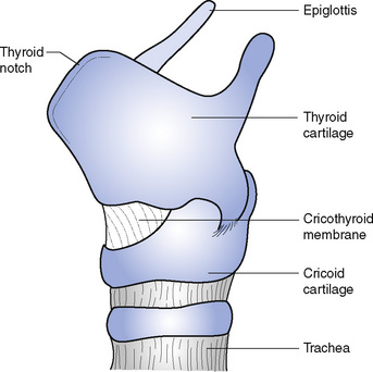
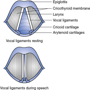
The hyoid bone and the laryngeal cartilages are joined together by ligaments and membranes. One of these, the cricothyroid membrane, is attached all round to the upper edge of the cricoid cartilage and has a free upper border, which is not circular like the lower border but makes two parallel lines running from front to back. The two parallel edges are the vocal ligaments (Fig. 17.4). They are fixed to the middle of the thyroid cartilage in front and to the arytenoid cartilages behind, and they contain much elastic tissue. When the intrinsic muscles of the larynx alter the position of the arytenoid cartilage the vocal ligaments are pulled together, narrowing the gap between them. If air is forced through the narrow gap, called the chink, during expiration, the vocal ligaments vibrate and sound is produced. The pitch of the sound produced depends on the length and tightness of the ligaments: an increased tension gives a higher note; a slacker tension a lower note. Loudness depends on the force with which the air is expired. The alteration of the sound into different words depends on the movements of the mouth, tongue, lips and facial muscles. An inflammation of the larynx is called laryngitis and this may be caused by infections or chemicals. Laryngitis interferes with speech and leads to a hoarse voice.


< div class='tao-gold-member'>

Stay updated, free articles. Join our Telegram channel

Jul 18, 2016 | Posted by in NURSING | Comments Off on The respiratory system

Full access? Get Clinical Tree

Get Clinical Tree app for offline access