25 The reproductive system
Reproduction
Meiosis
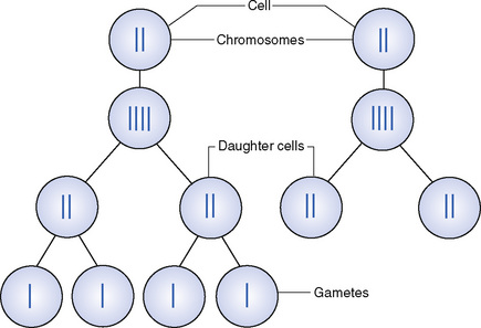
Reproduction in higher animals, including humans, depends on the fusion of a spermatozoon from the male parent and an ovum from the female. These reproductive cells are also called gametes. Each gamete must receive half as many chromosomes as somatic cells so that when they unite in fertilization the full complement of chromosomes is obtained. As the sex cells mature, two processes of cell division occur: the first is mitosis, in which each daughter cell receives a complete set of chromosomes. Following this a two-stage cell division occurs which is peculiar to reproductive tissue and is called meiosis (Fig. 25.1). The first division is similar to mitosis and gives rise to two daughter cells, each containing the original number of chromosomes; the second division follows the first very quickly and results in four gametes, each containing half the number of chromosomes. In humans normal body cells have 46 chromosomes (in 23 pairs) while each gamete has only 23 single chromosomes. Following fusion of the gametes the resulting cell, a zygote, has 46 chromosomes (in 23 pairs). Cell division of a zygote is by mitosis and the resulting multicelled organism is called an embryo.
Sex determination
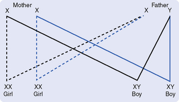
One pair of chromosomes from the father and one pair from the mother are the sex chromosomes which will determine the sex of the child. In the female the sex chromosomes are the same and are called XX. In the male they are different and are called XY. One chromosome from each pair will determine the sex of the child. If the child has an X chromosome from the mother and an X chromosome from the father, it will be a girl (XX). If the child has an X chromosome from the mother and a Y chromosome from the father, it will be a boy (XY) (Fig. 25.2).
The central dogma of genetics
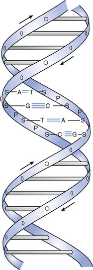
DNA consists of two strands held together in a double helix by hydrogen bonds (Fig. 25.3). The primary structure of the bases in DNA is the basis of the genetic code, which gives rise to all of our characteristics. The code consists of triplets (known as codons), each of which leads to the incorporation of an amino acid into a protein molecule.
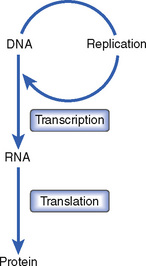
DNA can replicate and conserve the genetic code during cell division (see Chapter 2) and can also be transcribed into ribonucleic acid (RNA) (Fig. 25.4). RNA also contains the genetic code and is used as a template for the building of unique protein molecules. This process is, obviously, under very close regulation in living cells to ensure that the right protein molecules are made as required. Disturbance of this process and the unregulated production of particular cells are called ‘cancer’.
The male genital organs
The male genital organs (Fig. 25.5) consist of six components:
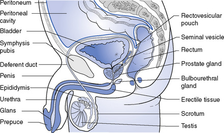
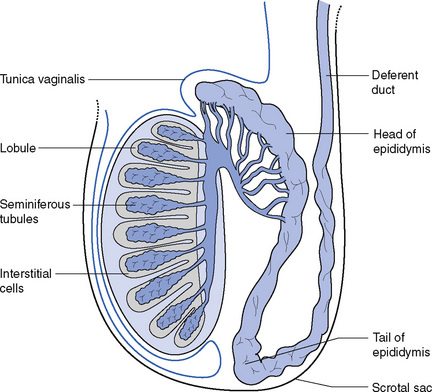
The testes (Fig. 25.6) are the reproductive glands in the male. They are suspended in the scrotum by the spermatic cords but they develop high up in the abdomen close to the kidneys and gradually descend through the inguinal canal into the scrotum shortly before birth. Occasionally one, or both, glands fails to descend and remains in the abdomen or in the inguinal canal; surgery may then be required to relocate it (or them).
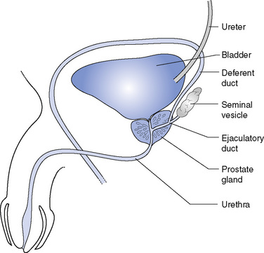
The seminal vesicles (Fig. 25.7) are two pouches lying between the base of the bladder and the rectum. They secrete an alkaline fluid containing nourishment, which forms a large part of the seminal fluid.
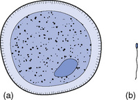
The spermatozoa are minute cells, each with a tail-like projection joined to the cell by a constricted portion called the neck (Fig. 25.8). The tail has a lashing movement that enables the cell to move after the semen leaves the male reproductive tract. As a result, when the spermatozoa are deposited in the vagina they can make their way up the uterus and uterine tubes in search of the ova. They are produced in enormous numbers and it is estimated that, on average, 300 000 000 are deposited in the vagina at one time, though only one is necessary to fertilize the ovum.
< div class='tao-gold-member'>
Stay updated, free articles. Join our Telegram channel

Full access? Get Clinical Tree


