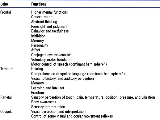
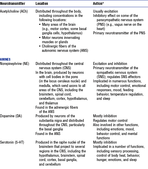
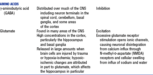
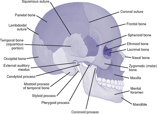
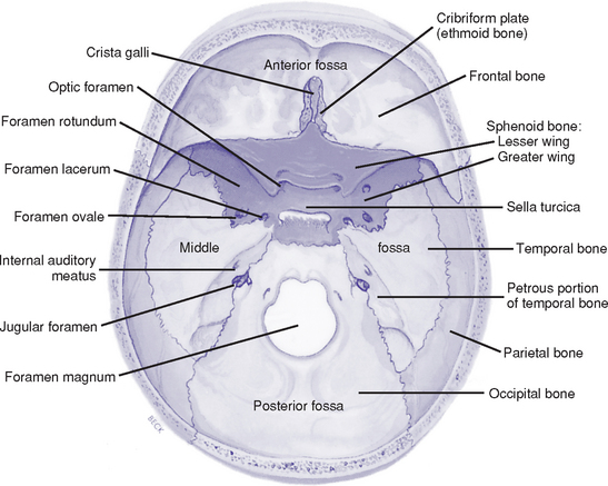
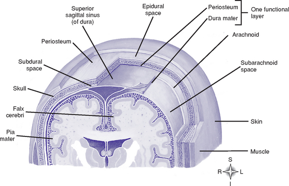
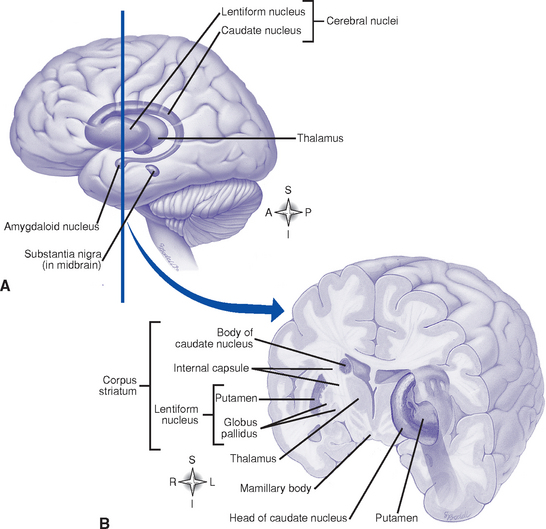
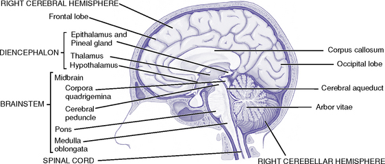
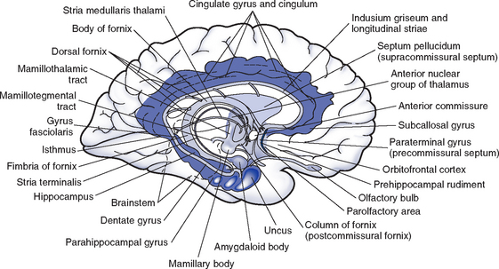
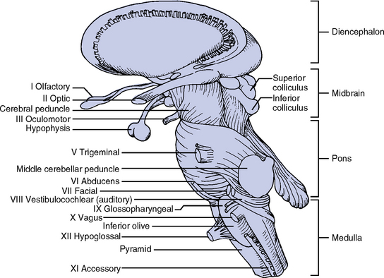
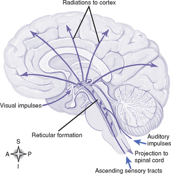
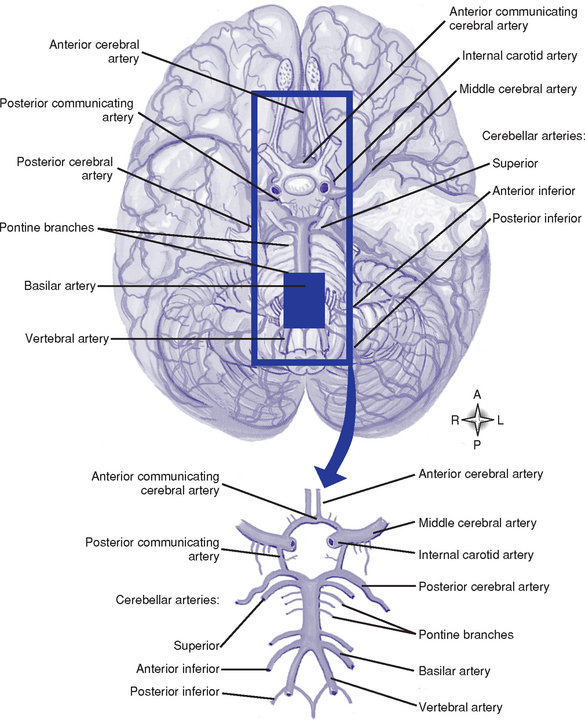
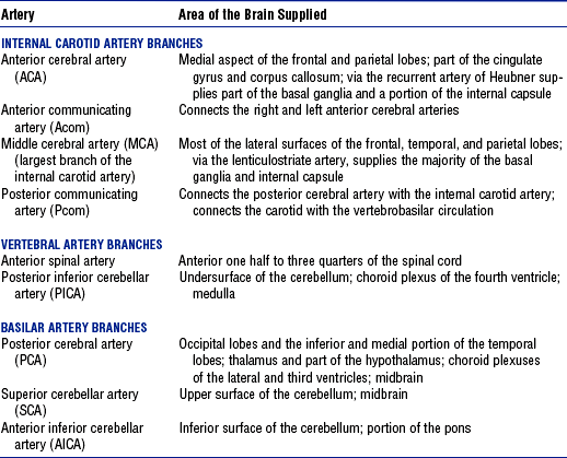
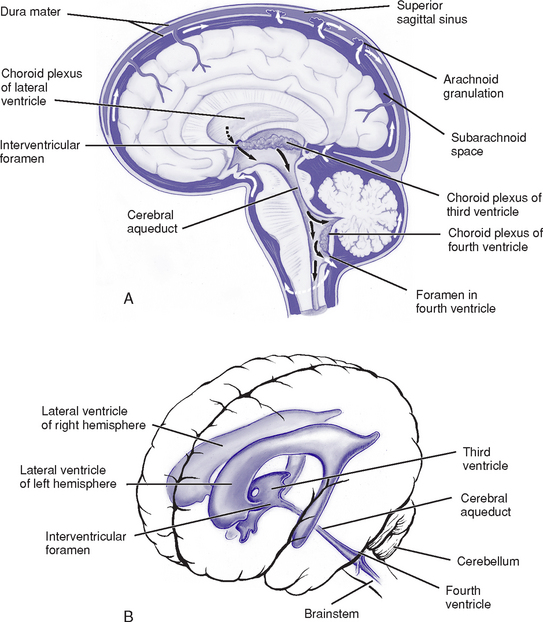
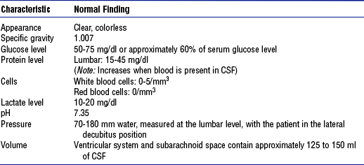
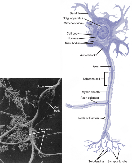
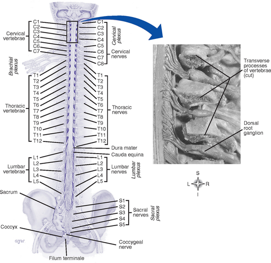
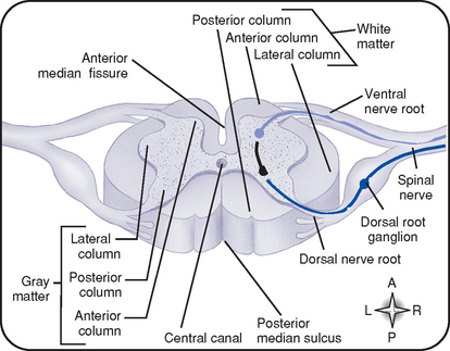
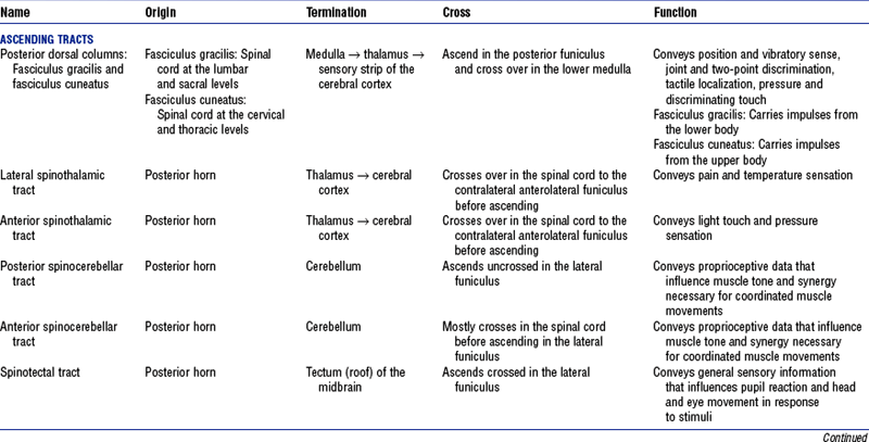
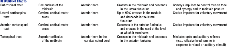
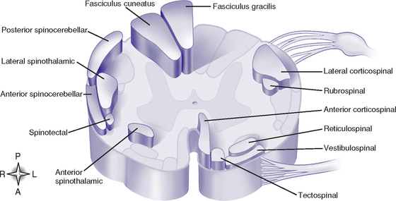
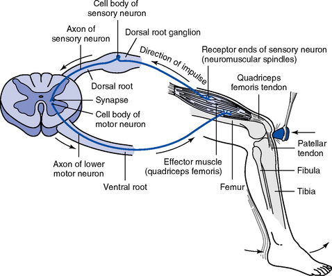
CHAPTER 4 (b) Subcutaneous fascia: Fibrous fatty layer between the skin and galea that contains blood vessels (c) Galea aponeurotica: Freely movable, tendinous tissue; covers the vertex of the skull; absorbs the force of external trauma (d) Subaponeurotic or subgaleal space: Contains the diploic and emissary veins (a) Part of the skull that houses and protects the brain (Figure 4-1) (b) Bones: Frontal, sphenoid, ethmoid, occipital, two temporal and two parietal bones (c) Basilar skull: Base of the skull has three depressions—the anterior, middle, and posterior fossae (Figure 4-2) (1) Outermost covering of the brain; consists of two layers of tough fibrous tissue (2) Outer layer forms the periosteum of the bone (3) Inner layer folds to form the falx cerebri, tentorium cerebelli, falx cerebelli, and diaphragma sella (4) Meningeal arteries and venous sinuses lie in clefts formed by the inner and outer layers of the dura mater (5) Subdural space: Lies between the inner dura mater and the arachnoid mater (1) Fine, fibrous, elastic layer between the dura mater and pia mater a) Lies between the arachnoid mater and pia mater; expanded areas of this space form cisterns at the base of the brain b) Contains blood vessels, including the circle of Willis c) Contains cerebrospinal fluid (CSF), which completely surrounds the brain and spinal cord; acts as a shock absorber d) Contains the arachnoid villi: Projections of the arachnoid mater that absorb CSF into the venous system (a) Telencephalon: Two cerebral hemispheres separated by a longitudinal fissure; joined by the corpus callosum (1) Functional localization in the cerebral cortex, including cerebral dominance (Table 4-1) TABLE 4-1 Functional Localization in the Cerebral Cortex *Cerebral dominance: In right-handed and most left-handed people, the left cerebral hemisphere is dominant for language, mathematical, and analytic functions. The opposite nondominant hemisphere is thought to be concerned with nonverbal, geometric, spatial, visual, and musical functions. (2) Corpus callosum: Commissural fibers that transfer learned discriminations, sensory experiences, and memory from one cerebral hemisphere to corresponding parts of the other (3) Basal ganglia (basal nuclei) (Figure 4-4) a) Masses of gray matter; includes the caudate, putamen, globus pallidus, claustrum, amygdaloid, and, functionally, the subthalamic and substantia nigra nuclei b) Functions: Exert regulating and controlling influences on the coordination of voluntary motion, motor integration, movement initiation, muscle tone, and postural reflexes. A major center of the extrapyramidal motor system. (1) Thalamus: Two egg-shaped masses of gray matter that abut the lateral walls of the third ventricle; subdivided into several nuclei a) Certain nuclei receive, integrate, and process sensory input for relay to the cerebral cortex b) Other nuclei participate in affective aspects of brain function; are functionally related to the association areas of the cortex; or have a role in conscious pain, temperature, and touch awareness, motor function, and the ascending reticular activating system (2) Hypothalamus: Below the thalamus; regulates c) Behavior: Part of the limbic system; concerned with aggressive and sexual behavior; elicits physical expressions associated with emotions; may be involved with sleep-wake cycles and circadian rhythm control d) Autonomic responses: Control center for the autonomic nervous system (ANS); controls numerous visceral and somatic activities (e.g., heart rate, pupil constriction and dilation) e) Hormonal secretion of the pituitary gland (see Chapter 6) 1) Posterior pituitary gland (neurohypophysis): Stores and releases antidiuretic hormone (ADH) and oxytocin, produced by the hypothalamus. ADH causes vasoconstriction and increases renal water reabsorption. Oxytocin stimulates uterine contraction and milk ejection. 2) Anterior pituitary gland (adenohypophysis): Secretes prolactin and growth-stimulating, thyroid-stimulating, adrenal-stimulating, follicle-stimulating, and luteinizing hormones; hormonal secretion is under the control of pituitary releasing and inhibiting factors produced in the hypothalamus and transported to the anterior pituitary via a pituitary portal system (see Chapter 6) (3) Subthalamus: Functionally related to the basal ganglia (4) Epithalamus: Dorsal part of the diencephalon (c) Limbic system (Figure 4-6) (1) Composed of the limbic lobe (cingulate and parahippocampal gyri) plus structures to which it is anatomically and functionally connected such as the amygdala, hippocampus, fornix, hypothalamus, olfactory tract, and thalamus (2) Responsible for emotional behavioral responses and accompanying visceral, endocrine, and somatic responses; has a role in basic instinctual drives (e.g., mating, hunger, motivation) and in some aspects of memory and learning ii. Brainstem (Figure 4-7; also see Figure 4-5) (a) Midbrain (mesencephalon): Located between the diencephalon and pons (1) Contains nuclei of cranial nerve (CN) III (oculomotor) and CN IV (trochlear) and some CN V (trigeminal) nuclei (2) Contains motor and sensory pathways (3) Holds respiratory control centers (4) Tectal region (inferior and superior colliculi): Concerned with the auditory and visual systems (5) Connects to the cerebellum via the superior cerebellar peduncles (b) Pons (metencephalon): Between the midbrain and medulla (1) Contains nuclei of CN V, VI (abducens), and VII (facial), and some CN VIII (acoustic) nuclei (2) Middle cerebellar peduncles on its basal surface provide extensive connections between the cerebral cortex and cerebellum, ensuring maximal motor efficiency (3) Contains motor and sensory pathways (4) Holds respiratory control centers that help coordinate breathing patterns (c) Medulla (myelencephalon): Between the pons and spinal cord (1) Contains nuclei of CN IX (glossopharyngeal), X (vagus), XI (spinal accessory), and XII (hypoglossal) and some nuclei from CN V, VII, and VIII. (2) Motor and sensory tracts of spinal cord continue into the medulla (3) Attaches to the cerebellum via inferior the cerebellar peduncles (4) Holds respiratory, cardiac, and vasomotor control centers (d) Reticular formation (RF) (Figure 4-8): Diffuse cellular network in the brainstem, with axons projecting to the thalamus and into the cortex; receives input from the cerebrum, spinal cord, other brainstem nuclei, and the cerebellum; has a role in the control of autonomic and endocrine functions, skeletal muscle activity, and visceral and somatic sensation. The reticular activating system is part of the RF. iii. Cerebellum: Lies in the posterior fossa behind the brainstem; separated from the cerebrum by the tentorium cerebelli (a) Influences muscle tone in relation to equilibrium, locomotion, posture, and nonstereotyped movements (b) Important in the synchronization of muscle action to enable coordinated movement (c) Input is from the spinal cord, brainstem, vestibular system, and cerebral centers; output to the brainstem and thalamus influences spinal and cerebral activities c. Cerebral circulation (Figure 4-9) i. Arterial system: Supplied by the internal carotid and vertebral arteries (a) Circle of Willis: Anastomosis of arteries at the base of the brain formed by a short segment of the internal carotid and anterior and posterior cerebral arteries, which are connected by an anterior communicating artery and two posterior communicating arteries. This anastomosis may permit collateral circulation if a carotid or vertebral artery becomes occluded. (b) Internal carotid system: Internal carotid arteries arise from the common carotid arteries. Table 4-2 shows the branches of this system and the areas they supply. (c) Vertebral system: Vertebral arteries arise from the subclavian arteries and join at the lower pontine border to form the basilar artery. Branches of this system and the areas they supply are summarized in Table 4-2. (d) Branches of the internal carotid, external carotid, and vertebral arteries (e.g., anterior, middle, posterior meningeal arteries) provide blood supply to the meninges (a) Normal CBF averages 50 ml/100 g of brain tissue per minute (b) Cerebral perfusion pressure (CPP) and intrinsic regulatory mechanisms affect CBF (1) CPP: Pressure gradient that drives blood into the brain; calculated as the difference between the mean arterial pressure (MAP) and the intracranial pressure (ICP): CPP = MAP − ICP (2) Regulatory mechanisms influence the diameter of the cerebrovasculature a) Pressure or myogenic autoregulation: Alteration in the diameter of the brain’s resistance vessels (arterioles) that maintains a constant CBF over a range of pressures between 50 and 150 mm Hg. Chronic hypertension can increase the upper and lower pressure ranges for the range of autoregulation. b) Elevated arterial partial pressure of carbon dioxide (Paco2) and hypoxemia (arterial partial pressure of oxygen [Pao2] of <50 mm Hg) cause vasodilatation and increased CBF; decreased Paco2 causes vasoconstriction and reduced CBF c) Metabolic autoregulation: CBF varies with metabolic activity. Factors that increase the metabolic rate (e.g., seizures, fever) increase CBF; reduced metabolic requirements (e.g., hypothermia) decrease CBF. (3) Inadequate CBF results in brain tissue ischemia (CBF <18 to 20 ml/100 g/min) and death (CBF <8 to 10 ml/100 g/min) iii. Venous system: Brain surface drains into the superficial veins; the central interior cerebrum drains into the internal veins beneath the corpus callosum (Figure 4-10). Veins have no valves. (a) Veins empty into venous sinuses between dural layers (Table 4-3) TABLE 4-3 Major Venous Drainage Structures, Their Locations, and Areas Drained (b) Internal jugular veins collect blood from the large dural venous sinuses and return blood to the heart iv. Blood-brain barrier: Specialized permeability of the brain capillaries that limits transfer of certain substances from blood into brain tissue. Barrier formed by tight junctions between brain capillary endothelial cells, reduced transport mechanisms of these cells, and footlike projections from the astrocytes that encase the capillaries. (a) Water, carbon dioxide, oxygen, glucose, and lipid-soluble substances cross the cerebral capillaries with ease. Uptake of other substances, such as dyes and ions (e.g., Na+, K+), is much slower. (b) Regulates the entry or removal of various substances to maintain a homeostatic environment for the central nervous system (CNS) (c) Clinically significant in treating and diagnosing CNS disease. Blood-brain barrier disruption and increased permeability occurs with brain injury, tumors, infections, and stroke. d. Ventricular system and CSF (Figure 4-11) i. Ventricles: Four cavities containing CSF (a) Lateral ventricles: Largest ventricles, one in each cerebral hemisphere. The anterior (frontal) horns lie in the frontal lobes; the bodies extend back through the parietal lobes to the posterior (occipital) horns, which project into the occipital lobes; the inferior (temporal) horns lie in the temporal lobes. (b) Third ventricle: Midline between the two lateral ventricles, surrounded by the diencephalon (c) Fourth ventricle: In the posterior fossa bordered by the pons, medulla, and superior cerebellar peduncles; continuous with the cerebral aqueduct (aqueduct of Sylvius) superiorly and the central spinal canal inferiorly (a) Cushions the brain and spinal cord from injury (b) Provides support and buoyancy for the brain, decreasing its effective weight on the skull (c) Its displacement out of the cranial cavity (and, to an extent, its increased reabsorption) compensates for increases in intracranial volume and pressure (d) Regulates the nervous system chemical environment to preserve homeostasis (e) Enables water-soluble metabolites to diffuse from the brain (f) Serves as a channel for neurochemical communication within brain iii. CSF properties: See Table 4-4 (a) Rate of synthesis estimated as 500 ml/day or 22 to 25 ml/hr (b) Choroid plexus: Tuft of capillaries covered by epithelial cells found in all ventricles; principal source of CSF; lateral ventricles produce most (c) Small amounts of CSF are produced by the blood vessels of the brain and meningeal linings v. Circulation and absorption of CSF (see Figure 4-11) (a) CSF circulates from the lateral ventricles through the interventricular foramina (foramina of Monro) to the third ventricle and to the fourth ventricle via the aqueduct of Sylvius; CSF then circulates to the subarachnoid space via the foramina of Luschka and Magendie (b) Most CSF is absorbed via the arachnoid villi into the dural sinuses (c) When CSF pressure exceeds venous pressure, CSF is absorbed through the unidirectional valves of the arachnoid villi vi. Blood-CSF barrier: Choroid plexus epithelium imposes a barrier analogous to the blood-brain barrier; permits selective transport of substances from the blood into the CSF i. Brain has high metabolic energy requirements; energy primarily used for neuronal conductive and metabolic activities ii. At rest, the brain consumes 25% of body glucose and 20% of body oxygen; cerebral oxygen consumption averages 49 ml/min iii. Brain utilizes glucose as its principal energy source iv. Minimal storage of oxygen and glucose in the brain necessitates a constant supply for normal neuronal function v. Anaerobic glucose metabolism (glycolysis) yields insufficient adenosine triphosphate (ATP) to meet cerebral energy demands. Rate of glycolysis increases markedly during hypoxia in an attempt to maintain functional neuronal activity. vi. Within seconds to minutes of anoxia, the energy-dependent sodium-potassium pump fails; cytotoxic cerebral edema results vii. Hypoglycemia causes neuronal dysfunction and may lead to convulsions, coma, and death f. Cells of the nervous system i. Neuron: Basic functional unit of the nervous system; transmits nerve impulses (a) Components of each cell (Figure 4-12) (1) Cell body: Carries out the metabolic functions of the cell; contains a nucleus, cytoplasm, and organelles surrounded by a lipoprotein cell membrane (2) Dendrites: Short branching extensions of the cell body; conduct impulses toward the cell body (3) Axon hillock: Thickened area of the cell body from which the axon originates (4) Axon: Long extension of the cell body; conducts impulses away from the cell body; usually myelinated. Outside the brain, axons are also covered with neurilemma. Branch into several processes at the terminal end. (5) Myelin sheath: White protein-lipid complex that surrounds some axons; laid down by oligodendrocytes in the CNS and by Schwann cells in the PNS (6) Nodes of Ranvier: Periodic interruptions in the myelin covering along the axon. Impulses are conducted from node to node (saltatory conduction), which makes conduction more rapid and efficient. (7) Synaptic knobs: At the terminal ends of the axon; contain vesicles that store neurotransmitter substances ii. Neuroglial cells: Support, nourish, and protect the neurons; about 5 to 10 times as numerous as neurons. Four types: (a) Microglia: Phagocytize tissue debris when nervous tissue is damaged (b) Oligodendroglia: Responsible for myelin formation on axons in the CNS (c) Astrocytes: Contribute to the structure of the blood-brain barrier. Provide nutrients for neurons. Constitute the structural and supporting framework for nerve cells and capillaries. Remove excess potassium and neurotransmitters. Contribute to scar formation in response to neuronal cell injury or death. (d) Ependyma: Line the ventricles of the brain and the central canal of the spinal cord. Regulate the flow of substances from these cavities into the brain. Aid in CSF production. g. Synaptic transmission of impulses: Unidirectional conduction of an impulse from a presynaptic neuron across a junction or synapse to a postsynaptic neuron i. Resting membrane potential (RMP): Voltage difference across the cell membrane when the neuron is resting. Determined by the difference in ion concentrations on either side of the membrane. At rest, cells are positively charged outside and negatively charged inside. ii. Depolarization: Stimulus causes sodium channels to open, which results in an intracellular influx of sodium ions (Na+) (a) These ionic fluxes decrease RMP (b) This depolarization is called the excitatory postsynaptic potential (EPSP) iii. Action potential: If a transient voltage change that occurs with depolarization is of sufficient magnitude (threshold level), an action potential is produced and transmitted (conducted as an impulse) along the nerve fibers in an active, self-propagating process iv. Summation: Simultaneous excitation of numerous excitatory presynaptic terminals (or rapidly successive discharges from the same presynaptic terminal) can add together to cause a progressive increase in the postsynaptic potential that may eventually reach threshold to generate an action potential v. Neurotransmitters (Table 4-5): Chemicals secreted by presynaptic knobs or vesicles (usually located at the axon terminal) that excite, inhibit, or modify the response of a postsynaptic neuron. When an action potential reaches the synaptic knob, calcium channels are opened, allowing Ca++ influx into the knob, which triggers neurotransmitter release. TABLE 4-5 Major Neurotransmitters: Type, Location, and Action *Action is determined by the postsynaptic receptor rather than the neurotransmitter. (a) Transmitter diffuses across the synapse and binds with postsynaptic membrane receptors, which causes certain ion channels to open (b) Excitatory neurotransmitters: Open sodium and potassium channels, which results in postsynaptic membrane depolarization (a) Absolute refractory period: Membrane is unresponsive to any stimulus, so that the neuron is incapable of producing an action potential. Occurs for a fraction of a second after the membrane surpasses the threshold potential. Limits the frequency of the impulses that a cell can generate. (b) Relative refractory period: Neuron can be excited again but only by a very strong stimulus (i.e., summation above threshold); occurs during membrane repolarization (a) At the peak of an action potential, the cell membrane again becomes impermeable to Na+; potassium channels open and allow rapid efflux of K+ from the cell, which thereby reestablishes the RMP (b) RMP returns with the aid of the sodium–potassium–adenosine triphosphatase (ATPase) pump, which pumps Na+ out of the cell and K+ into the cell (a) Inhibitory postsynaptic potential (IPSP) (1) Inhibitory neurotransmitters open potassium and/or chloride channels; this causes increased negativity of the membrane potential, which results in hyperpolarization of the cell membrane (2) Decreases excitability and inhibits impulse transmission (b) Presynaptic inhibition: Reduced amount of neurotransmitter is released; this reduces the magnitude of the EPSP to subthreshold levels a. Vertebral column (Figure 4-13) (1) Support the head and neck; smallest vertebrae (2) Atlas (first cervical vertebra): Supports the head; articulates with the occipital bone superiorly and the axis inferiorly (3) Axis (second cervical vertebra) (b) Thoracic: Twelve vertebrae; articulate with the ribs; support the chest muscles (c) Lumbar: Five vertebrae; support the lower back muscles; the largest and strongest vertebrae (d) Sacral: Five fused vertebrae; form a large triangular bone, the sacrum ii. Anatomic features of a typical vertebra (a) Body: Flat round, solid portion; lies anteriorly (b) Arch: Posterior part of the vertebra. Consists of: (1) Pedicles: Two short bony projections that extend posterior from the body (2) Lamina: Join each pedicle and fuse posteriorly at the midline to complete the arch; processes project from the laminae (3) Spinous process: Midline projection protruding posteriorly from the laminae (4) Transverse processes: Projections from the laminae on each side of the vertebrae (5) Articular processes (facets): Projections from the laminae that protrude upward or downward (superior or inferior articulating processes); inferior processes articulate with the superior processes of the vertebra directly below (c) Intervertebral foramina: Openings between the vertebrae through which spinal nerves pass (d) Spinal foramina: Opening between the arch and the body through which the spinal cord passes (a) Fibrocartilage layer between the bodies of adjoining vertebrae (c) Composed of the annulus fibrosus (tough outer layer) and nucleus pulposus (gelatinous inner layer) iv. Spinal ligaments: Hold the vertebrae and discs in alignment; prevent excessive spinal flexion or extension i. Location: Extends from the superior border of the atlas to the first or second lumbar vertebra (a) Continuous with the medulla oblongata (b) Conus medullaris: Caudal end of the spinal cord (c) Central canal: In the center of the spinal cord; contains CSF and is continuous with the fourth ventricle (d) Filum terminale: Nonneural filament that extends downward from the conus medullaris and attaches to the coccyx; helps maintain the position of spinal cord during trunk movement ii. Meninges: Continuous with the layers covering the brain iii. Gray matter (Figure 4-14) (a) An H-shaped, internal mass of gray substance surrounded by white matter; consists of cell bodies and their dendrites and axons (b) Anterior gray column (anterior horn): Contains cell bodies of efferent motor fibers (c) Lateral column: Contains preganglionic fibers of the ANS (d) Posterior gray column (posterior horn): Contains cell bodies of afferent sensory fibers iv. White matter (see Figure 4-14) (a) Composed of three longitudinal columns (funiculi): Anterior, lateral, and posterior (b) Contains mostly myelinated axons (c) Funiculi contain tracts (fasciculi): Composed of axons with similar origin, course, and termination that perform specific functions; clinically significant tracts are summarized in Table 4-6; classified as follows (Figure 4-15): (1) Ascending or sensory tracts: Pathways to the brain for impulses that enter the cord via the dorsal roots of the spinal nerves (2) Descending or motor tracts: Transmit impulses from the brain to the motor neurons of the spinal cord that exit via the ventral root of the spinal nerves (d) Most tracts are named to indicate the column in which the tract travels, the location of its cells of origin, and the location of axon termination v. Upper and lower motor neurons (a) Lower motor neurons (LMNs): Spinal and cranial motor neurons that directly innervate muscles. LMN lesions cause flaccid paralysis, muscular atrophy, absent reflexes. (b) Upper motor neurons (UMNs): Located completely in the CNS; regulate LMN activity. UMN lesions are associated with spastic paralysis, clonus, increased tone, hyperactive reflexes, Babinski’s sign. i. Reflex arc: Requires a receptor, sensory neuron, motor neuron, and effector (e.g., muscle or gland) (Figure 4-16) ii. Monosynaptic reflex arc: Direct synapse between the afferent and efferent neurons (a) Stimulation of afferent nerve fibers sends impulses to the spinal cord through the dorsal roots of spinal nerves (b) Impulse synapses with anterior motor neurons, sending out an efferent discharge confined to the axons supplying the muscle from which the afferent impulse originated (a) More than one synapse required to complete the reflex arc (b) Most reflexes are polysynaptic; may involve interneurons (neurons in the CNS that transmit impulses from a sensory neuron to or toward a motor neuron) and multiple spinal segments and/or areas of brain iv. Reciprocal innervation: Impulses that excite motor neurons supplying a particular muscle also inhibit motor neurons of antagonistic muscles 3. Peripheral nervous system (PNS) i. Thirty-one symmetrically arranged pairs of nerves, each possessing a sensory (dorsal) root and a motor (ventral) root: 8 cervical pairs, 12 thoracic pairs, 5 lumbar pairs, 5 sacral pairs, 1 coccygeal pair (see Figure 4-13) ii. Fibers of the spinal nerves (a) Motor fibers: Originate in the anterior gray column of the spinal cord, form the ventral root of the spinal nerve, and pass to skeletal muscles (b) Sensory fibers: Originate in the spinal ganglia of the dorsal roots; peripheral branches distribute to visceral and somatic structures as mediators of sensory impulses to the CNS
The Neurologic System
SYSTEMWIDE ELEMENTS
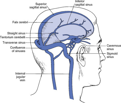
Venous Structure
Location and Area Drained
Superior sagittal sinus
Courses along the midline at the superior border of the falx cerebri; superior cerebral veins empty into it
Straight sinus
Lies in the midline attachment of the falx cerebri and the tentorium; drains the system of internal cerebral veins (including the inferior sagittal sinus and great cerebral vein of Galen)
Transverse sinuses
Lie in the bony groove along the fixed edge of the tentorium cerebelli; drain the straight sinus and the superior sagittal sinus
Sigmoid sinuses
Lie on the mastoid process of the temporal bone and jugular process of the occipital bone; receive blood from the transverse sinuses and empty into the internal jugular veins
Inferior sagittal sinus
Lies along the free inferior border of the falx cerebri just above the corpus callosum; receives blood from the medial aspects of the hemispheres
Emissary veins
Connect the dural sinuses with veins outside the cranial cavity

The Neurologic System


Get Clinical Tree app for offline access