4 The nervous system
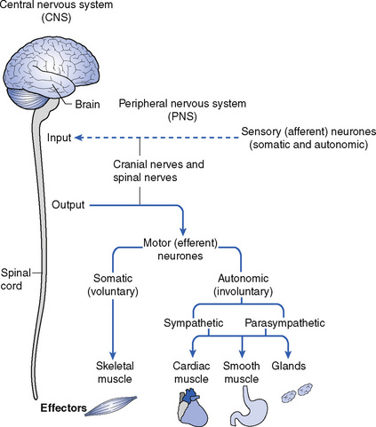
Figure 4.1 shows how the nervous system is organized into the central and peripheral nervous systems and also shows what these divisions of the nervous system are responsible for.
Nervous tissue
When nervous tissue is examined under the microscope it is seen to be composed of:
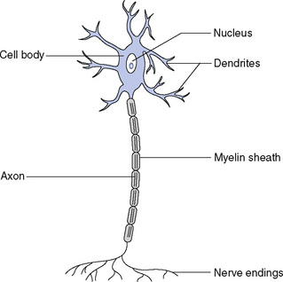
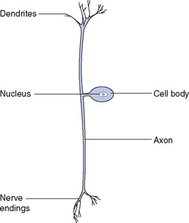
A neurone with many processes arising from the cell body is termed a multipolar neurone (Fig. 4.2). Other types of neurone have one process arising from the cell body, which then divides into two branches, one – the axon – conveying impulses towards the central nervous system, and one conveying impulses from an organ to the cell; these are unipolar neurones (Fig. 4.3). Bipolar neurones have two processes, one at each end of the cell; one is a dendrite carrying impulses to the cell and one is an axon conveying impulses from the cell.
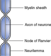
Axons and some dendrites are surrounded by a thin fatty sheath composed of myelin, which lies inside the outer covering of connective tissue, called the neurilemma (Fig. 4.4). The myelin sheath is compressed at intervals and here the neurilemma dips in towards the nerve fibre; these constrictions are called the nodes of Ranvier. At these points the nerve fibre has contact with the surrounding tissue fluids, allowing exchange of nutrients and waste materials.
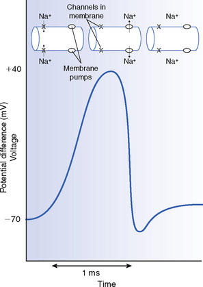
The action potential
When an action potential is generated in a nerve cell by the arrival of an electrical impulse from another nerve cell, positively charged sodium ions (Na+) enter the cell and reverse the potential difference across the membrane to a slightly positive potential. This process is called depolarization. The positive potential difference is rapidly reversed (in less than a millisecond), but not before the process is repeated along the nerve cell, leading to conduction of the electrical impulse. The changes in potential difference are shown in Figure 4.5.
Central nervous system
The brain
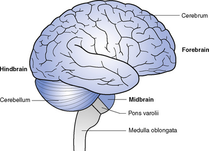
The brain, when fully developed, is a large organ which fills the cranial cavity. Early in its development the brain becomes divided into three parts, known as the forebrain, midbrain and hindbrain (Fig. 4.6).
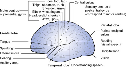
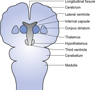
The forebrain is the largest part and comprises the cerebrum; it is divided into right and left hemispheres by a deep longitudinal fissure (Fig. 4.7). The separation is complete at the front and the back but in the centre the hemispheres are joined by a broad band of nerve fibres called the corpus callosum. The outer layer of the cerebrum is called the cerebral cortex and is composed of grey matter (cell bodies) thrown into numerous folds or convolutions called gyri, separated by fissures called sulci. This enables the surface area of the brain, and therefore the number of cell bodies, to be greatly increased. The general pattern of the gyri and sulci is the same in all humans: three main sulci divide each hemisphere into four lobes, each named after the skull bone under which it lies. The central sulcus runs downwards and forwards from the top of the hemisphere to a point just above the lateral sulcus; the lateral sulcus runs backwards from the lower part of the front of the brain; and the parieto-occipital sulcus runs downwards and forwards for a short way from the upper posterior part of the hemisphere. The lobes of the hemispheres are the frontal lobe, lying in front of the central sulcus and above the lateral sulcus; the parietal lobe, lying between the central sulcus and the parieto-occipital sulcus and above the line of the lateral sulcus; the occipital lobe, which forms the back of the hemisphere; and the temporal lobe, lying below the lateral sulcus and extending back to the occipital lobe (Fig. 4.8).
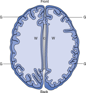
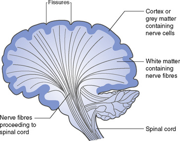
A longitudinal section of a hemisphere shows grey matter (cell bodies) on the outside and white matter (nerve fibres) forming the interior (Fig. 4.9). The nerve fibres connect one part of the brain with other parts and with the spinal cord; however, within the white matter groups of nerve cells can be seen forming areas of grey matter. These areas of grey matter are called cerebral nuclei (Fig. 4.10). The main function of these areas is the co-ordination of movement and posture of the body; disorders affecting these areas cause jerky movements and unsteadiness.
The hindbrain has three parts:
Spinal cord
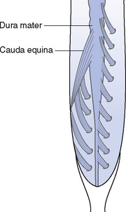
The spinal cord is continuous with the medulla oblongata above and constitutes the central nervous system below the brain. It commences at the foramen magnum and terminates at the level of the first lumbar vertebra; it is approximately 45 cm long. At its lower end, it tapers off into a conical shape called the conus medullaris, from the end of which the filum terminale descends to the coccyx, surrounded by nerve roots called the cauda equina (Fig. 4.11). The cord gives off nerves in pairs throughout its length. It varies somewhat in thickness, swelling out in both the cervical and lumbar regions, where it gives off the large nervous supply to the limbs. The cord is deeply cleft back and front, so that it is almost completely divided into right and left sides like the cerebrum.
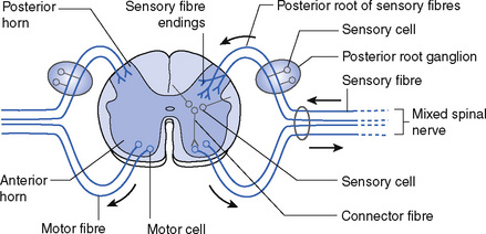
The spinal cord, like the medulla, consists of white matter on the surface and grey matter in the centre. The white matter consists of fibres running between the cord and the brain only, not to the body tissues (Fig. 4.12). The cord contains:
The motor system
The motor system is concerned with the movement of various parts of the body. The motor area is situated in front of the central sulcus in an area called the precentral gyrus. Here the body is represented upside-down. At the bottom of the gyrus is a large area for the head and eye; above that is a large area for the hand and arm, then a small area for the trunk and a large area for the leg extending over the top of the cerebral hemisphere (see Fig. 4.8). There is considerable overlap between these areas. It will be apparent that an area that can undertake a great deal of fine movement, such as the hand and arm, will require a larger area of cell bodies than an area such as the trunk which, although greater in extent, does not carry out as much detailed movement. In front of the motor area lies the premotor area which is concerned with a whole pattern of movement.
Beginning from cells in the motor area the corticospinal fibres pass downwards in a fan shape (see Fig. 4.9) and then pass through the internal capsule, which lies between the thalamus and the basal ganglia. All the motor fibres serving one side of the body are gathered together in the internal capsule so injury there will cause paralysis in the affected side (hemiplegia). The fibres then pass through the pons to the medulla oblongata where they form a long narrow projection called a pyramid. In the pyramids, most of the motor fibres cross over to the other side, at the decussation of the pyramids, so that fibres that arose in the left cerebral hemisphere will now be on the right side of the tract and will supply the right side of the body. The fibres then pass down the spinal cord as the lateral corticospinal tract. The fibres that did not cross to the opposite side at the decussation of the pyramids pass down the spinal cord in the anterior corticospinal tract and eventually cross to the opposite side in the spinal cord.
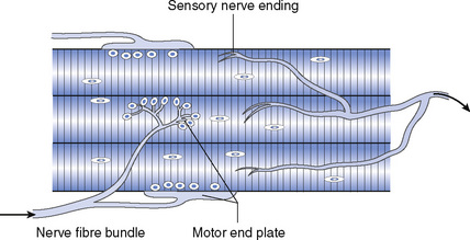
The fibres pass to the anterior horn of the spinal cord, where they form a synapse with the cell bodies situated there, and then emerge from the front of the spinal cord as the anterior root and join the corresponding posterior root of sensory fibres to form a mixed spinal nerve (see Fig. 4.12). As peripheral nerves, these end in branches to various areas, including muscles. The motor fibres divide into branches and each branch ends in a motor end plate attached to an individual muscle fibre (Fig. 4.13). Sensory fibres have their cells in the posterior root ganglion on the spinal nerves and have endings of various types within the muscles.
< div class='tao-gold-member'>
Stay updated, free articles. Join our Telegram channel

Full access? Get Clinical Tree


