The heart and blood vessels

14 The heart and blood vessels


The purpose of this chapter is to consider the anatomical location and structure of the heart and major blood vessels. Some aspects of the function of the heart are included, such as the cardiac cycle, the pathway of blood around the heart and the generation of electrical activity at the sinoatrial node.


The heart


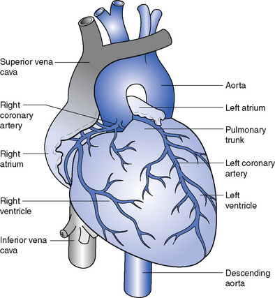
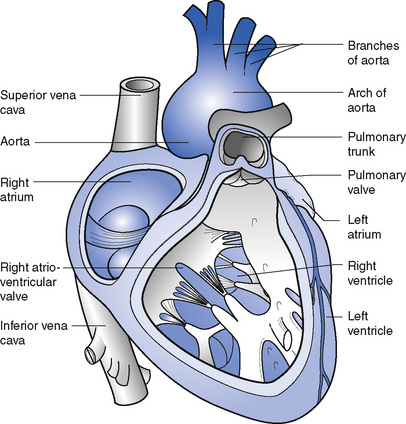
The heart is a hollow, muscular, cone-shaped organ (Fig. 14.1). It lies between the lungs in an area called the mediastinum, behind the body of the sternum with two-thirds of its bulk on the left side. The circular base of the cone is directed upwards and to the right and the apex points downwards, forwards and to the left. It is usually on the level of the fifth intercostal space about 9 cm from the midline. The heart measures about 12 cm from base to apex and about 9 cm in width; it is about 6 cm thick.


General structure


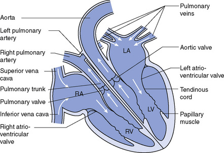
The heart is divided from base to apex by a muscular partition called the septum. The two sides of the heart have no communication with each other in health. Additionally, each side is subdivided into an upper and lower chamber. The upper chamber on each side, the atrium, is smaller and is a receiving chamber into which the blood flows through veins. The lower chamber, the ventricle, is the discharging chamber from which the blood is driven into the arteries. Each atrium communicates with the ventricle below it on the same side of the heart through an opening, guarded by a valve called the atrioventricular valve (Fig. 14.2).


The heart is composed of cardiac muscle, the myocardium, on the action of which the circulation of the blood depends. The myocardium varies in thickness, being thickest in the left ventricle, thinner in the right ventricle and thinnest in the atria.


The atria and ventricles are lined with a thin, smooth, glistening membrane called the endocardium, which consists of a single layer of endothelial cells and which is continuous with the valves and the lining of the blood vessels.


The pericardium covers the heart and the roots of the great vessels and has two layers. The outer layer of fibrous pericardium is securely anchored to the diaphragm, the outer coats of the great vessels and the posterior surface of the sternum and therefore maintains the heart in its position in the chest. Because of its fibrous nature it also prevents overdistension of the heart. The inner layer, the serous pericardium, lines the fibrous pericardium and is invaginated by the heart; it therefore also has two layers. The inner layer is known as the visceral portion, or epicardium, and it is reflected back to form the outer or parietal layer. The layers are normally all in close contact and are moistened by fluid that exudes from the serous membrane; this prevents any friction as the heart continually contracts and relaxes. In inflammatory conditions, such as pericarditis, the amount of fluid within the pericardium may embarrass the action of the heart and may be aspirated.


Valves


The heart is provided with valves to prevent the blood from flowing in the wrong direction. There are four main valves.


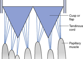
The right atrioventricular (or tricuspid) valve lies between the right atrium and the right ventricle. It consists of three triangular flaps or cusps, each consisting of a double layer of endocardium strengthened with fibrous tissue. The undersurface of the cusps provides attachment for a number of fine, tendinous cords, called the chordae tendinae, which originate in the papillary muscles in the wall of the ventricle. When the ventricle contracts, the blood is pushed back towards the atrioventricular opening but is prevented from entering the atrium by the cusps of the valve, which close because of the increased pressure in the ventricle. The contraction of the papillary muscles exerts tension on the chordae tendinae, which prevent the valve cusps from being carried into the right atrium (Figs 14.3 and 14.4).


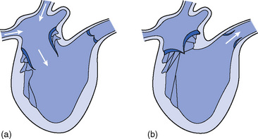
The left atrioventricular valve is also called the mitral valve (due to its resemblance to a bishop’s mitre) (Fig. 14.5). The structure is similar to that of the right atrioventricular valve. It prevents the backflow of blood into the left atrium during contraction of the left ventricle.


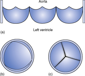
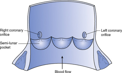
The aortic valve consists of three cusps, which surround the entrance into the aorta from the left ventricle (Figs 14.6 and 14.7). The cusps are half-moon-shaped and are fixed by their curved edges to the wall of the aorta, the straight edge being free so that pockets are formed facing into the aorta. As the blood flows from the left ventricle into the aorta the cusps lie flat against the vessel wall; as the ventricle relaxes the pockets fill with blood and bulge out, meeting in the centre and blocking the opening completely, thus preventing blood flowing back into the ventricle. The coronary arteries, which supply the heart muscle with oxygenated blood, arise from the aorta just above the attached edges of the cusps of the aortic valve.


The pulmonary valve guards the opening from the right ventricle into the pulmonary trunk. It is similar in structure and action to the aortic valve.


The blood that returns to the heart from the myocardium passes through the coronary sinus and pours directly into the right atrium. The opening of the coronary sinus is protected by a thin, semicircular valve, called the valve of the coronary sinus, which prevents backflow of blood into the sinus during contraction of the right atrium. There is also an imperfect valve guarding the opening from the inferior vena cava into the right atrium; it is called the valve of the inferior vena cava.


The blood supply to the heart muscle is by the right and left coronary arteries, which are the first branches from the aorta. There are a number of places within the heart muscle where these two arteries join, or anastomose, but most of the blood returns from the myocardium in veins that empty into the coronary sinus. Blockage of the left coronary artery by a blood clot leads to a reduction in the supply of blood to the left ventricles: this is a myocardial infarction. Depending on where the infarction takes place the outcome is more or less serious. An infarction near the point where the left coronary artery leaves the aorta can be fatal.


Function


The heart is a pump whose purpose is to drive the blood into and through the arteries, but the right and left sides of the heart function quite separately from one another.


Blood from all parts of the body is returned to the right atrium through the two large veins, the superior and inferior venae cavae. When it is full, the right atrium contracts and drives the blood through the right atrioventricular valve into the right ventricle which, in turn, contracts sending the blood through the pulmonary valve and into the pulmonary trunk. The pulmonary trunk divides into right and left pulmonary arteries, which carry the blood to the lungs where gaseous exchange occurs. The blood is finally collected into four pulmonary veins, which return the blood to the left atrium. When it is full the left atrium contracts, simultaneously with the right atrium, and the blood is driven through the left atrioventricular valve into the left ventricle. This chamber contracts, simultaneously with the right ventricle, and sends the blood into the aorta, the main artery of the body.


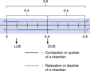
The heart contracts about 70–80 times each minute throughout life, though the rate varies with age, emotion, exercise and other influences. Each beat is a cycle of events that lasts about 0.8 second (Fig. 14.8).


Blood pours into the atria from the great veins until both are full and they then contract simultaneously, emptying their contents into the ventricles. Atrial contraction lasts about 0.1 second. Rising pressure in the ventricle as blood enters forces the atrioventricular valves to close and causes the first heart sound, which can be heard through a stethoscope placed over the apex of the heart and has been likened to the word ‘lub’. Ventricular contraction follows, lasting 0.3 second, and forcing open the pulmonary and aortic valves. Blood is forced into the aorta and pulmonary trunk and as the ventricles relax the pressure in the great vessels forces the aortic and pulmonary valves to close and causes the second heart sound, which can be heard best over the second right rib and has been likened to the word ‘dub’ because it is a sharper sound. During ventricular contraction the atria are relaxed. Following ventricular contraction the whole heart is relaxed for approximately 0.4 second. The word for contraction is systole and that for relaxation is diastole.

< div class='tao-gold-member'>

Stay updated, free articles. Join our Telegram channel

Jul 18, 2016 | Posted by in NURSING | Comments Off on The heart and blood vessels

Full access? Get Clinical Tree

Get Clinical Tree app for offline access