6 The eye
The structure of the eye
The eye is spherical and is embedded in fat. It has three coats: an outer fibrous coat, a vascular, pigmented coat and an inner nervous coat (Fig. 6.1).
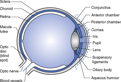
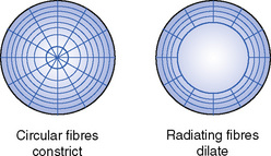
The vascular, pigmented coat has three parts. The choroid lines all but the front part of the eye. It is dark brown and supplies blood to the other layers of the eye, particularly the retina. The ciliary body is a thickened part of the middle coat containing muscular and glandular tissue. The ciliary muscles control the shape of the lens, enabling it to focus light rays from near or far away as required. They are known as the muscles of accommodation. The ciliary glands produce a watery fluid, the aqueous humour, which fills the eye in front of the lens and passes into the veins through small openings in the angle between the iris and the cornea. The iris is the coloured part of the eye. It lies between the cornea and the lens and divides the space between them into anterior and posterior chambers. The iris contains muscular tissue arranged in circular and radiating fibres; the circular fibres contract the pupil and the radiating fibres dilate it. There is a circular opening in the centre called the pupil which is contracted in bright light to prevent too much light entering the eye and dilated in poor light to allow as much light as possible to reach the retina (Fig. 6.2).
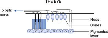
The inner lining of the eye is called the retina. It is a delicate membrane adapted for the reception of light rays and contains many nerve cells and fibres. It is made of rods and cones (Fig. 6.3), which are thought to have separate functions. The cones are more numerous in the centre of the retina and are responsible for detailed vision and colour perception; the rods are more numerous around the outer edge of the retina and are sensitive to the movements of objects within the field of vision. They contain a pigment called visual purple for the synthesis of which vitamin A is required; lack of vitamin A in the diet may result in night blindness. Near the centre of the back of the retina there is an oval yellowish area called the macula lutea where only cones are present. This is the area where an image is formed by light from an object being looked at and is the point where vision is clearest. About 3 mm to the nasal side of the macula lutea the optic nerve leaves the eye; this area is called the optic disc and, because it is insensitive to light, it is called the blind spot. An object can only be opposite the blind spot in one eye at a time. To find a blind spot, mark a piece of paper as shown here:
< div class='tao-gold-member'>
Stay updated, free articles. Join our Telegram channel

Full access? Get Clinical Tree


