The ear

5 The ear


The ear is the organ of hearing and also plays an important part in maintaining balance. The external ear, the middle ear and the cochlea of the internal ear are concerned with hearing; the semicircular canals, the utricle and the saccule of the internal ear are concerned with balance.


The structure of the ear


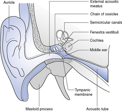
The external ear has two parts: the auricle and the external acoustic meatus (Fig. 5.1). The auricle projects from the side of the head. It is composed of a thin piece of elastic fibrocartilage, covered with skin, which funnels sound waves towards the external acoustic meatus. The external acoustic meatus is a tubular passage approximately 4 cm long leading into the temporal bone. The outer one-third has walls of cartilage and the inner two-thirds walls of bone; the canal is curved, running first forwards and upwards, then backwards and upwards and finally forwards and slightly downwards. These curves may be straightened by gentle traction on the auricle: in adults the pull is upwards and backwards; in children backwards only; and in infants downwards and backwards. The inner end of the meatus is closed by the tympanic membrane. The skin lining the cartilaginous meatus contains hair follicles and numerous glands, which secrete cerumen. These protect the canal from foreign bodies by entangling dust and other particles but the cerumen may itself block the canal if it accumulates and will then require removal.


The middle ear is a small space within the temporal bone. The tympanic membrane separates it from the external ear and its further (medial) wall is formed by the lateral wall of the internal ear. The cavity is lined with mucous membrane and is filled with air, which enters from the pharynx through the acoustic tube. This equalizes air pressure on both sides of the tympanic membrane. It contains a chain of three tiny bones, the ossicles, which transmit the vibrations of the tympanic membrane across to the internal ear. The tympanic membrane is thin and semitransparent and the handle of the malleus, the first of the ossicles, is firmly attached to the inner surface. The incus articulates with the malleus and with the stapes, the base of which is attached to the fenestra vestibuli, which leads to the internal ear. The posterior wall of the middle ear has an irregular opening leading into the mastoid antrum and this in turn leads to a number of mastoid air cells. These are air-filled cavities within the bone which, like the nasal sinuses, may become infected.


The internal ear lies in the petrous part of the temporal bone. It consists of two parts: the bony labyrinth and the membranous labyrinth.


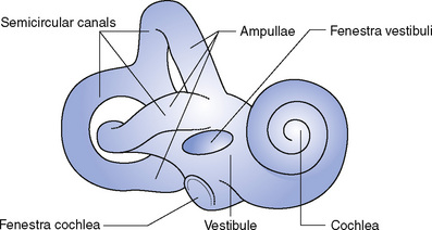
The bony labyrinth is again divided into three parts: the vestibule, the cochlea and the semicircular canals (Fig. 5.2).


< div class='tao-gold-member'>

Stay updated, free articles. Join our Telegram channel

Jul 18, 2016 | Posted by in NURSING | Comments Off on The ear

Full access? Get Clinical Tree

Get Clinical Tree app for offline access