18 The digestive system
The digestive system (Fig. 18.1) consists of all the organs that are concerned in the chewing, swallowing, digestion and absorption of food and in the elimination from the body of indigestible and undigested food. It consists of the digestive tube or alimentary canal and the accessory organs of digestion.
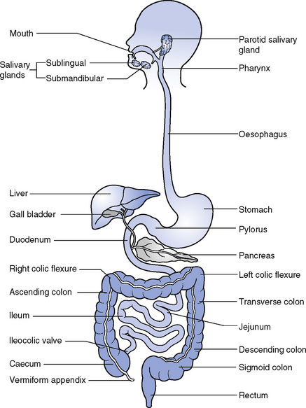
The digestive tract is about 9 m long and consists of six parts:
The accessory organs of digestion are:
The mouth
The mouth is a cavity bounded externally by the lips and cheeks and leading into the pharynx. The roof is formed by the hard and soft palates, and the anterior two-thirds of the tongue fills the floor of the mouth (Fig. 18.2). The walls are formed by the muscles of the cheeks. The mucous membrane, which lines the mouth, is continuous with the skin of the lips and with the mucous lining of the pharynx. The lips enclose the orbicularis oris muscle, which keeps the mouth closed. The hard palate is formed by parts of the palatine bones and the maxillae; its upper surface forms the floor of the nasal cavity. The soft palate is suspended from the posterior border of the hard palate and extends downwards between the oral and nasal parts of the pharynx. Its lower border hangs like a curtain between the mouth and the pharynx and a small conical process, called the uvula, hangs down from it. Two curved folds of mucous membrane extend sideways and downwards from each side of the base of the uvula, called the palatoglossal and palatopharyngeal arches, between which lie the masses of lymphoid tissue known as the palatine tonsils.

Tongue
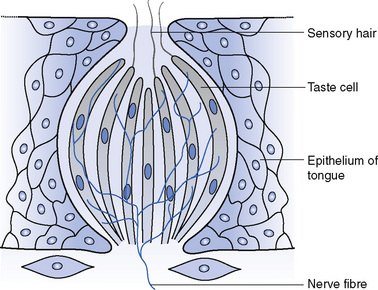
The tongue is a muscular organ that is attached to the hyoid bone and the mandible. It is covered in certain areas with modifications of the mucous membrane, which appear as projections called papillae, to increase the surface area. In addition, specialized areas called taste buds (Fig. 18.3) are widespread over almost the entire area of the tongue. The undersurface of the anterior part of the tongue is connected to the floor of the mouth by a fold of mucous membrane called the frenulum. The functions of the tongue are:
Teeth
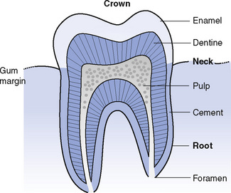
Each tooth consists of three parts (Fig. 18.4):
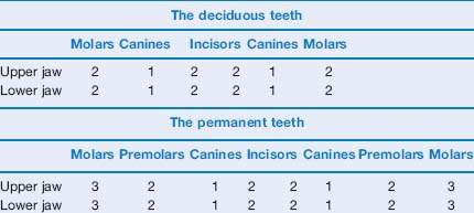
There are four types of teeth:
There are 20 deciduous teeth and 32 permanent teeth (Table 18.1).
The pharynx
When food is well chewed and moistened the tongue rolls it into a bolus and carries it towards the oropharynx. The soft palate rises up to occlude the nasopharynx and the epiglottis moves upwards and forwards so that the bolus passes over the closed inlet of the larynx and on into the laryngopharynx and then to the oesophagus. This is an excellent example of muscular co-ordination and if it is not achieved correctly, choking will result (see Fig. 17.2).
The oesophagus
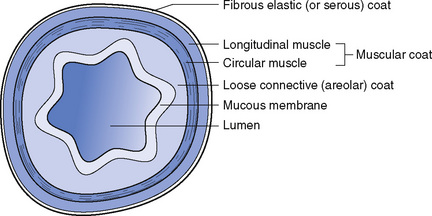
The oesophagus has four layers and is similar in structure to the remainder of the alimentary canal (Fig. 18.5):
The stomach
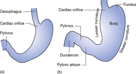
The stomach is the most dilated part of the digestive tube and is situated between the end of the oesophagus and the beginning of the small intestine. Its shape and position are altered by changes within the abdominal cavity and by the stomach contents. It lies below the diaphragm, slightly to the left of the midline (Fig. 18.6).
The wall of the stomach consists of four layers (Fig. 18.7):
Stay updated, free articles. Join our Telegram channel

Full access? Get Clinical Tree


