15 The circulation
After reading this chapter you should understand:
The circulation of the blood is divided into three main parts:
The systemic circulation
The vessels carrying the blood from the left ventricle through the body generally to the right atrium are those that constitute the systemic circulation. Arteries may subdivide into several branches at the same point or several branches may be given off in succession. Arteries do not always end in capillaries but may unite with one another, forming anastomoses. Examples are found in the brain where two vertebral arteries anastomose to form the basilar artery, and where two anterior cerebral arteries are connected by the anterior communicating artery (see Fig. 14.2). An enlarged anastomosis may provide a collateral circulation if a vessel is occluded by accident or disease; sudden occlusion may be followed by death of the tissue supplied by the vessel whereas gradual occlusion may allow dilation of the anastomosis and adequate nourishment of the tissue. Some arteries have no anastomosis with other arteries and these are called end-arteries. Occlusion of an end-artery will cause death (necrosis) of the tissue supplied by the vessel; an example is the central artery of the retina, following occlusion of which permanent blindness will occur.
Arteries
Aorta
The aorta is the main artery of those that carry oxygenated blood to the tissues of the body (Fig. 15.1). It arises from the upper part of the left ventricle, passes upwards and to the right (the ascending aorta) and then arches backwards to the left (the arch of the aorta) and passes down through the thorax on the left side of the spine (the descending thoracic aorta). It enters the abdominal cavity through an opening in the diaphragm called the aortic hiatus and is then called the abdominal aorta. It ends at the lower border of the fourth lumbar vertebra by dividing into the right and left common iliac arteries.
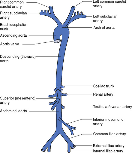
The arch of the aorta has three branches, which arise from the top of the arch:
The vertebral arteries arise from the first part of the subclavian arteries and pass upwards in the foramina of the transverse processes of the cervical vertebrae; they enter the skull through the foramen magnum (see Fig. 9.3) and join together to form the basilar artery.
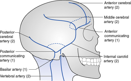
An anastomosis named the circulus arteriosus connects the vertebral arteries and the two internal carotid arteries. This circle is situated at the base of the brain (Fig. 15.2). In front the two anterior cerebral arteries are joined by the anterior communicating artery (Fig. 15.3). Behind, the basilar artery, formed by the junction of the two vertebral arteries, divides into two posterior cerebral arteries, each of which is joined to the internal carotid arteries by the posterior communicating artery. This anastomosis increases the likelihood of an adequate blood supply being maintained to the brain if one of the vessels is injured or occluded.
< div class='tao-gold-member'>
Stay updated, free articles. Join our Telegram channel

Full access? Get Clinical Tree


