Inspect sclerae, conjunctivae, buccal mucosa, tongue, lips, nail beds, and palms. Palpate with dorsal surface of hand or fingers. Measure all dimensions. Use Wood’s lamp to distinguish fluorescing lesions. Transilluminate to determine presence of fluid. Note color, odor, amount, and consistency of lesion. Check lesion for annular, grouped, linear, arciform, or diffuse arrangement. Check lesion for generalized/localized, body region, patterns, or discrete/confluent.
Skin, Hair, and Nails
Examination
Technique
Findings
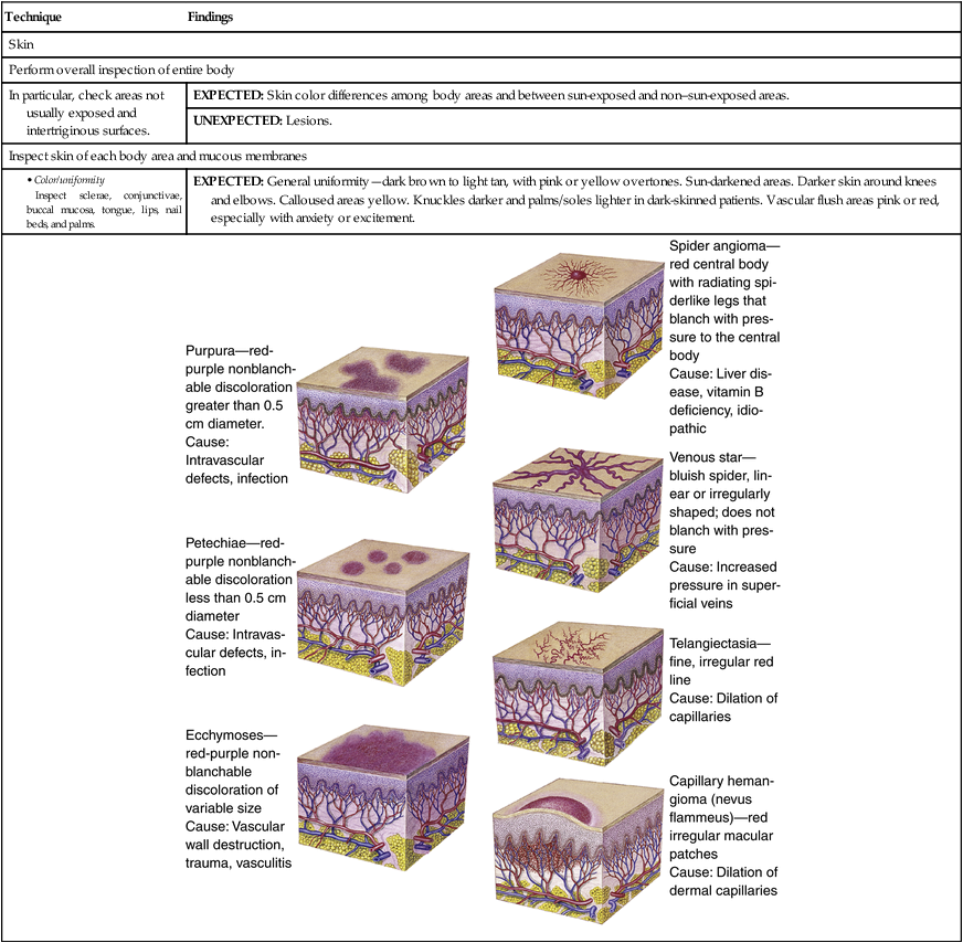
Skin
Perform overall inspection of entire body
In particular, check areas not usually exposed and intertriginous surfaces.
EXPECTED: Skin color differences among body areas and between sun-exposed and non–sun-exposed areas.
UNEXPECTED: Lesions.
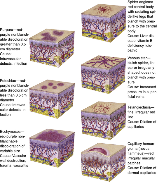
Inspect skin of each body area and mucous membranes
EXPECTED: General uniformity—dark brown to light tan, with pink or yellow overtones. Sun-darkened areas. Darker skin around knees and elbows. Calloused areas yellow. Knuckles darker and palms/soles lighter in dark-skinned patients. Vascular flush areas pink or red, especially with anxiety or excitement.
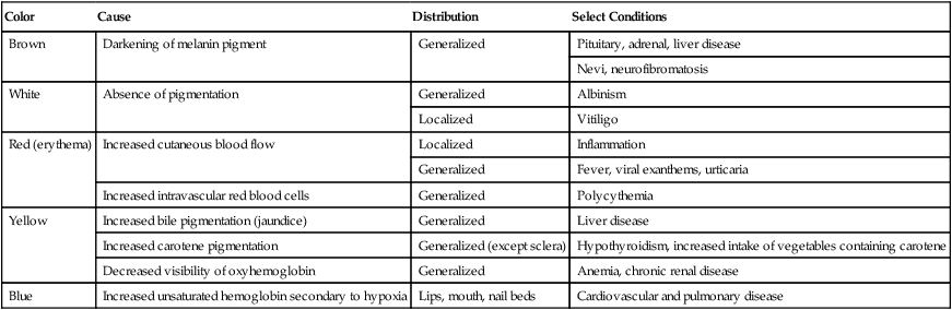
Color
Cause
Distribution
Select Conditions
Brown
Darkening of melanin pigment
Generalized
Pituitary, adrenal, liver disease
Nevi, neurofibromatosis
White
Absence of pigmentation
Generalized
Albinism
Localized
Vitiligo
Red (erythema)
Increased cutaneous blood flow
Localized
Inflammation
Generalized
Fever, viral exanthems, urticaria
Increased intravascular red blood cells
Generalized
Polycythemia
Yellow
Increased bile pigmentation (jaundice)
Generalized
Liver disease
Increased carotene pigmentation
Generalized (except sclera)
Hypothyroidism, increased intake of vegetables containing carotene
Decreased visibility of oxyhemoglobin
Generalized
Anemia, chronic renal disease
Blue
Increased unsaturated hemoglobin secondary to hypoxia
Lips, mouth, nail beds
Cardiovascular and pulmonary disease
Technique
Findings
Pigmented nevi. Nonpigmented striae. Freckles. Birthmarks.
UNEXPECTED: Dysplastic, precancerous, or cancerous nevi. Chloasma. Unpigmented skin. Generalized or localized color changes. Vascular skin lesions. Vascular changes.
EXPECTED: Thickness variations, with eyelids thinnest, areas of rubbing thickest. Calluses on hands and feet.
UNEXPECTED: Atrophy. Hyperkeratosis. Corns.
EXPECTED: Bilateral symmetry.
EXPECTED: Clean.
Palpate skin.
EXPECTED: Minimal perspiration or oiliness. Increased perspiration (associated with activity, environment, obesity, anxiety, excitement) noticeable on palms, scalp, forehead, axillae.
UNEXPECTED: Damp intertriginous areas.
EXPECTED: Cool to warm. Bilateral symmetry.
EXPECTED: Smooth, soft, and even. Roughness resulting from heavy clothing, cold weather, or soap.
UNEXPECTED: Extensive or widespread roughness.
EXPECTED: Resilience.
Gently pinch skin on forearm or in sternal area, and release.
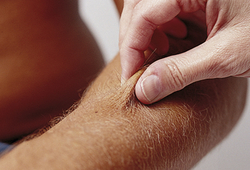
UNEXPECTED: Failure of skin to return to place quickly.
Inspect and palpate lesions
UNEXPECTED: See table on pp. 40-43.
Description
Examples
Macule
Flat, circumscribed area that is a change in skin color; <1 cm in diameter
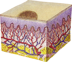
Freckles, flat moles (nevi), petechiae, measles, scarlet fever
Papule
Elevated, firm, circumscribed area <1 cm in diameter 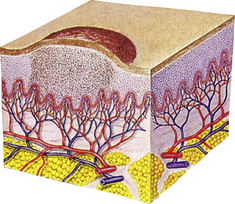
Wart (verruca), elevated moles, lichen planus
![]()
Stay updated, free articles. Join our Telegram channel

Full access? Get Clinical Tree

 Get Clinical Tree app for offline access

Skin, Hair, and Nails

Get Clinical Tree app for offline access