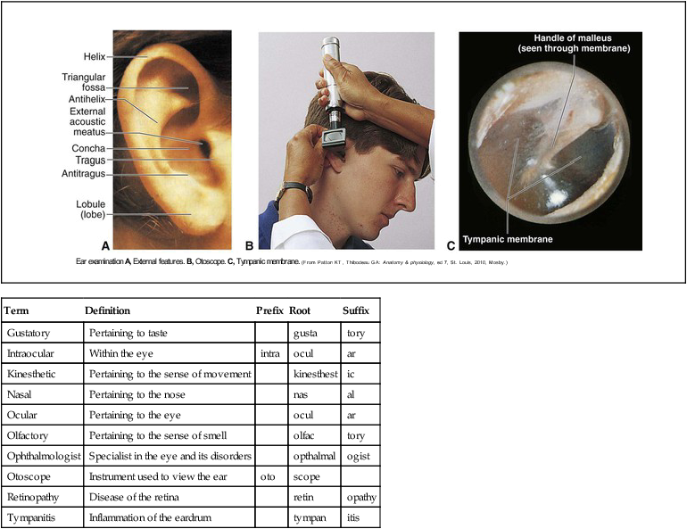
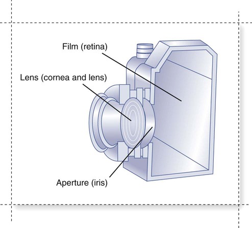
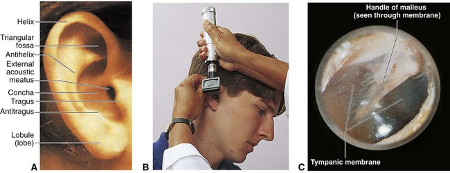
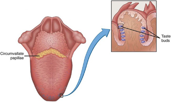
Chapter 20 *A transition syllable or vowel may be added to or deleted from the word parts to make the combining form. The sensory system consists of receptors in specialized cells and organs that perceive changes (stimuli) in the internal and external environment. The stimuli cause nerve impulses that are sent to the brain for interpretation. Environmental stimuli are perceived with the senses of vision, hearing, touch, taste, position, and balance. Specialized organs of the senses include the eye, ear, tongue, nose, and skin. The eyes are considered the most important sensory organ because 90% of the information about the environment reaches the brain from the eyes. The external structures of the eye are shown in Fig. 20-1. The orbital cavity is composed of bones that protect and adipose tissue that cushions the eye. When the eyelids and eyelashes close or “blink,” they protect the eye from injury. The conjunctiva is a mucous membrane that protects and lubricates the eyelids and part of the eye. The lacrimal apparatus forms tears that keep the eye moist and lubricated. Movement of the eye is controlled by the extrinsic muscles. Only one fifth of the eye is actually exposed to the environment. The internal structures of the eye are also shown in Fig. 20-1. The sclera is a tough, white tissue that supports and gives structure to the eye. The sclera is continuous with the transparent cornea that covers the iris and pupil. The cornea focuses light rays on the retina at the back of the eye. The blood supply for the eye originates in the choroid, iris, and ciliary muscles. The iris and ciliary muscles are the intrinsic muscles of the eye. Vision is similar to the action of a camera, beginning with refraction, the process of the lens bending light rays as they enter the eye to focus on the retina (Fig. 20-2). The eye changes the shape of the lens to focus near and far through the process of accommodation. The pupil constricts to focus the object on the retina and protect it from receiving too much light. The eyes converge on the object so that single binocular vision occurs and only one object is seen. Taste, or the gustatory sense, is perceived by specialized cells located in projections (papillae) on the tongue called taste buds. Taste buds are chemoreceptors. The five tastes perceived by the tongue are sweet, sour, bitter, salty, and umami (Fig. 20-4). Umami, described as “meatiness,” is a response to glutamic acid found in processed meats, cheese, and many Asian foods. Each taste bud has 50 to 100 taste cells. These cells respond to all five types of taste. All areas of the tongue sense all five of the tastes. As more research is completed, new “tastes” may be identified. It is possible that “metallic” is also a primary taste. Before a taste can be sensed, the substance must be dissolved in fluid or saliva. The particular flavor of an item is identified by smell as well as taste.
Sensory System
 Define at least 10 terms relating to the sensory system.
 Describe the function of the sensory system.
 Identify at least 10 sensory system structures and the function of each.
 Identify at least three methods of assessment of the sensory system.
Term
Definition
Prefix
Root
Suffix
Gustatory
Pertaining to taste
gusta
tory
Intraocular
Within the eye
intra
ocul
ar
Kinesthetic
Pertaining to the sense of movement
kinesthest
ic
Nasal
Pertaining to the nose
nas
al
Ocular
Pertaining to the eye
ocul
ar
Olfactory
Pertaining to the sense of smell
olfac
tory
Ophthalmologist
Specialist in the eye and its disorders
opthalmal
ogist
Otoscope
Instrument used to view the ear
oto
scope
Retinopathy
Disease of the retina
retin
opathy
Tympanitis
Inflammation of the eardrum
tympan
itis
Structure and Function of the Sensory System
Eye
Tongue
![]()
Stay updated, free articles. Join our Telegram channel

Full access? Get Clinical Tree


Sensory System

 Brain Byte
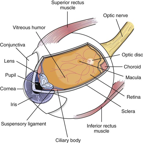
 Brain Byte
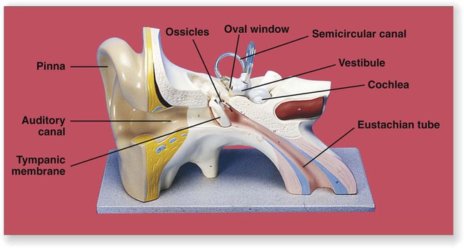
Get Clinical Tree app for offline access
