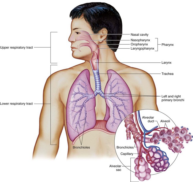
Chapter 13 Respiratory System Terminology* *A transition syllable or vowel may be added to or deleted from the word parts to make the combining form. The respiratory system functions in three ways: There are three processes of respiration: Both the voluntary and involuntary nervous systems control respiration. The body chemically regulates the rate (how fast the breaths are) and depth (how deep they are). If the concentration of gases in the blood changes, the brain adjusts respiratory rate and depth to counteract the changes to maintain homeostasis (Table 13-1). TABLE 13-1 Air enters the respiratory system (Fig. 13-1) through the nose (nasal cavity). The nasal cavity is lined with hairs called cilia to help filter out any foreign particles. The diaphragm is a large, flat muscle that separates the thoracic cavity from the abdominal cavity. The diaphragm contracts and moves downward during inhalation. This creates suction, and air is pulled in from outside the body. Exhalation occurs when the diaphragm relaxes (Fig. 13-2).
Respiratory System
 Define at least 10 terms referring to the respiratory system.
 Describe the three functions of the respiratory system.
 Identify at least 10 respiratory system structures and the function of each.
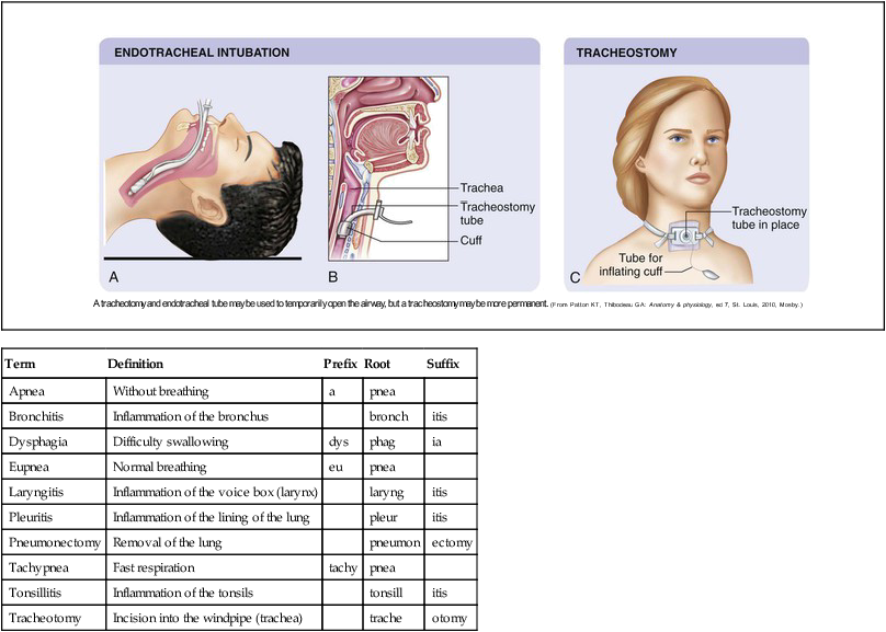
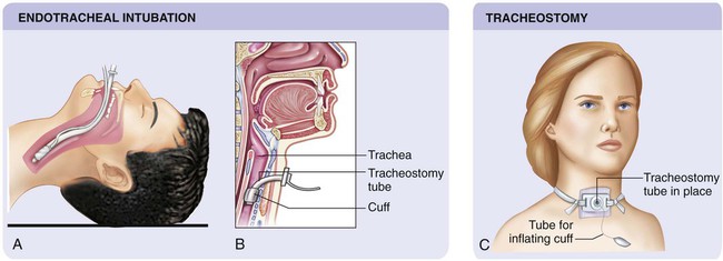
 Describe at least three methods of assessment of the respiratory system.
Term
Definition
Prefix
Root
Suffix
Apnea
Without breathing
a
pnea
Bronchitis
Inflammation of the bronchus
bronch
itis
Dysphagia
Difficulty swallowing
dys
phag
ia
Eupnea
Normal breathing
eu
pnea
Laryngitis
Inflammation of the voice box (larynx)
laryng
itis
Pleuritis
Inflammation of the lining of the lung
pleur
itis
Pneumonectomy
Removal of the lung
pneumon
ectomy
Tachypnea
Fast respiration
tachy
pnea
Tonsillitis
Inflammation of the tonsils
tonsill
itis
Tracheotomy
Incision into the windpipe (trachea)
trache
otomy
Structure and Function of the Respiratory System
 It exchanges gases between the blood and the lungs.
 It helps regulate body temperature by cooling or warming the blood.
 External respiration, or ventilation, brings oxygen into the lungs.
 Internal respiration exchanges oxygen and carbon dioxide between blood and body cells.
 Cellular respiration changes acid produced during metabolism into harmless chemicals in the cells.
Reflex
Cause
Action
Cough
Irritation to the trachea or bronchi
Epiglottis and glottis close, muscles raise pressure in lungs, and then they open, releasing a burst of air to remove foreign matter
Diving
Submersion in cold water
Heart and breathing rate slow, blood diverted to the brain, heart, and lungs until breathing resumes
Gasp
Sudden sensation (cold, surprise, pain, sleep apnea)
Deep inhalation of air through mouth
Hiccup
Cause unknown, lack of sleep, overeating or drinking, stress, chronic hiccups may indicate tumor or laryngitis
Rib muscles and diaphragm contract, causing rapid inhalation
Sneeze
Irritation of nasal passages, respiratory illness, allergy, can be exposure to bright light (genetic)
Eyes and nasal passages secrete fluid, diaphragm causes large breath to be inhaled, followed by contraction of chest muscles to push air out of nose and mouth
Sniff
Identify smell, nasal congestion
Small audible inhalations with little exhalation
Yawn
Bored, tired, stress (hypotheses include low oxygen content in blood, regulation of brain temperature, muscle stretching)
Mouth opens wide to take in deep breath; air is released after lungs are expanded to capacity
![]()
Stay updated, free articles. Join our Telegram channel

Full access? Get Clinical Tree


Nurse Key
Fastest Nurse Insight Engine


 Brain Byte
 Brain Byte
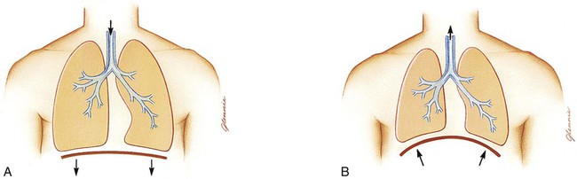
Get Clinical Tree app for offline access
