CHAPTER 2 Respiratory Disorders
Section One Acute Respiratory Disorders
Acute respiratory disorders are short-term diseases or acute complications of chronic conditions. They can occur once and respond to treatment or recur to further complicate an underlying disease process.
Atelectasis
Overview/Pathophysiology
Atelectasis is a spontaneous collapse of alveolar lung tissue secondary to persistent hypoinflation. It is most common following major abdominal or thoracic surgery and results from hypoventilation of dependent portions of the lungs or inadequate clearing of secretions. Atelectasis can be an acute or a chronic condition and occurs most often in individuals with COPD. Postoperatively, atelectasis can be precipitated by the effects of anesthesia, sedation, and decreased mobility. Other precipitating factors include mucus plugs, foreign objects in the airways, pleural effusion, bronchogenic carcinoma, history of smoking, and obesity. Atelectasis can lead to pulmonary infection.
Assessment
The clinical picture is determined by the site of collapse, rate of development, and size of the affected area.
Signs and symptoms/physical findings
Pleuritic chest pain, tachypnea, shortness of breath, fever, dyspnea, decreased chest wall movement on affected side, dullness to percussion, decreased or absent breath sounds, crackles (rales) persisting after deep inspiration or cough, restlessness, agitation, change in level of consciousness (LOC), cyanosis.
Collaborative Management
Management is aimed at preventing this condition in all patients. If atelectasis occurs and is left untreated, the affected lung area eventually may become infected, fibrotic, and functionless.
Deep-breathing and coughing exercises
Expand alveoli deep in the lungs and mobilize/clear secretions.
Hyperinflation therapy
Expands partially collapsed lung areas and thereby improve gas exchange. Incentive spirometry may be used at the bedside.
Bronchoscopy
Patient is intubated and a fiberoptic scope is passed into the bronchi to visualize the area and remove mucous plugs, retained secretions, or foreign objects.
Nursing Diagnoses and Interventions
For Patients at Risk for Atelectasis
Ineffective breathing pattern
related to decreased lung expansion secondary to inactivity or omission of deep breathing
Desired outcomes
Patient demonstrates deep breathing and effective coughing at least hourly and is eupneic (respiratory rate [RR] 12-20 breaths/min with normal depth and pattern) at all other times. Auscultation of patient’s lungs reveals no adventitious sounds.
Nursing Interventions
Deep breathing expands the alveoli and aids in mobilizing secretions to the airways, and coughing further mobilizes and clears the secretions. Monitor patient’s progress and document in nurses’ notes.
Patient-Family Teaching and Discharge Planning
Provide verbal and written information about the following:
Pneumonia
Overview/Pathophysiology
Pneumonia is an acute bacterial or viral infection that causes inflammation of the lung parenchyma (alveolar spaces and interstitial tissue). As a result of the inflammation, involved lung tissue becomes edematous and air spaces fill with exudate (consolidation), gas exchange cannot occur, and nonoxygenated blood is shunted into the vascular system, with resulting hypoxemia. Bacterial pneumonias involve all or part of a lobe, whereas viral pneumonias appear diffusely throughout the lungs.
Influenza, which can cause pneumonia, is the most serious viral airway infection for adults. Patients more than 65 yr old, residents of extended care facilities, and individuals with chronic health conditions have the highest mortality from influenza.
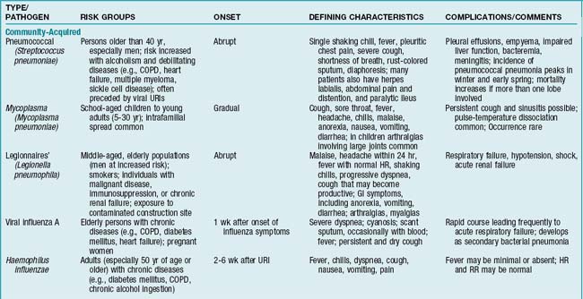
Pneumonias usually are classified into two general types: community acquired and hospital associated (nosocomial). A third type now recognized is pneumonia in the immunocompromised individual.
Community acquired
Individuals with community-acquired pneumonia, the most common type, generally do not require hospitalization unless an underlying medical condition, such as chronic obstructive pulmonary disease (COPD), cardiac disease, or diabetes mellitus, or an immunocompromised state complicates the illness.
Hospital associated (nosocomial)
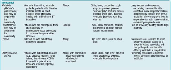
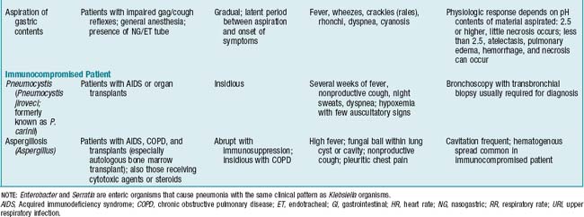
Nosocomial pneumonias usually occur following aspiration of oropharyngeal flora or stomach contents in an individual whose resistance is altered or whose coughing mechanisms are impaired (e.g., a patient who has decreased level of consciousness (LOC), dysphagia, diminished gag reflex, or a nasogastric tube or who has undergone thoracoabdominal surgery or is on mechanical ventilation). Bacteria invade the lower respiratory tract via three routes: (1) gastric acid aspiration (the most common route), which causes toxic injury to the lung and makes it susceptible to bacterial growth; (2) partial obstruction of airway by a foreign body or fluids contaminated with bacteria; and (3) outright infection (rare occurrence). Gram-negative pneumonias are associated with a high mortality rate, even with appropriate antibiotic therapy. If the alveolar-capillary membrane is affected, acute respiratory distress syndrome (ARDS) (formerly known as adult respiratory distress syndrome) may develop.
Pneumonia in the immunocompromised individual
Immunosuppression and neutropenia are predisposing factors in the development of nosocomial pneumonias from both common and unusual pathogens. Severely immunocompromised patients are affected not only by bacteria but also by fungi (Candida, Aspergillus), viruses (cytomegalovirus), and protozoa (Pneumocystis jiroveci, formerly known as P. carinii). Most commonly, P. jiroveci is seen in persons with human immunodeficiency virus (HIV) disease or in persons who are immunosuppressed therapeutically following organ transplants.
Assessment
Findings are influenced by patient’s age, extent of the disease process, underlying medical condition, and pathogen involved. Generally, any factor that alters integrity of the lower airways, thereby inhibiting ciliary activity, increases the likelihood of developing pneumonia (TABLE 2-1).
Signs and symptoms/physical findings
Cough (productive and nonproductive), increased sputum production (rust colored, discolored, purulent, bloody, or mucoid), fever, pleuritic chest pain (more common in community-acquired bacterial pneumonias), dyspnea, chills, headache, myalgia, restlessness; anxiety; decreased skin turgor and dry mucous membranes secondary to dehydration; presence of nasal flaring and expiratory grunt; use of accessory muscles of respiration (scalene, sternocleidomastoid, external intercostals); decreased chest expansion caused by pleuritic pain; dullness on percussion over affected (consolidated) areas; tachypnea (respiratory rate [RR] more than 20 breaths/min); tachycardia (heart rate [HR] more than 90 beats per minute [bpm]); increased vocal fremitus; egophony (“e” to “a” change) over area of consolidation; decreased breath sounds; high-pitched and inspiratory crackles (rales) (increased by or heard only after coughing); low-pitched inspiratory crackles (rales) caused by airway secretions; and circumoral cyanosis (a late finding). Older adults may be confused or disoriented and have low-grade fevers but may present with few other signs and symptoms.
Diagnostic Tests
Sputum for Gram stain and culture and sensitivity tests
Sputum is obtained from lower respiratory tract by endotracheal aspiration, protected catheter brush, or bronchoalveolar lavage (BAL) before initiation or change of antibiotic therapy to identify causative organism in cases of hospital-acquired pneumonia. For community-acquired pneumonias, this approach is recommended only if organism resistant to usual antibiotic therapy is suspected.
WBC count
Increased (more than 12,000/mm3) in the presence of bacterial pneumonias. Normal or low WBC (less than 4000/mm3) count may be seen with viral or mycoplasma pneumonias.
Blood culture and sensitivity
Determine presence of bacteremia and aid in identification of causative organism. Used in cases of hospital-acquired pneumonia and in seriously ill patients with community-acquired pneumonia.
Arterial blood gas (ABG) values
May vary, depending on presence of underlying pulmonary or other debilitating disease. May demonstrate hypoxemia (PaO2 less than 80 mm Hg) and hypocarbia (PaCO2 less than 32-35 mm Hg), with resultant respiratory alkalosis (pH more than 7.45), in the absence of underlying pulmonary disease.
Serologic studies
Acute and convalescent antibody titers drawn to diagnose viral pneumonia. A relative rise in antibody titers suggests viral infection.
Collaborative Management
O2 therapy
Administered when O2 saturation or ABG results demonstrate hypoxemia. Goal is to maintain PaO2 at 60 mm Hg or higher. Special consideration must be given to patients with chronic CO2 retention because their respiratory drive is stimulated by low/decreasing PaO2 levels and not by increasing PaCO2 levels as is normal. Therefore high concentrations of O2 can depress respiration in these patients so O2 is delivered in low concentrations initially, and O2 saturations or ABG levels are monitored closely. If O2 saturation or PaO2 does not rise to acceptable levels (greater than 92% or 60 mm Hg or more, respectively), fraction of inspired O2 (FIO2) is increased in small increments, with concomitant checks of ABG values or O2 saturations.
Antibiotic agents
Prescribed empirically based on presenting signs and symptoms, clinical findings, and chest x-ray results until sputum or blood culture results are available. Many organisms responsible for nosocomial pneumonias are resistant to multiple antibiotics. Proper identification of the organism and determination of sensitivity to specific antibiotics are critical for determining appropriate therapy.
A macrolide or levofloxacin is used for empirical out-patient treatment of community-acquired pneumonia; cephalosporins plus a macrolide and doxycycline are used for patients who are not in the ICU; IV beta-lactam and an IV quinolone or IV azithromycin are used for ICU patients.
Hospital-acquired pneumonia is empirically treated with IV ceftriaxone, or levofloxacin, or ampicillin/sulbactam or ertapenem if it is early onset and the patient has no risk factors for multidrug-resistant (MDR) disease; all other cases are treated initially with multiagent regimens. Vancomycin is added if risk of methicillin-resistant Staphylococcus aureus (MRSA) exists.
Hydration
IV fluids may be necessary to replace fluids lost from insensible sources (e.g., tachypnea, diaphoresis, fever) and decreased oral intake.
Percussion and postural drainage
Indicated if deep breathing and coughing are ineffective in mobilizing secretions.
Antitussives
Given in the absence of sputum production if coughing is continuous and exhausting to the patient.
Antipyretics and analgesics
Prescribed to reduce fever and provide relief from pleuritic pain or pain from coughing.
Vaccines
Pneumococcal vaccine
Administered to patients who have chronic health conditions and to those who are more than 65 yr old and/or are residents of an extended care facility and who have not received the vaccine within the last 5 yr. Vaccine history should be assessed on admission, and vaccine should be given to patients who meet criteria without contraindications (allergy).
Influenza vaccine
Administered to patients with chronic health conditions and to those who are more than 50 yr old and/or are residents of an extended care facility and who have not received the vaccine within the year. Influenza vaccines are routinely administered from October through March. Vaccine history should be assessed on admission, and vaccine should be given to patients who meet criteria without contraindications (e.g., allergy, history of Guillain-Barré syndrome).
Nursing Diagnoses and Interventions
For Patients with Pneumonia
Impaired gas exchange
related to altered O2 supply and alveolar-capillary membrane changes secondary to inflammatory process in the lungs
Desired outcomes
Hospital discharge based on patient exhibiting at least five of the following indicators: temperature 37.8° C or less, HR 100 bpm or less, RR 24 breaths/min or less, SBP 90 mm Hg or more, O2 saturation 90% or more, and ability to maintain oral intake.
Nursing Interventions
Ineffective airway clearance
related to presence of tracheobronchial secretions secondary to infection or related to pain and fatigue secondary to lung consolidation
Desired outcomes
Patient demonstrates effective cough. Following intervention, patient’s airway is free of adventitious breath sounds.
Nursing Interventions
Deficient fluid volume
related to increased insensible loss secondary to tachypnea, fever, or diaphoresis
Desired outcomes
At least 24 hr before hospital discharge, patient is normovolemic as evidenced by urine output 30 mL/hr or more, stable weight, HR less than 100 bpm, SBP greater than 90 mm Hg, fluid intake approximating fluid output, moist mucous membranes, and normal skin turgor.
Nursing Interventions
Desired outcome
Patient is free of infection as evidenced by normothermia, WBC count 12,000/mm3 or less, and sputum clear to whitish.
Nursing Interventions

Patient-Family Teaching and Discharge Planning
Provide verbal and written information about the following:
Pleural Effusion
Overview/Pathophysiology
A pleural effusion is an accumulation of fluid (blood, pus, chyle, serous fluid) in the pleural space. Generally, fluid gravitates to the most dependent area of the thorax, and the adjacent lung becomes compressed. Pleural effusion is rarely a disease in itself, but rather it is caused by a number of inflammatory, circulatory, or neoplastic diseases. Transudate effusion results from changes in hydrodynamic forces in the circulation and usually is caused by heart failure (increased hydrostatic pressure) or cirrhosis (decreased colloidal osmotic pressure). Exudate effusion results from irritation of the pleural membranes secondary to inflammatory, infective, or malignant processes. More exact nomenclature can be used once the nature of the fluid in the pleural effusion has been identified, that is, hydrothorax (a transudate or exudate of serous fluid), pyothorax or empyema (collection of purulent material), hemothorax (bloody fluid), or chylothorax (effused chyle).
Assessment
Clinical indicators of pleural effusion are related to the underlying disease. Patients with a small effusion (less than 300 mL) may be asymptomatic.
Signs and symptoms/physical findings
Pleuritic chest or shoulder pain, diaphoresis, cough, fever, dyspnea, orthopnea, decreased breath sounds, dullness to percussion, decreased tactile fremitus, egophony (“e” to “a” change) over effusion site, tracheal deviation away from affected side, and pleural friction rub.
Diagnostic Tests
Chest x-ray examination
Shows evidence of effusion if more than 300 mL of fluid is in the pleural space. With effusion greater than 1000 mL, the x-ray film may show mediastinal shift away from the affected lung.
Chest computed tomography (CT)
Enables imaging of the entire pleural space, pulmonary parenchyma, and mediastinum simultaneously. CT assists in differentiation between lung consolidation and pleural effusion.
Thoracentesis
Removal of fluid from the pleural space for examination to provide definitive diagnosis and determine type of effusion.
Collaborative Management
Therapeutic thoracentesis
Removes fluid and thereby allows lung to reexpand. Rate of recurrence and time span for return of symptoms are recorded.
Chest tube insertion
Provides continuous drainage of larger effusions through 26F-30F catheter connected to closed chest-drainage system.
Nursing Diagnoses and Interventions
Ineffective breathing pattern
related to decreased lung expansion secondary to fluid accumulation in the pleural space
Nursing Interventions
Patient-Family Teaching and Discharge Planning
Provide verbal and written information about the following:
Pulmonary Embolism
Overview/Pathophysiology
The most common pulmonary perfusion abnormality is pulmonary embolism (PE). PE is caused by passage of a foreign substance (blood clot, fat, air, or amniotic fluid) into the pulmonary artery or its branches, with resulting obstruction of the blood supply to lung tissue and subsequent collapse. The most common source is a dislodged blood clot from the systemic circulation, typically the deep veins of the legs or pelvis. Thrombus formation is the result of the following factors: blood stasis, alterations in clotting factors, and injury to vessel walls. A fat embolus is the most common nonthrombotic cause of pulmonary perfusion disorders (see p. 74).
Assessment
Signs and symptoms/physical findings
Often nonspecific and variable, depending on extent of obstruction and whether patient has infarction as a result of the obstruction.
Sudden onset of dyspnea and sharp chest pain, restlessness, anxiety, nonproductive cough or hemoptysis, palpitations, nausea, syncope, tachypnea, tachycardia, hypotension, crackles (rales), decreased chest wall excursion secondary to splinting, S3 and S4 gallop rhythms, transient pleural friction rub, jugular venous distention, diaphoresis, edema, and cyanosis.
If infarction has occurred, fever, pleuritic chest pain, and hemoptysis are common.
History and risk factors
Immobility
Especially significant when it coexists with surgical or nonsurgical trauma, carcinoma, or cardiopulmonary disease. Risk increases as duration of immobility increases.
Cardiac disorders
Atrial fibrillation, heart failure, myocardial infarction, rheumatic heart disease.
Surgery
Risk increases in postoperative period, especially for patients with orthopedic, pelvic, thoracic, or abdominal surgery and for those with extensive burns or musculoskeletal injuries of the hip or knee.
Chronic pulmonary and infectious diseases
Trauma
Especially lower extremity fractures and burns. Degree of risk is related to severity, site, and extent of trauma.
Carcinoma
Particularly neoplasms involving the breast, lung, pancreas, and genitourinary and alimentary tracts.
Diagnostic Tests
Arterial blood gas (ABG) values
Hypoxemia (PaO2 less than 80 mm Hg), hypocarbia (PaCO2 less than 35 mm Hg), and respiratory alkalosis (pH more than 7.45) usually are present. Normal values do not rule out PE.
D-dimer
A degradation product produced by plasmin-mediated proteolysis of cross-linked fibrin and measured by enzyme-linked immunosorbent assay (ELISA). The higher the result (with less than 250 ng/mL considered negative in most laboratories), the more likely it is patient has PE. This test is not sensitive or specific enough to diagnose PE, but it may be used in conjunction with other diagnostic tests.
Cardiac troponin level
Elevated in PE as a result of right ventricular dilation and myocardial injury.
Chest x-ray examination
Initially findings are usually normal, or elevated hemidiaphragm may be present. After 24 hr, x-ray examination may reveal small infiltrates secondary to atelectasis that result from the decrease in surfactant. If pulmonary infarction is present, infiltrates and pleural effusions may be seen within 12-36 hr.
Spiral or helical computed tomography (CT)
Images pulmonary arteries (PAs) during a single breath. Spiral CT is rapidly becoming the test of choice in diagnosing PE because of its higher specificity and sensitivity.
Pulmonary ventilation/perfusion scan
Used to detect abnormalities of ventilation or perfusion in the pulmonary system. Radiopaque agents are inhaled and injected peripherally. Images of distribution of both agents throughout the lung are scanned. If the scan shows a mismatch of ventilation and perfusion (i.e., pattern of normal ventilation with decreased perfusion), vascular obstruction is suggested.
Pulmonary angiography
Definitive study for PE: An invasive procedure that involves catheterization of right side of the heart and injection of dye into the PA to visualize pulmonary vessels. Abrupt vessel “cutoff” may be seen at the site of embolization. Usually, filling defects are seen. More specific findings are abnormal blood vessel diameters (i.e., obstruction of right PA would cause dilation of left PA) and abnormal blood vessel shapes (i.e., affected blood vessel may taper to a sharp point and disappear).
Collaborative Management
The three goals of therapy are (1) prophylaxis for individuals at risk for development of PE, (2) treatment during acute embolic event, and (3) prevention of future embolic events in individuals who have experienced PE.
O2 therapy
Delivered at appropriate concentration to maintain a PaO2 of more than 60 mm Hg or O2 saturation greater than 90%.
Anticoagulation
Low molecular weight heparin (LMWH) or unfractionated heparin (UFH) therapy
Started immediately in patients without bleeding or clotting disorders and in whom PE is strongly suspected with the aim of inhibiting further thrombus growth, promoting resolution of the formed thrombus, and preventing further embolus formation. Continued for at least 5 days to allow for depletion of thrombin.
LMWH
Preferred to UFH because of more predictable dosing, fewer side effects, once- or twice-daily subcutaneous administration, and lack of need to monitor activated partial thromboplastin time. Dose is weight based and differs for various LMWH preparations. Dose must be adjusted for individuals with renal impairment because most LMWH is excreted by the kidneys. LMWH has been shown to be safe if given during pregnancy.
UFH
Has shorter half-life than LMWH and effect is completely reversible with protamine. Ideally, dosage is weight based (e.g., IV bolus of 80 units/kg followed by a maintenance dose of 18 units/kg/hr). Alternatively, an initial IV bolus of 5000-10,000 units followed by continuous infusion of 1000 units/hr may be given. Effect is monitored by activated partial thromboplastin time (aPTT) measurements every 6 hr after initial dose until the goal of 1.5 to 2.5 × control value is consistently established.

Stay updated, free articles. Join our Telegram channel

Full access? Get Clinical Tree