REPRODUCTIVE HEALTH

Chapter 40 REPRODUCTIVE HEALTH


KEY TERMS/CONCEPTS


SPERMATOGENESIS


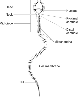
Sperm cells, or spermatozoa (Figure 40.2), are produced continuously throughout life from puberty onwards under the influence of the hormones testosterone and follicle-stimulating hormone (FSH). The anterior pituitary gland secretes FSH. Sperm cells originate from primordial cells that appear in the embryo early in gestation but are not functional until puberty. Before puberty the testes produce stem cells called spermatogonia. These cells multiply by mitotic division. At puberty, under the influence of FSH, spermatogonia undergo both mitotic and meiotic cell division, which results in sperm production. A mature sperm consists of a head, mid-piece and tail. The head contains the nucleus, with its genetic material (DNA). The mid-piece contains mitochondria to provide energy for the rapid tail movements. The tail propels the sperm with a lashing movement through the female reproductive tract.


DISORDERS OF THE MALE REPRODUCTIVE SYSTEM


Many male reproductive disorders are developmental or age related. Abnormalities of the genitourinary system may be present at birth. Congenital or structural defects of the genitourinary tract can cause physical and psychological effects for a child. Both urinary and reproductive function can be compromised. Early corrective surgical treatment is necessary to minimise the possibility of these effects occurring. Nursing interventions aim to provide support for the child and family before and after surgery. The nurse can assist the child and family by educating them about procedures and allowing them time and opportunity to express any fears or concerns. Fact sheets that explain the condition without overuse of medical jargon can be given to parents to reinforce information given by the health care team.


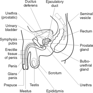
Disorders of the male reproductive system are referred to as genitourinary disorders because of the shared anatomical structures of the urinary and reproductive systems. Common problems, which vary according to the type and severity of the disorder, include alterations in:


CANCER OF THE TESTES


Cancer of the testes generally affects males between the ages of 15 and 40. Most cancers are of germ-cell origin. The aetiology of testicular cancer is unknown and therefore precludes preventive strategies being implemented. Hypotheses include early puberty, exposure to environmental oestrogens, occupational exposure and smoking. Known risk factors include a history of cryptorchidism, genitourinary abnormalities, a history of mumps orchitis, white race, high socioeconomic status and family history. Early diagnosis and improved treatment modalities have given a cure rate of over 90%. However, 50% are only diagnosed in the advanced stage, when the cure rate is about 70%. An early stage indicator is the presence of a smooth painless lump in the scrotum. Later metastatic symptoms include general abdominal and inguinal aching. Other metastatic symptoms include bowel or urinary obstruction and abdominal pain.


Diagnostic evaluation is based on physical examination, blood serum markers and scrotal ultrasound. Elevated levels of alpha-fetoprotein (AFP) and human chorionic gonadotrophin (hCG) are present in testicular cancer. Ultrasonography differentiates between a solid and a cystic lesion. Tests to detect potential metastases include chest X-ray and computerised tomography (CT) scans of the chest, abdomen and pelvis.


Management of the client depends on the type and stage of the disease. Chemotherapy is used to treat primary and metastatic tumours. Surgical intervention generally involves a unilateral orchidectomy to remove the testis, tunica and spermatic cord, and retroperitoneal lymph node dissection. Radiotherapy to lymph node pathways is often used after surgery.


Testicular cancer affects young, mostly well men during prime reproductive years. Some treatments can lead to infertility. Sensitivity to the individual’s loss and fears about sexuality and mortality are key components of nursing management. The nurse can discuss possible sperm banking and reassure the client that surgery does not diminish virility. The client may be referred to support services outside the hospital and should be educated in testicular self-examination. It is recommended that he carry out periodic examinations, as there is an increased risk of a second tumour occurring.


DISORDERS OF THE PROSTATE GLAND


The prostate gland frequently becomes enlarged as a man ages. The three most common prostate problems are prostatitis, benign prostatic hyperplasia and prostate cancer.


Benign prostatic hyperplasia


Benign prostatic hyperplasia (BPH) is enlargement of the prostate gland as a result of small non-cancerous growths inside the prostate that may be related to hormonal changes that occur with ageing. BPH usually does not affect sexual function but does affect urination. As the prostate enlarges it presses against the bladder and the urethra, blocking the flow of urine. Problems include difficulty initiating a urine stream or maintaining more than a dribble, frequency or urgency and nocturia. These problems can be highly distressing because of the embarrassment of hesitancy, frequency, soiling and smell.


Straining to empty the bladder can cause thickening of the bladder wall and loss of elasticity. This can lead to urinary tract infections and eventual renal failure. A completely blocked urethra is a medical emergency requiring immediate catheterisation. Other serious potential complications of BPH include bladder calculi, hydronephrosis and bleeding. Men with mild symptoms may simply be checked at regular intervals and treated if symptoms become worse. More severe symptoms require surgical intervention.


The type of surgery depends on the size of prostate, location of the enlargement, whether surgery on the bladder is also needed, and the client’s age and physical condition. The most common surgical procedure is transurethral resection of the prostate (TURP), used in more than 90% of cases. Surgery relieves symptoms quickly, typically doubling the urinary flow within weeks. A fibre-optic scope is passed through the urethra to the prostate. Using either a tiny blade or an electric loop, the surgeon pares away the lining of the urethra and bits of excess prostate tissue to expand the passageway.


Postoperatively the individual is usually hospitalised for several days with a urinary catheter in situ to irrigate the bladder. TURP does not usually affect a man’s ability to have an erection or an orgasm, since the nerves that control erection lie outside the prostate and are not touched by the operation. A more common side effect is dry or retrograde ejaculation, which occurs when the neck of the bladder fails to close properly during ejaculation. The result is that semen spurts backwards into the bladder rather than through the penis. Men who experience this side effect still have the sensation of an orgasm but are unable to ejaculate during intercourse.


Transurethral needle ablation (TUNA), which can be done with a local anaesthetic on an outpatient basis, uses radio-frequency energy delivered through needles to kill excess prostate tissue. A catheter that directs the needles towards the obstructing prostate tissue is inserted into the urethra. Some clinical studies have reported that TUNA improves the urine flow, with minimal side effects, compared with other procedures.


Researchers are working to develop BPH treatments that are more effective, less traumatic and have fewer side effects. These include using laser surgery, powerful electric currents and microwaves. Balloon urethroplasty and insertion of a stent into the urethra are other possible options.


PROSTATE SURGERY— PROSTATECTOMY


Open prostatectomy may involve either a radical or partial procedure. A radical prostatectomy that removes the whole prostate is the treatment for cancer of the prostate. The incision is made through either the lower abdomen or the perineum. Impotence is a result of radical prostatectomy, as nerves and muscular tissue that function in penile erection are severed. When possible, nerve-sparing surgery is done to prevent this. The client is hospitalised for about 5–7 days. Partial prostatectomy, which leaves the posterior portion of the prostate intact, is used to treat BPH. The incision for a partial prostatectomy is usually through the suprapubic area of the abdomen. Open prostatectomy is used only when the prostate is extremely large.


Nursing management preoperatively requires establishment of adequate hydration and monitoring of the client’s fluid status. Insertion of an indwelling or suprapubic catheter may be necessary to establish urinary output. Alterations in urinary elimination and pain and discomfort related to bladder spasms may have caused the client to limit fluids. The client should be encouraged to drink fluids freely. Bowel preparation may include drinking 2–3 L of a cathartic and an evacuant enema. Postoperatively the nurse should maintain bladder irrigation and drainage, and monitor for clots and haemorrhage.


The client is encouraged to talk about concerns related to urinary control and sexual functioning before discharge from the health care facility. Nurses working in this area need to adopt a holistic approach, including the man’s partner in discussions if necessary. Nurses need to recognise that sexual activity does not necessarily stop with age, and nursing care planning and delivery should take account of this. Discharge education needs to take physical and psychosocial factors into consideration.


DISORDERS OF MULTIPLE CAUSE


ASSESSMENT AND DIAGNOSTIC TESTS


The client may be anxious or embarrassed about the physical examination and discussion of his sexual history. A calm insightful approach is required, as is preservation of the client’s individuality and dignity throughout. Cultural and religious customs such as circumcision should be considered.


EXAMINATION OF EXTERNAL GENITALIA


Before the physical examination the client’s genitourinary history is obtained. A history of urinary problems may be significant because of the anatomical relationship between reproductive and urinary systems. The external male genitalia and the inguinal canal are inspected for evidence of abnormalities such as penile discharge, tenderness, lesions, swelling, hard lumps or asymmetry. Abnormalities may indicate underlying disorders such as cancer of the testes or the presence of a sexually transmitted infection.


Testicular examination


Testicular self-examination (TSE [see Clinical Interest Box 40.1]) performed on a regular basis is the inspection and palpation of the testes to detect any changes. The testes are examined for size, shape, symmetry and texture. The earlier a change is discovered, the sooner it can be investigated and treated. It is important that the correct technique is taught to men in the target age group (15–40 years). Given that testicular cancer is most widespread in an age group in which men are generally healthy, there may be limited openings to teach TSE. Health promotion and screening in primary care, such as well-man clinics, affords an opportunity for education. Nurses working in the areas of adolescent, student and sexual health are seen as strategic professionals in educating men about TSE. Occupational health nurses involved in work-based screening may also have valuable opportunities to discuss health issues with men.


INTERNAL AND SPECIMEN TESTING


NURSING INTERVENTIONS IN MALE REPRODUCTIVE HEALTH


Men have specific health needs, experiences and concerns related to their gender as well as their biological sex. This is a relatively new concept in medical and nursing literature. Psychosocial factors related to male health are poorly understood and frequently ignored by many health practitioners. Men engage in less healthy lifestyles and adopt fewer health-promotion strategies than women, for reasons that are complex. An awareness of men’s health issues and poor health practices has led to the establishment of an international men’s health movement — the International Society for Men’s Health (www.ismh.org/). The aim of the movement is to increase awareness about current men’s health issues and promote men’s health initiatives.


A man with a reproductive system disorder may experience anxiety about sexual dysfunction, infertility, urinary problems and other implications of the disorder. He may suffer low self-esteem and encounter difficulties in accepting his condition and will often delay seeking health interventions. Nurses need to actively explore opportunities to promote health and to encourage and support men to make healthier lifestyle choices. To make appropriate health and lifestyle choices an individual must be provided with adequate information about his condition and its possible outcomes. He should also be offered opportunities to express his feelings, explore treatment options and, if necessary, be referred for sex counselling.


THE FEMALE REPRODUCTIVE SYSTEM


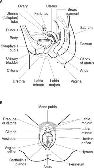
The female reproductive system functions to secrete hormones, produce ova, receive sperm and allow for fertilisation, implantation, development and birth of the baby. The female reproductive system (Figure 40.3) consists of essential and accessory organs. The essential organs, or gonads, are the ovaries. Accessory organs consist of a series of ducts, additional sex glands and external structures.


OOGENESIS


The ovaries function to produce ova (oogenesis) and hormones. The cells of the developing ovarian follicle are stimulated to secrete oestrogen by follicle-stimulating hormone (FSH) from the anterior pituitary gland. Oestrogen enhances the growth and maturation of the follicle. As it matures, the follicle becomes distended with fluid and bulges onto the surface of the ovary. A sharp increase in the level of luteinising hormone (LH) from the anterior pituitary gland acts to trigger ovulation and the mature follicle ruptures to release an ovum. The ovum is released into the peritoneal cavity near the distal end of the uterine tube, where it is captured by the waving fimbriae. Usually one mature ovum is released each month. After ovulation the ovary undergoes structural and chemical changes. Under the influence of LH the ruptured follicle, or corpus luteum, secretes oestrogen and progesterone. If fertilisation does not occur the corpus luteum regresses to a non-functional state, the corpus albicans.


Production of ovarian hormones begins at puberty. Oestrogen influences the development of the female secondary sexual characteristics, including:


Oestrogen also stimulates the lining of the uterus to prepare for a fertilised ovum.


The effects of progesterone are to:


ACCESSORY ORGANS


Uterine (fallopian) tubes, each about 10 cm long, extend from either side of the upper uterus to the ovaries. The distal end of each tube curves over an ovary and fans out in a trumpet shape with finger like projections (fimbriae). An outer covering of peritoneum, a middle layer of involuntary muscle and an inner lining of ciliated mucous membrane make up the walls of the tubes. Ova are propelled through the tube by peristalsis and the sweeping action of cilia. Fertilisation of the ovum generally occurs in the distal one-third of the uterine tube.


The uterus is a hollow pear-shaped organ located in the pelvic cavity posterior to the urinary bladder and anterior to the rectum. It is suspended in the pelvis by a peritoneal fold (the broad ligament). Normally the uterus is in an anteverted position so that the upper portion rests on the bladder. The uterus consists of an upper rounded body and a lower narrow neck, the cervix. Above the point where the fallopian tubes enter is the fundus of the uterus. The walls of the uterus consist of an outer covering of peritoneum (perimetrium), a middle layer of thick involuntary muscle (myometrium) and an inner lining of mucous membrane (endometrium). The cervix is lined with mucus-secreting glands that provide lubrication. The functions of the uterus are to:


The vagina, an expandable passageway about 10 cm long, is situated between the urinary bladder and the rectum. It extends from the uterus to the vulva, where it opens to form the vaginal opening. The cervix projects into the upper part of the anterior vaginal wall. The vagina is acidic from menarche to menopause, and its surface moist from fluid secreted by the vaginal epithelium. Functions of the vagina are to receive the penis during sexual intercourse, provide a passageway for menstrual flow to leave the body, and provide a passageway through which the fetus is expelled from the uterus.


ACCESSORY GLANDS


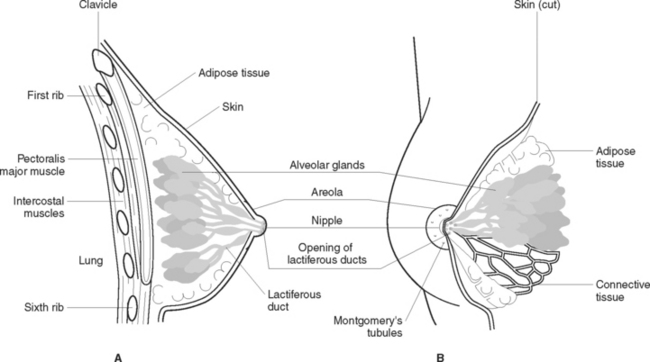
Mammary glands (Figure 40.4) are accessory glands of the reproductive system. Their reproductive function is lactation. They are situated over the pectoral muscles and are attached to them by Cooper’s ligaments. The breast is a complex structure composed of a glandular and ductal network, fat, connective tissue, fascia, blood vessels, nerves and lymphatic vessels. In the centre of the breast a pigmented area, the areola, surrounds the nipple. Each breast contains 15–20 lobes that radiate around the nipple and are separated from each other by adipose tissue. Within the lobes are lobules that contain clusters of milk-producing cells called alveoli. Lactiferous ducts drain the alveoli into openings in the nipple. Breast development at puberty is influenced by ovarian hormones.


EXTERNAL GENITALIA (VULVA)


The external genitalia consists of the mons pubis, labia majora, labia minora, clitoris, perineum, urethral and vaginal orifices, duct of Bartholin’s gland and hymen. The mons pubis is a rounded pad of fatty tissue over the symphysis pubis that is covered by pubic hair after puberty. The labia majora are two rounded folds of tissue that shape the lateral boundaries of the vulva to form a protective covering. Their inner surface is smooth and hairless. Surrounded by the labia majora are the labia minora, two long thin folds of tissue. Their surface is smooth and does not contain hair follicles. The urethral and vaginal orifices are enclosed by the labia minora. The hymen, a membranous fold of tissue, surrounds and covers the vaginal orifice. It has an opening to allow menstrual flow to escape. It is usually ruptured when sexual intercourse first occurs. After rupture the hymen appears as irregular projections into the vaginal orifice.


The clitoris is an area of erectile tissue about 5–6 mm in length and 6–8 mm in diameter situated between and partly covered by the anterior junction of the labia minora. It contains a large number of free nerve endings, which make it very sensitive to stimulation. Between the vaginal opening and the anus is a muscular layer covered with skin, the perineum.


Bartholin’s glands, two small glands situated on either side of the vaginal orifice, secrete mucus to keep the vagina moist. Ducts from the glands open into the space between the labia minora and the vaginal orifice.

Stay updated, free articles. Join our Telegram channel

Feb 12, 2017 | Posted by in NURSING | Comments Off on REPRODUCTIVE HEALTH

Full access? Get Clinical Tree

Get Clinical Tree app for offline access