Chapter 4 Renal physiology and the pathology of renal failure
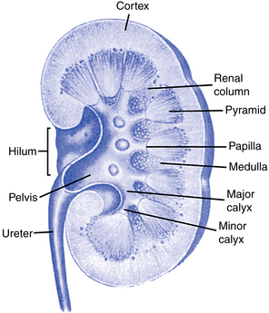
Before any discussion of the pathology of renal failure can begin, it is important to review the following functions that normal kidneys perform (Fig. 4-1):
In addition, the kidneys have several endocrine functions, including the following:
• Production of renin, which affects sodium, fluid volume, and blood pressure
• Formation of erythropoietin, which controls red cell production in the bone marrow
A normal kidney is also a receptor site for several hormones:
• Antidiuretic hormone (ADH), produced by the pituitary, reduces the excretion of water.
• Aldosterone, produced by the adrenal cortex, promotes sodium retention and enhances secretion of potassium and hydrogen ion.
• Parathyroid hormone increases phosphorus and bicarbonate excretion and stimulates conversion of vitamin D to the active 1,25-dihydroxycholecalciferol vitamin D3 form.
Renal physiology
How is blood supplied to the kidneys?
The kidneys are highly vascular organs that receive 20% to 25% of the resting cardiac output, which is greater than 1000 mL/min. Cardiac output is the volume of blood pumped per minute by each ventricle of the heart. Each kidney receives blood from a renal artery that originates from the abdominal aorta, and blood leaves the kidney through the renal vein. The renal artery branches out to form the afferent arterioles, which in turn form the glomerular capillaries of individual glomeruli. The glomerular capillaries then join to form the efferent arterioles, which in turn diffuse into peritubular capillaries and the vasa recta (Fig. 4-2).

Figure 4-2 The venous vessels of the kidney parallel the arterial vessels and are similarly named.
(From Copstead LC, Banasik JL: Pathophysiology, ed 4, St. Louis, 2010, Saunders.)
Blood flow to the kidney is dependent on hydration and cardiac output. Dehydration, blood loss, congestive heart failure, and myocardial infarction are examples of situations that would compromise blood flow to the kidney.
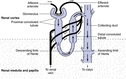
What is the difference between the peritubular capillaries and the vasa recta?
The peritubular capillaries surround the proximal and distal convoluted tubules and allow tubular secretion and reabsorption to occur. The vasa recta capillaries and branches surround the loops of Henle of the juxtamedullary nephrons and are located in the renal medulla. They play a major role in the concentration of urine as it moves through the tubules.
What is a nephron?
The nephron is the main functional unit of the kidney. There are more than a million such units in each of the two kidneys. Each nephron is a complex structure and has two main components: vascular and tubular. The vascular component consists of the afferent arteriole, glomerulus, efferent arteriole, and peritubular capillaries. The tubular portions of the nephron include Bowman’s capsule, proximal tubule, loop of Henle, and the distal tubule.
The glomerulus consists of a network of thin-walled capillaries supplied by the afferent arteriole and is closely surrounded by a pear-shaped epithelial membrane called Bowman’s capsule. The glomerulus and Bowman’s capsule combined are called the renal corpuscle. The space between the two layers of Bowman’s capsule opens into the proximal tubule, which makes a series of convolutions in the cortex of the kidney. It straightens out and then makes a U-turn, known as the loop of Henle, in the kidney medulla. It becomes convoluted again adjacent to its own glomerulus and finally joins other distal tubules to form a collecting duct to carry the freshly formed urine to the kidney pelvis (Fig. 4-3). Each kidney pelvis funnels the urine into its ureter, which connects with the urinary bladder. The urethra conveys the urine from the bladder to the exterior.
What is the first step in urine formation?
Blood enters the glomerulus through an afferent arteriole. Because of the blood pressure in the capillaries and because of their thin walls, filtration of blood occurs. Water and dissolved solutes of molecular weight less than 68,000 Da (albumin) pass freely into Bowman’s space. This essentially protein-free fluid is the glomerular filtrate, and its rate of production is the glomerular filtration rate (GFR). The GFR is the amount of filtrate the kidneys produce each minute. A man of average size produces about 180 L of filtrate per day, or 125 mL/min. Ninety-nine percent of this filtrate is reabsorbed as it passes through the tubules.
Glomerular filtration is dependent on sufficient blood circulation to the glomerulus and maintenance of normal filtration pressures. The filtration of molecules depends on their shape, size, and ionic charge. As the molecular weight and size increase, the degree of filtration decreases. The glomerular basement membrane exerts a net negative charge. Substances carrying a negative charge will be repelled by the basement membrane, and its filtration will be prohibited.
What happens to the filtrate as it moves through the tubules?
The main function of the tubules is reabsorption and secretion. Tubular reabsorption is the process of the filtrate moving back into the blood of the peritubular capillaries or vasa recta (Fig. 4-4). This process is very selective and depends on the body’s needs at the time. Materials that are reabsorbed into the bloodstream are such ions as sodium, potassium, chloride, bicarbonate, and calcium.

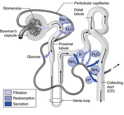
Figure 4-4 Path of filtrate as it moves through different parts of a nephron.
(From Thibodeau GA, Patton KT: Anatomy & physiology, ed 7, St. Louis, 2010, Mosby.)
Of the 180 L of glomerular filtrate produced each day, about 2 L remain as the final urine. The rest of the water is reabsorbed along with glucose, amino acids, small proteins, and most electrolytes. The remaining filtrate becomes concentrated and begins to resemble the ultimate urine as it progresses down the tubule. Final adjustments of water-to-solute load occur in the distal tubule under the influence of ADH. The tubules conserve water and electrolytes by returning them to the blood. Hydrogen ions and metabolic wastes are excreted along with a volume of water appropriate to the total body need. The majority of reabsorption occurs in the proximal tubule; however, some reabsorption does occur in the distal tubule.
Tubular secretion adds materials to the filtrate from the blood. Tubular secretion helps to remove toxic substances from the blood and to restore blood pH by excreting excessive hydrogen ions. Substances secreted into the tubules include potassium, hydrogen, ammonia, creatinine, and some drugs.
Renal failure
What happens in kidney failure?
The normal urinary system maintains fluid volumes and the levels of many chemicals in the body. When the urinary system is not working properly, normal blood composition is disrupted and the patient will experience symptoms. Renal failure may be acute or chronic. In both types of renal failure there is enough loss of nephron function to upset the normal steady state of the body’s internal environment. Waste products of protein metabolism accumulate and will necessitate treatment of some kind.
This accumulation of waste products of protein metabolism is termed azotemia, indicating retention of nitrogenous products (azote = nitrogen). Azotemia is a major component of the uremic syndrome.
What is urea?
Urea is the waste product of protein metabolism and has a molecular weight of 60 Da. Urea is the most abundant organic waste and is freely filtered at the glomerulus. Most urea is produced during the breakdown of amino acids. Its normal value in the blood is 15 to 40 mg/dL. Blood urea level is influenced by many things, which is why it is not the best indicator of renal function or dysfunction. Increased levels may be seen with increased dietary consumption of protein, bleeding into the gastrointestinal tract, steroid use, and any hypercatabolic state. Decreased levels may be seen with a low dietary consumption of protein, liver disease, and overhydration.
What is creatinine?
Creatinine is a protein produced by muscle and released into the blood. The creatinine level in the blood is determined by the rate that creatinine is removed in the urine.
What is uremia?
Uremia, or the uremic syndrome, encompasses a complex of symptoms and findings resulting from disordered biochemical processes when kidney function fails.
Is retention of urea the cause of uremia?
Severity of the uremic symptoms roughly parallels the rise in blood urea. Urea clearly contributes to some of the symptoms—malaise, lethargy, anorexia, insomnia—but it is not the primary toxin of uremia. Numerous other substances are retained in the body when kidney function fails. More than 200 potential uremic toxins have been identified.

Stay updated, free articles. Join our Telegram channel

Full access? Get Clinical Tree

