CHAPTER 27 Red eye
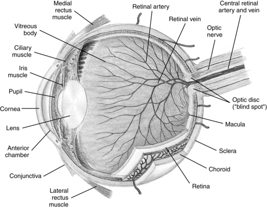
The term red eye is used to denote a cardinal sign of ocular inflammation. The anatomical location in and around the eye and the probable cause of the eye disorder provide an important framework to use in assessment. General anatomical locations are the ocular adnexa, conjunctiva, cornea and anterior segment, and posterior eye (Figure 27-1).

FIGURE 27-1 Anatomical structures of the human eye.
(From Seidel HM, Ball JW, Dains JE, Flynn J, Solomon B, Stewart R: Mosby’s guide to physical examination, ed 7, St Louis, 2011, Mosby.)
Diagnostic reasoning: focused history
Recent sinus infection
Can I rule in or rule out trauma?
Key question
Blunt trauma to the ocular adnexa can cause lid swelling and/or discoloration. Rupture of the globe, fractures of the orbital bones, and internal bleeding may also be possible. Sharp trauma to the area can cause lacerations of the lid and underlying lacerations of the globe. Internal bleeding may be subconjunctival (between the conjunctiva and sclera) or intraocular (hyphema). The cornea may have a foreign body and/or abrasions.
Recurrence
Can I narrow the problem by location?
Key question
Unilateral redness is more likely to indicate trauma or infection, whereas bilateral redness is more likely to indicate an allergy or an underlying systemic process. Blepharitis, inflammation of the eyelids, causes itching and crusting of the lash line and is usually bilateral. A hordeolum (sty) produces redness at the base of eyelashes and is usually unilateral. A chalazion is a chronic granulomatous inflammation of the meibomian gland. It is found in the mid eyelid, often on the conjunctival side, and is usually unilateral. Some conditions can present with either unilateral or bilateral symptoms. Conjunctivitis often starts in one eye and then spreads to the other eye, sparing the limbal area of the eyes. Subconjunctival hemorrhage is often unilateral but may involve both eyes. Herpetic infection may be unilateral or bilateral.
Vision loss
Distinguish visual loss from blurry vision caused by the discharge associated with conjunctivitis. No decrease in vision is seen with bacterial and allergic conjunctivitis, beyond that reasonably related to blurring from the heavy discharge. Vision is mildly decreased in iritis but markedly decreased in acute glaucoma and with corneal abrasions or ulcers. Box 27-1 lists symptom patterns of pain and visual loss (also see Chapter 35).
Box 27-1 Symptom Patterns of Pain and Visual Loss
| Red Eye (No Pain or Visual Loss) | Red Eye (Painful) | ||
| Vision Normal | Vision Impaired | ||
| Conjunctivitis | Episcleritis | Iritis | |
Blurring
True blurring is caused by an ocular problem. When the cornea, lens, aqueous humor, or vitreous is hazy, vision blurs and often there is dazzle in bright light. Some patients describe both refractive errors and double vision as blurred vision. Heavy discharge associated with conjunctivitis can also produce perceived blurring of vision.


