A woman experiences significant alterations in physical and psychosocial status after childbirth. The postpartum period is a time of transition involving physiologic changes, adaptation to the maternal role, and modification of the family system by the addition of the baby. Nurses have a unique opportunity to promote and support maternal and family adaptation. Inpatient postpartum care routines are ideally focused on the needs of the mother, baby, and family, rather than on arbitrary rules and unit traditions. Postpartum nursing care should be as individualized and flexible as needed. The perinatal nurse recognizes women and their families as integral members of the healthcare team and encourages them to enter into decision-making processes and planning of care (Association of Women’s Health, Obstetric and Neonatal Nurses [AWHONN], 2009). Open visiting and family interaction with the new mother and newborn are supported. The father of the baby and other support persons should be encouraged to be present, according to the desires of the woman, and actively involved in postpartum and newborn care. Family-centered maternity care is based on the philosophy that the physical, social, psychological, and spiritual needs of the family are included in all aspects of the nursing care provided (AWHONN, 2009).
Implementation of family-centered maternity care requires collaboration among childbearing women, families, and healthcare providers. The family is defined by the woman and frequently extends beyond traditional definitions. Cultural beliefs and values of the woman and her family should be respected and accommodated. Chapter 2 and the March of Dimes Nursing Module Cultural Competence: An Essential Journey for Perinatal Nurses (Moore, Moos, & Callister, 2010) provide a comprehensive review and discussion of various cultural and religious perspectives and can be helpful in developing nursing care for specific women.
A woman experiences significant changes in physical and psychosocial status following childbirth. Nurses have a unique opportunity to promote and support maternal and family adaptation during this time. The chapter begins with a discussion of planning the transition to parenthood, which ideally occurs during pregnancy. Physiologic changes during the postpartum period, appropriate nursing assessments, and interventions for healthy women, as well as common complication and care for these are reviewed as well. Physiologic and psychosocial adaptation are reviewed as well as educational needs and follow-up after discharge.
PLANNING FOR THE TRANSITION TO PARENTHOOD
According to current trends, length of stay (LOS) for childbirth is now more realistically in line with the physical and psychosocial needs of the new mother and her family rather than with the previous arbitrary mandates from health insurance companies in an effort to control costs. However, although LOS has slightly increased over the past several years, it is still relatively short in all settings. One observation of the effect of longer LOS is a significant decline in neonatal readmissions but not in 1-year mortality (Datar & Sood, 2006). Thus, discharge planning cannot be delayed until admission for childbirth. Women in active labor and women who have just given birth are not candidates for a discussion of learning needs or for typical classroom education about self-care or infant care. The woman’s focus during the intrapartum period is on safe passage through labor and on a healthy, positive childbirth experience.
Rubin’s (1961a) classic research suggests that during the immediate postpartum period, the new mother is not physically or emotionally ready to listen to extensive presentations of how to care for herself and her newborn. Postpartum women have transient deficits in cognition, particularly in memory function, the first day after giving birth. According to Rana, Lindheimer, Hibbard, and Pliskin (2006), normal pregnant women can temporarily develop mild cognitive defects during labor and after birth when compared with nonpregnant women. Women in their study reported problems with attention, concentration, and memory throughout pregnancy and in the early postpartum period (Rana et al., 2006). This decline was not attributed to depression, anxiety, sleep deprivation, or other physical changes associated with pregnancy. Causes suggested included the increased levels of pregnenolone and allopregnanolone that are correlated with negative effects on memory, as well as the high levels of endogenous and exogenous cortisol, which affect hippocampal integrity and, therefore, explicit memory (Rana et al., 2006). Based on available evidence, traditional discharge planning that includes verbal transmission of information or instruction in child care techniques immediately after birth or on the first postpartum day will be poorly remembered. These findings underscore the importance of providing the family with appropriate written material they can review after discharge. Priorities for most women in the first 24 hours postpartum are rest; time to touch, hold, and get to know their baby; and an opportunity to review and discuss their labor and birth (Rubin, 1961a).
New mothers recognize their babies by olfactory and/or tactile cues, senses that do not primarily require cognitive function. In a recent study, 90% of women tested identified their newborns by olfactory cues after only 10 minutes to 1 hour exposure to their babies. All of the women tested recognized their babies’ odor after exposure periods greater than 1 hour. Interestingly, women holding nonrelated infants for at least 1 hour could also recognize the infant, suggesting that this may be a human trait (Jacob, 2006). The childbearing experience is very different today than it was nearly 50 years ago when Rubin (1961a, 1961b) began her research. Major changes include the availability of prenatal classes; women and families as active participants in all aspects of perinatal care; fathers, siblings, and other support persons present for labor and birth; epidural anesthesia/analgesia; open visiting; single-room maternity care; couplet care models; and shorter hospitalizations. However, despite the progress made toward a healthcare environment where childbearing women have more participation and control, much of Rubin’s work about the taking-in and taking-hold phases of the postpartum period is still valid today.
The discharge planning process should be ongoing during pregnancy, labor, and the postpartum period. Although education about maternal and infant care can occur during the inpatient stay, information should be offered during pregnancy and reinforced after discharge in many different settings using a variety of teaching methods. The prenatal period provides a window of opportunity to prepare families not only about pregnancy and birth but also about the postpartum and newborn periods. Prenatal visits to the primary healthcare provider are an ideal time for the perinatal nurse to assess family learning needs and concerns and to offer information, support, and resources in a personalized way. Prenatal classes should provide education about pregnancy, health promotion, labor and birth, postpartum, maternal and infant care, and the transition to parenthood. Critical concepts can then be reviewed and reinforced during the postpartum stay.
Follow-up home visits or visits to outpatient clinics and offices offer the perinatal nurse another opportunity to assess knowledge, skills, and learning needs, as well as to provide individualized education, support, and referrals (Ladewig, London, & Davidson, 2009). If home visits are not possible, follow-up phone calls after discharge provide the perinatal nurse an opportunity to clarify information and answer additional questions. Finally, classes and support groups for new parents are another way for families to continue learning about maternal and infant care, parenting, and how to nurture the couple relationship.
Healthcare providers continue to struggle to find innovative ways to provide safe, cost-effective, and comprehensive perinatal care in response to economic pressures. Although consumer pressures resulted in state and federal laws mandating minimal LOS coverage, costs remain a very real issue for institutions and individual healthcare providers. Prenatal education in healthcare provider offices and in the classroom combined with written materials and/or videos are some methods that prepare women and their families for discharge (Brown, 2006; Roudebush, Kaufman, Johnson, Abraham, & Clayton, 2006). Women feel more in control and express greater feelings of maternal confidence and competence when they have adequate knowledge about how to care for themselves and their newborns after discharge (Brown, 2006; Roudebush et al., 2006).
PRENATAL PATIENT DATABASE
Early identification and entry into the system can facilitate prenatal preparation for hospitalization and postpartum discharge. Successful programs require communication among primary healthcare providers, prenatal educators, community resources, and the perinatal center. A simple, user-friendly system that incorporates demographic data, estimated date of birth, significant clinical history, family assessment and learning needs, participation in prenatal education programs, and a mechanism to communicate all pertinent information to the inpatient unit is critical. A computerized database is ideal, but traditional file systems in addition to communication in person or by phone, facsimile (fax), and e-mail also work well for women with access to the Internet. Healthcare providers at each institution can develop a prenatal patient database to meet specific needs. For example, when a woman registers her intent to give birth at the institution early in pregnancy, demographic and insurance data can be entered into the system. Primary healthcare providers and those in community prenatal clinics that refer women for inpatient care can be encouraged to send notification about women who have selected the institution for childbirth.
Some hospitals have developed Internet Web sites where women can register for childbirth classes, seek prenatal care information, download patient education materials, and take a virtual tour of the facilities. Summaries of the educational and clinical backgrounds, services, office hours, and insurance plan participation of healthcare providers that have privileges at the institutions can offer valuable information to women who are in the process of selecting a healthcare provider and an institution for birth. Links to offices of obstetricians, family practice physicians, and nurse midwives on staff can be helpful as well. Information about financial criteria for eligibility to participate in programs such as Medicaid and the Special Supplemental Nutrition Feeding Program for Women, Infants, and Children (WIC) are useful for selected families. As more families of childbearing age gain access to the Internet, these Web sites will meet the needs of both patients and healthcare institutions.
Healthcare providers can provide mothers with a childbirth and parenting preparation book (or suggest one to be purchased) in early pregnancy, supply expectant families with information about the institution, and urge them to take a tour of the perinatal unit. Brochures describing all perinatal services available at the institution, including telephone numbers, are especially helpful and ideally should be available in the offices of all healthcare providers affiliated with the institution. Written materials should be available in the languages of the population served. Families should be informed about prenatal classes and encouraged to attend. Selected women and their families can be referred for case management, especially when the pregnancy and the social or economic situation are complex.
By the 36th week of pregnancy, a record of prenatal care should be sent by the primary healthcare provider to the perinatal institution to be available when the woman is admitted for labor and birth (American Academy of Pediatrics [AAP] & American College of Obstetricians and Gynecologists [ACOG], 2008). Specific individualized care plans or guides developed by the case manager, social worker, or perinatal nurse for the woman’s inpatient stay also should be added to the patient database. With this type of system, when the woman is admitted for childbirth, the perinatal nurse has valuable information about current maternal-fetal health status, family learning needs, and the education programs the family attended. The quality and quantity of prenatal data about the childbearing family enhance the perinatal nurse’s ability to provide individualized care and teaching.
PRENATAL CLASSES
In the past, most prenatal classes consisted of a series lasting 6 to 8 weeks in the last trimester of pregnancy focused primarily on preparation for labor and birth and, to a lesser extent, on pregnancy, infant care, and the transition to parenthood. For many working couples, making the time commitment to attend a series of six classes is often difficult. Multiple factors such as perceived knowledge, support systems, transportation issues, work schedules, childcare availability, and previous newborn and childbirth experience influence the decision to attend prenatal classes. Many institutions and perinatal educators have responded to consumer needs by streamlining content to decrease program length and offering alternatives such as weekend programs, flexible hours, informative Web sites, and antepartum home visits.
Information about health promotion should be presented early in pregnancy to have the opportunity to make a difference in pregnancy outcomes. Topics of early pregnancy classes may include:
Fetal growth and development
Expected physical changes during pregnancy
Normal discomforts of pregnancy
Lifestyle modifications
Nutrition
Activity and exercise
Effects of smoking cigarettes, drinking alcohol, and using illegal drugs
The need to ask primary healthcare providers about use of over-the-counter medications and medications prescribed by other healthcare providers prior to use
Warning signs of pregnancy complications including a comprehensive discussion about preterm labor signs and symptoms and when to call their primary healthcare provider if these signs and symptoms should occur
Many hospitals have developed courses that cover early pregnancy content and have systems in place that prompt referral to these classes when women are seen for their first prenatal visit. Close partnerships with healthcare providers who are affiliated with the institution are essential to encourage women and their partners to attend classes that focus on early pregnancy health promotion.
Traditional prepared childbirth classes are offered during the third trimester when the mother and her partner are intent on learning about how to cope with labor pain and what to expect during labor and birth. Although the process of labor and birth is valuable content, information about parenthood and infant care is also important for expectant parents. (Display 17-1 displays course content for classes that include prepared childbirth and infant care information for a 6-week course.) For couples unable to meet the 6-week time commitment, classes that meet less frequently can be designed. Options can include a 1-, 2-, or 3-week series covering critical content that is prioritized based on class time limitations. (Display 17-2 lists sample curricula for prenatal classes based on a 1-, 2-, or 3-week series.) Classes about parenting issues and infant care have been developed by many institutions. These classes are more commonly attended during pregnancy; however, some institutions have been successful in offering these classes to new parents during evening hours and inviting them to bring their newborn.
DISPLAY 17-1 Prepared Childbirth and Infant Care Class Curriculum Overview
Prepared Childbirth
Infant Care
Class I
Introduction
Selecting a pediatrician
Discomforts of pregnancy
Immunizations
Preterm labor
Child care
Exercises
Baby time
Relaxation and breathing patterns
Slow-paced breathing and progressive relaxation
Class II
Preview of labor
Bathing video or bathing baby demonstration
Relaxation and breathing patterns
Changing or diapering
Favorite place, slow-paced, and bridged breathing
Holding baby
Position changes for labor
Class III
Birth video
Breastfeeding versus bottle-feeding
Goody bag
Burping
Relaxation and breathing patterns
Sleeping patterns
Transition
Pacifiers
Class IV
Labor, birth, and nursery tour
Newborn characteristics
Medical interventions
Class V
Labor rehearsal
Infant CPR
Review breathing and relaxation
Choking demonstration
Emergency childbirth
Illness and when to call pediatrician
Cesarean birth video
Parents as teachers video
Medication, analgesia, anesthesia
Class VI
Postpartum discussion
Safety discussion
Postpartum care
Car seat safety
Party
CPR, cardiopulmonary resuscitation.
Adapted from Harper, J. (2006). Prenatal Class Content Outline. St. Louis, MO: St. John’s Mercy Medical Center.
Although there is a significant need for information to assist in preparing couples for labor and birth, pregnant women and their partners can benefit from a comprehensive educational program that includes preconception health promotion, healthy behaviors during the prenatal period, breastfeeding, the postpartum period, and infant care content. (Display 17-3 shows suggested course content from early pregnancy to the postpartum period.)
Prenatal education classes should be designed to meet the needs of the population served and based on the knowledge of what information and skills are useful and relevant to expectant parents at various stages of pregnancy. Classes should be made available to a variety of women including teenagers, women who have previously given birth but desire a review class, women requesting private instruction, women who are hospitalized or on bed rest at home for much of their pregnancy, and women who speak a language other than English. Classes focusing on breastfeeding, sibling preparation, grandparents, car seat safety, exercise during pregnancy, labor preparation for women who desire a vaginal birth after cesarean birth (VBAC), and families expecting more than one baby complement the core curriculum.
A comprehensive education program developed by parent and childbirth educators in partnership with healthcare providers, perinatal nurses, and parents is ideal. An education curriculum designed using a team approach ensures that educators provide families with important information about pregnancy, childbirth, infant and postpartum care, and the transition to parenthood. Parent and birth educators play a significant role in preparing families for the postpartum period. It is important that their work is valued and that there is ongoing communication between the educators and the perinatal unit staff, especially if the educators are not formally affiliated with the institution. Disappointments and unmet expectations for the labor, birth, and postpartum experience can be avoided if childbirth educators are fully familiar with the policies of the perinatal unit. In some institutions, educators are invited to attend staff meetings and retreats and are offered the opportunity to spend time on the unit with a labor nurse periodically to maintain an awareness of the daily realities of the childbirth experience and unit routines.
DISPLAY 17-2 Sample Curricula for Prenatal Classes
Class Content: One 3-Hour Prenatal Class
Essentials to bring to the hospital
Early signs of labor and when to come to the hospital
What to expect during labor and birth
Formulating a birth plan
Anesthesia and analgesia options
Visiting policies
How to anticipate length of stay (LOS), third-party payer issues, precertification, deductibles, and copayments
Choosing a pediatrician
Warning signs of pregnancy complications, including preterm labor and preeclampsia
Maternal care issues: episiotomy care, normal lochia, afterpains, incision care, breast care, nutrition, rest, “baby blues”
Newborn care issues: umbilical cord care, circumcision care, breastfeeding, formula feeding, diapering, bathing, behavioral and satiation cues, crying, comforting, car seats, sleep positioning, and other safety issues
Videotapes and booklets to reinforce class content
Parent hotline number for additional questions
Community and institutional resources
Class Content: Two 3-Hour Prenatal Classes
Class I
Essentials to bring to the hospital
Formulating a birth plan
Early signs of labor, relaxation, and breathing techniques
When to come to the hospital
The admission process
Ambulation in early labor
Electronic fetal monitoring and intermittent auscultation
Labor induction and augmentation
Amniotomy
Active labor
Transition
Second stage of labor
Anesthesia and analgesia options, review of pain relief and comfort measures, nonpharmacologic and pharmacologic
The unanticipated cesarean birth
Visiting policies
How to anticipate LOS, third-party payer issues, precertification, deductibles, and copayments
Warning signs of pregnancy complications, including preterm labor and preeclampsia
Class II
Choosing a pediatrician
Maternal care issues: episiotomy care, normal lochia, afterpains, incision care, breast care, nutrition, rest, baby blues, sexuality issues, contraception, and family planning
Newborn care issues: umbilical cord care, circumcision care, breastfeeding, formula feeding, diapering, bathing, behavioral and satiation cues, sleep-awake state, crying, comforting, car seats, sleep positioning, other safety issues
Videotapes and booklets to reinforce class content
Parent hotline number for additional questions
Community and institutional resources
Class Content: Three 3-Hour Prenatal Classes
Class I
Brief overview of fetal development
Changes during pregnancy
Nutrition and lifestyle modification
Sexuality during pregnancy
Formulating a birth plan
Relaxation and breathing techniques
Childbirth options
Visiting policies
How to anticipate LOS, third-party payer issues, precertification, deductibles, and copayments
Choosing a pediatrician
Tour of perinatal unit
Warning signs of pregnancy complications, including preterm labor and preeclampsia
Class II
Essentials to bring to the hospital
Early signs of labor and when to come to the hospital
The admission process
Ambulation in early labor
Electronic fetal monitoring and intermittent auscultation
Labor induction and augmentation
Amniotomy
Active labor
Transition
Second stage of labor
Anesthesia and analgesia options, review of pain relief and comfort measures, nonpharmacologic and pharmacologic
Reinforcement of relaxation and breathing techniques
The unanticipated cesarean birth
Class III
Maternal care issues: episiotomy care, normal lochia, afterpains, incision care, breast care, nutrition, rest, baby blues, sexuality issues, contraception, and family planning
Newborn care issues: umbilical cord care, circumcision care, breastfeeding, formula feeding, diapering, bathing, behavioral and satiation cues, sleep-awake state, crying, comforting, car seats, sleep positioning, other safety issues
Videotapes and booklets to reinforce class content
Parent hotline number for additional questions
Community and institutional resources
DISPLAY 17-3 Becoming Parents Course
The Becoming Parents course is designed to provide expectant families with the knowledge and skills needed to promote health during pregnancy and to prepare for childbirth and parenting. It reflects our philosophy that birth is one of life’s most special events, and that the role of family maternity education is to enhance the joy of this experience by providing families with support and education throughout the childbearing years in partnership with healthcare providers and hospital staff.
Celebrating Your Pregnancy—First Trimester
This informative and entertaining evening seminar focuses on how to grow a healthy baby and have a healthy pregnancy. You will learn about fetal growth and development, optimal lifestyle choices for pregnancy, workplace and environmental hazards to avoid, common changes in family relationships during pregnancy, how to be an informed healthcare consumer, and resources for education and support at our hospital and in the community. 2-hour seminar
Growing the Baby of Your Dreams—Second Trimester
In the middle trimester, most families begin to imagine what their baby will look like and be like. You’ll be amazed to discover the unique physical characteristics and capabilities of newborns and how able they are to respond to your love. We’ll talk about what it means to be a parent and how becoming a parent affects your family relationships. You’ll also learn about baby care and what supplies you’ll need, and you will begin to master some of the labor coping skills such as relaxation and the first level of breathing. This series is also offered as Christian Growing the Baby of Your Dreams, with time to discuss the spiritual aspects of pregnancy, birth, and parenting.
Two 2-hour classes or one 4-hour class, small group
Breastfeeding Basics—Second or Third Trimester
This class is recommended for all expectant families. You will learn about the health benefits of breastfeeding for mothers and infants; how to get off to a good start; how fathers, partners, and family can support breastfeeding; and resources for support. A panel of families shares their breastfeeding experiences and valuable insights.
2-hour seminar
Breastfeeding and the 21st Century Family: Beyond the Basics—Second or Third Trimester (can be repeated after the birth)
Expectant mothers, fathers, partners, and family are encouraged to attend this class to learn about how breastfeeding fits into a busy lifestyle. You will learn how to express or pump breast milk, which pumps are best, how to store breast milk and for how long, and how to feed your baby when mother is away or at work. Tips for working mothers are provided. Breastfeeding Basics is a prerequisite.
2-hour seminar
Sneak Peek: A Look at Postpartum—Early Third Trimester
Take a peek at the realities of life with a newborn baby—the joys and the challenges. You will learn about physical recovery from birth and self-care to enhance healing and increase comfort. Also discussed are emotional and lifestyle changes to expect after delivery and resources to help you cope. A new-parent panel offers suggestions for a smooth transition to parenthood.
2-hour seminar
Birth Basics—Middle to Late Third Trimester
This class prepares you for childbirth. What to expect in labor and birth, how to develop an individual approach to birth, the partner’s role in labor, breathing and relaxation techniques, methods of pain management, medical procedures, common variations in childbirth, and cesarean birth will be discussed. Included in this class is a tour of the Family Maternity Center. This series is also offered as Christian, Spanish language, and Teen Birth Basics courses.
Four 2-hour classes or two 4-hour weekend classes, small group
OTHER CLASSES RELATED TO CHILDBIRTH AND EARLY PARENTING
During Pregnancy
Vaginal Birth After Cesarean (VBAC)
Labor and Birth Refresher
Becoming Grandparents
For Dads Only
Sibling Preparation
Car-Safe Kids
Maternity Fitness and Education
Yoga for Pregnancy
Family Maternity Center Tours
After Birth
Parent-Baby Classes—a weekly class for parents and babies
through the first year
Yoga for New Moms
Infant Massage
Infant and Child CPR
Early parenting seminars and series:
Guiding Children’s Behavior
Toddler Development
Parenting with Love and Logic
Breastfeeding the Older Baby
Starting Solids
Infant Sleep and the Conflict with American Culture
Young Moms Support Group
This Is Not What I Expected—emotional support for new families in postpartum
During Pregnancy or After
IMET: Keys for Couples—a weekend seminar to strengthen couple relationships
In Special Circumstances
Private classes for childbirth preparation are available.
CPR, cardiopulmonary resuscitation.
EDUCATIONAL METHODS AND MATERIALS
Whether education is provided at the bedside, in a large auditorium, or in a small classroom, the teaching method must complement the information presented and must be appropriate for the learner. Skills such as comfort measures and pain relief during labor, infant bathing, or cord care are most effectively taught by demonstration and return demonstration. Feelings and beliefs are best addressed individually or in a small group setting. Consideration needs to be given to personal learning styles and basic education level and knowledge. Including parents as part of the prenatal education team enhances the likelihood that topics offered will be what parents want and need to know. Some institutions periodically hold new parent focus groups or use telephone or mailed surveys to validate the content of their educational materials and classes and to make sure they are consistent with the needs of their patients.
Written materials support and reinforce interactive learning. Books, pamphlets, brochures, and other handouts must have a consistent message and support the philosophy of the perinatal institution. There are a number of excellent resources in print, or the prenatal educators may develop their own education materials. Selection or development of written materials should be based on the education level of the population served and the financial resources available. The healthcare provider, educator, and representatives from the perinatal institution should collectively choose written materials and be familiar with the content. Parents should be asked to evaluate the usefulness and helpfulness of the written materials. Providing families with standardized written materials can be a timesaving and cost-effective strategy. If the institution chooses to use materials purchased from companies that specialize in developing childbirth and parenting educational materials, it is important to thoroughly review the content for accuracy before distributing to patients. Standardized materials (either purchased or developed by the institution) provide a ready resource for parents at any time and potentially decrease the volume of calls to the perinatal center and to the primary healthcare provider. Written materials should be developed or chosen with consideration to the basic language level of the family and to the language they speak. Families who do not speak English should be supplied with language-appropriate materials.
Some institutions use in-hospital television channels with programs on newborn bath, cord and circumcision care, and breastfeeding and formula feeding. Purchased videos or those produced by the institution are another method of providing important information. The videos can be given as gifts, loaned to families, or purchased in the hospital gift shop. As with written materials, television instruction and videos need to be evaluated for consistent, correct information that is learner appropriate and reflects the philosophy of the perinatal institution.
FAMILY PREFERENCE PLAN
Involving women and their families in decisions about their perinatal care increases satisfaction and promotes a collaborative relationship between healthcare providers and families. A preference plan helps to individualize the family’s care (Display 17-4). Women who are asked about care preferences feel their unique needs will be met by nurses and other healthcare providers. Advantages of methods that encourage women and their families to define their individual approach to birth such as a birth plan or a list of family preferences include validation of the family’s knowledge of available labor and birth options and perinatal center policies. The family preference plan can be given to all pregnant women registered for birth or given to families during prenatal classes. The preference plan provides the healthcare provider with information about the family’s special needs, concerns, and requests and allows the healthcare provider to have a meaningful discussion with the family about their expectations. This discussion should occur during the pregnancy with the primary healthcare provider and with the perinatal nurse on admission to the unit for labor and birth.
Preference plans can be helpful in clarifying unit protocols and avoiding unmet expectations when women have plans for techniques or procedures that are not available on the unit. For example, the woman may have learned about the benefits of hydrotherapy in a Jacuzzi tub and wish to labor in the tub, but the unit may not have a Jacuzzi tub available; the woman may intend to use candles for aromatherapy; however, the unit may not allow burning candles for safety reasons; or the woman may plan to use a birthing ball during labor, but the unit may not have birthing balls. If these limitations are known in advance of admission, alternative plans can be made. In the examples described here, hydrotherapy can be provided in the shower, aromatherapy can be used with methods that do not include candles, and a birthing ball can be obtained on loan from a childbirth educator or purchased before admission.
The preference plan can be sent to the hospital with the prenatal care records or brought to the unit by the woman on admission for childbirth. It is important that any record-keeping process about childbirth preferences includes a system to ensure that it is available to all healthcare providers when the woman is admitted for labor and birth. The initial interaction during the admission process is used to develop rapport with the woman and her family and to get a sense of their expectations for their birth experience. Ideally, the amount of childbirth preparation and type of pain management anticipated during labor are covered prior to or during the admission assessment. A review of preferences for childbirth, including reinforcement of options that are available at the institution, works best to facilitate a positive experience. Although some nurses and physicians have negative feelings about written birth plans, a birth plan helps the nurse meet the couple’s expectations and indicates that the woman has given considerable thought to how she would like labor and birth to proceed. Every effort should be made to meet the expectations and wishes of the woman. The woman’s desires for positioning, ambulation, and method of fetal assessment should be honored in ways that are consistent with safe care. If maternal and/or fetal status is such that the woman’s wishes cannot be met within reason, a thorough discussion with adequate explanation of the rationale for the decision should occur. The woman should be allowed and encouraged to ask any questions and be given appropriate answers to those questions. It may be necessary for the primary healthcare provider to talk to the woman in person or by telephone about her concerns. The nurse should acknowledge the woman’s disappointment and assure her that every attempt to meet her expectations will be made if the clinical situation changes.
DISPLAY 17-4 Family Preference Plan
My name: ___________________________
My doctor’s name: _____________________________
1. I would like to have these persons visit during labor:
Arbitrary rules prohibiting more than one support person during labor and birth are contrary to the philosophy that the birth experience belongs to the woman and her family rather than to those providing clinical care. Although healthcare providers are sometimes inclined to attempt to control this aspect of the birth process using various arguments for safety and convenience, when examined critically, these arguments have little scientific merit. Women should be able to choose who will be with them during this very special and unique life experience. Family-centered care supports the concept that the “family” is defined by childbearing women. Families should be free to take still pictures and record video and/or audiotapes during labor and birth. Policies restricting cameras are in conflict with the philosophy that the birth experience should be what the woman and her family desire within reason. Concerns about safety, liability, privacy, and space limitations can be adequately addressed without restrictive policies about visitors and use of cameras or filming during labor and birth if care providers are committed to meeting the needs of child-bearing women and their support persons.
LEARNING NEEDS ASSESSMENT
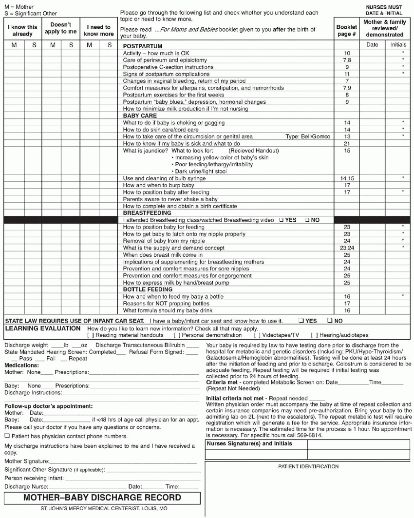
Needs assessment tools, care paths, or teaching lists can assist nurses and families in identifying learning needs and in documentation of the type and timing of prenatal education (Brown, 2006). A learning needs assessment can be initiated at various times during the perinatal period depending on when a woman has first contact with the hospital system. Opportunities include prenatal classes, prenatal visits, hospital tours, and telephone contact with a case manager during admission to the hospital, following birth, and at the mother-infant follow-up visit or contact. Many families attending prenatal classes are first-time parents. A detailed assessment of individual learning needs discussed during the first prenatal class alerts prospective parents to information and skills they need to acquire by the time they are discharged from the hospital. The learning needs assessment tool or the defined curriculum and supporting written materials document the information, instruction provided, and the skills taught. When a formal needs assessment tool is used during pregnancy, the tool is forwarded to the perinatal unit from the prenatal instructor to be stored with the woman’s prenatal data. Display 17-5 is an example of a tool that can be used on admission (if not in active labor) to assess learning needs and continued after birth to document teaching of self-care and baby care, and it then becomes the discharge teaching record. Content with an asterisk is reviewed with all women prior to hospital discharge. Referencing specific content to written materials provides reinforcement and promotes the use of materials as a reference for both families and perinatal nurses.
For women who have not attended prenatal classes or completed a learning needs assessment during a prenatal visit, the process begins on admission to the hospital. With the help of the labor nurse, families identify specific learning needs they want to address during the inpatient stay. Whether the needs assessment is completed prior to admission, during early labor, or after birth, the education process begins as soon as possible for each woman and family.
Primary responsibility for patient and family education varies with the institution. Patient education may be coordinated by the case manager, clinical nurse specialist, or perinatal educator; however, in any practice model, the perinatal staff nurse plays a key role. Critical concepts and essential information that families need have been identified. They are presented regardless of the family’s past experience or self-assessment. Critical concepts include the following:
Maternal care
Activity and rest
Pain relief and comfort measures
Care of the perineum and care of lacerations or episiotomy
Breast care for breastfeeding women and lactation suppression for women who are formula feeding
Postoperative cesarean birth instructions
Expected emotional adaptations
Signs of postpartum complications to report to the nurse in the hospital or to the healthcare provider after discharge
DISPLAY 17-5 Mother-Baby Discharge Record
Newborn care
Newborn adaptation to extrauterine life: need to be held, need for thermoregulation, need for comfort
Newborn feeding cues
Breastfeeding basics
Formula feeding basics
Care of an infant who is spitting up or choking
Use of the bulb syringe
Umbilical cord care
Circumcision care and care of the uncircumcised penis
Position for sleep: Back to Sleep campaign to reduce the risk of sudden infant death syndrome (SIDS)
Information about immunizations and newborn screening tests
Signs of newborn complications to report to the nurse in the hospital or to the healthcare provider after discharge and contact telephone numbers
Safe use of infant and child car seats
Appointment made for follow-up clinic or home visit offered by the hospital or community nursing agency
When to schedule the first mother and newborn visits with their primary care provider
Written materials provided to the woman and her family should contain information about all critical concepts. Some institutions have adopted interactive documentation forms signed by the mother and the nurse providing the education (Brown, 2006; Roudebush et al., 2006).
Before discharge, knowledge and skills about self-care and infant care are validated. Validation can be accomplished by discussion with the new mother during which understanding is verbalized or by demonstration of critical skills such as feeding, sleeping position, or umbilical cord care. No one method of validation is superior; rather, nurses in each institution can develop a system with enough flexibility to meet the needs of the population served. Validation ensures that women who indicate they need no additional information are, in reality, prepared and knowledgeable. The goal is for all women to verbalize understanding or demonstrate skills related to all critical concepts. Women with special needs who have not demonstrated knowledge of critical concepts or have not acquired the skills to care for themselves or their infants are referred for follow-up support and care. Referrals are made to the clinical nurse specialist, lactation consultant, social worker, dietitian, and/or home care agency. Follow-up contacts to ensure that the critical concepts have been learned and to verify that the woman can safely care for her infant and herself can occur at a clinic or home visit, during a phone assessment, through involvement in support groups and community programs, or at healthcare provider office visits. Assessment of maternal knowledge and skills is documented on the discharge teaching record or other appropriate medical record form.
ANATOMIC AND PHYSIOLOGIC CHANGES DURING THE POSTPARTUM PERIOD
Perinatal nurses should have knowledge of normal anatomic and physiologic changes that occur during the postpartum period in order to plan comprehensive assessments and interventions for new mothers. Many changes are apparent immediately after birth and require inpatient nursing assessment and intervention. However, over the course of the first 12 weeks postpartum, there are ongoing alterations as the woman’s body returns to its nearly prepregnant state.
UTERUS
Involution results from a decrease in myometrial cell size, not in the number of myometrial cells. This decrease is the result of ischemia, autolysis, and phagocytosis. Ischemia occurs when the retraction of uterine musculature necessary for hemostasis after placental separation results in decreased blood flow to the uterus. Proteolytic enzymes are released, and macrophages migrate to the uterus, resulting in autolysis or self-digestion and subsequent reduction in myometrial cell size. Some of the excess elastic and fibrous tissue is removed by phagocytosis, but the incomplete process results in a uterus that does not return to its nulliparous size. Within 24 hours, the uterus is approximately the size it was at 20 weeks’ gestation (Cunningham et al., 2009). Immediately after birth, the uterus weighs approximately 1,000 g (2 lb, 4 oz). As involution occurs, the uterine weight continues to decrease to 500 g (1 week), 300 g (2 weeks), and by 6 weeks postpartum, it weighs 100 g or less. Immediately after birth, the uterine fundus can be palpated midway between the umbilicus and symphysis pubis. During the first 12 hours after birth, the muscles relax slightly, and the fundus returns to the level of the umbilicus. Beginning on postpartum day 2 or 3, the usual progression of uterine descent into the pelvis is 1 cm/day (Display 17-6).
DISPLAY 17-6 Uterine Involution
Time
Location of Fundus
Immediately
At the level of the umbilicus
1-2 hours
Midline, midway between umbilicus and symphysis
12 hours
1 cm above umbilicus
24 hours
1 cm below umbilicus
3 days
3 cm below umbilicus
7 days
Just palpable at symphysis
14 days
Not palpable
During the first few days after birth, oxytocin secretion causes strong uterine contractions and a further reduction in size, especially after breastfeeding and in multiparas. Multiparity, multiple gestation, polyhydramnios, and bladder distention can influence uterine size and the progression of uterine involution.
PLACENTAL SITE AND LOCHIA
The placenta separates spontaneously from the uterus within 15 minutes of birth in 90% of women and within 30 minutes after birth in 95% of women. Separation of the placenta and membranes includes the spongy layer of the endometrium, leaving the decidua basalis in the uterus. This remaining layer reorganizes into basal and superficial layers. The superficial layer becomes necrotic and is sloughed in the lochia, and the basal layer becomes the source of new endometrium. The endometrium is regenerated by 2 to 3 weeks after birth, except at the site of placental attachment (Blackburn, 2012; Cunningham et al., 2009). Immediately after delivery of the placenta, the placental site is approximately 8 to 10 cm, and by end of the second week, it is about 3 to 4 cm. Exfoliation, the process of placental site healing, occurs over the first 6 weeks after birth by necrotic sloughing of the infarcted superficial tissues. A reparative process follows in which the endometrium regenerates from the margins and base. This process prevents the formation of a fibrous scar in the decidua. At 7 to 14 days’ postpartum, the infarcted superficial tissue over the placental site sloughs. At this time, the woman may notice an episode of increased vaginal bleeding, which is usually self-limited. Bleeding lasting more than 1 to 2 hours should be evaluated for late postpartum hemorrhage (PPH). Ultrasonography can be useful in determining the presence of retained placental tissue (Thorpe, 2009).
Lochia is the postpartum uterine discharge. Although lochia varies in amount, the total volume lost usually is 150 to 400 mL. Initially, lochia rubra is reddish and continues for 3 to 4 days. Lochia serosa, a pinkish discharge, continues from day 4 to day 10. Lochia alba, a yellow-white discharge, follows lochia serosa (Table 17-1). The choice of feeding method for the baby and the use of oral contraceptives do not affect duration of lochia (Cunningham et al., 2009).
Table 17-1. TYPES OF LOCHIA
Rubra
Serosa
Alba
Normal color
Red
Pink, brown tinged
Yellowish-white
Normal duration
1-13 days
3-110 days
10-14 days, but not abnormal to last longer
Normal discharge
Bloody with clots; fleshing odor; increased flow on standing or breastfeeding, or during physical activity
Serosanguineous (blood and mucus) consistency; fleshy odor
Mostly mucus, no strong odor
Abnormal discharge
Foul smell; numerous and/or large clots; quickly saturates perineal pad
Foul smell; quickly saturates perineal pad
Foul smell; saturates perineal pad; reappearance of pink or red lochia; discharge lasts far too long (past 4 wk)
CERVIX, VAGINA, AND PELVIC FLOOR
The cervix and lower uterine segment are thin and flaccid immediately postpartum. Cervical lacerations can occur during any birth; however, women with precipitous labor and operative procedures are at increased risk for lacerations. At 2 to 3 days, the cervix has resumed its customary appearance but remains dilated 2 to 3 cm. By the end of the first week, the cervical os narrows to a diameter of 1 cm. The external cervical os remains wider than its pregravid state, and bilateral depressions typically are seen at the site of lacerations. Cervical edema may persist for several months (Cunningham et al., 2009). The vagina and vaginal outlet are smooth walled and may appear bruised early in the puerperium. The apparent bruising, caused by pelvic congestion, disappears quickly after birth. Rugae reappear in the distended vagina by the third week. The voluntary muscles and supports of the pelvic floor gradually regain tone during the first 6 weeks postpartum. These changes occur in response to the reduced amount of circulating progesterone. For some women, vaginal tone may be improved by perineal tightening exercises, such as Kegel exercises (Ladewig et al., 2009; Weber & Richter, 2005). In the lactating woman, the hypoestrogenic state resulting from ovarian suppression may cause the vagina to appear pale and without rugae. This may result in dyspareunia.
OVARIAN FUNCTION AND RETURN OF MENSES
Although the return of menses and ovulation varies, the first menstrual period usually occurs within 7 to 9 weeks postpartum in nonnursing mothers. There are great variations in the return of menses for women who are nursing because of depressed estrogen levels. In nursing mothers, menstruation usually returns between months 2 and 18.
Estrogen and progesterone levels decrease suddenly after placental delivery. For the first 2 to 3 weeks after birth, there is minimal gonadotropin activity, possibly because of a transient pituitary insensitivity to luteinizing hormone-releasing factor. As sensitivity returns, hormonal function returns to normal levels. The first menstrual cycle is usually anovulatory, but 25% of women may ovulate before menstruation. The mean for the return of ovulation is 10 weeks postpartum for women who are not lactating and approximately 17 weeks postpartum for women who are breastfeeding. The delay in the resumption of menses in lactating women in part may result from elevated prolactin levels (Cunningham et al., 2009).
METABOLIC CHANGES
Prolactin, a pituitary hormone, is responsible for stimulating and sustaining lactation. Like estrogen and progesterone, prolactin levels decrease with placental delivery, although they remain elevated over nonpregnant levels. The decrease in estrogen and progesterone stimulates the anterior pituitary to produce prolactin. Between the third and fourth week postpartum, the prolactin level returns to normal in women who formula-feed their infants. For those who breastfeed, prolactin levels increase with each nursing episode (Cunningham et al., 2009).
Thyroid function returns to prepregnant levels within 4 to 6 weeks after birth. Because immunosuppression is a normal physiologic consequence of pregnancy, there is an increased risk of developing transient autoimmune thyroiditis, followed by hypothyroidism. This depression of thyroid function may cause depression, carelessness, and impairment of memory and concentration. There is a slightly increased risk of recurrence of autoimmune hypothyroidism or hyperthyroidism postpartum (Nader, 2009; Cunningham et al., 2009).
Low levels of placental lactogen, estrogen, cortisol, growth hormone, and the placental enzyme insulinase reduce their anti-insulin effect in the early puerperium. This results in lower glucose levels for women during this period and a reduction in insulin requirements for insulin-dependent diabetic women (Cunningham et al., 2009). Breastfeeding may precipitate hypoglycemic episodes in women with insulin-dependent diabetes. Women with gestational diabetes often have normal glucose levels immediately postpartum. Nutritional needs must be reassessed during this period. The basal metabolic rate (BMR) increases 20% to 25% during pregnancy because of fetal metabolic activity. The BMR remains elevated for 7 to 14 days after giving birth.
In the first 2 hours postpartum, plasma renin and angiotensin II levels (involved in blood pressure [BP] maintenance) fall to normal, nonpregnant levels and then rise again and remain elevated for up to 14 days (Roberts & Funai, 2009). BP should remain stable during the postpartum period, but lowered vascular resistance in the pelvis may result in orthostatic hypotension when a woman moves from a supine to a sitting position. An increase in BP, especially if accompanied by headaches or visual changes, may indicate postpartum preeclampsia and should be evaluated. In the past, an incremental increase of 30 mm Hg systolic or 15 mm Hg diastolic above baseline values was used as diagnostic criteria. This is no longer recommended, as research shows those women are not likely to have adverse outcomes; however, patients with BP increases should be closely observed, especially if the BP is above 140/90 mm Hg on two or more occasions at least 6 hours apart (Roberts & Funai, 2009; Cunningham et al., 2009).
KIDNEYS AND BLADDER
Mild proteinuria (1+) may exist for 1 to 2 days after birth in 40% to 50% of women. Nonpathology can be assumed only in the absence of the symptoms of infection or preeclampsia (Blackburn, 2012).
If a urine specimen is necessary, it should be obtained through catheterization or as a clean-catch technique. These methods avoid contamination by protein-laden lochia (Gilbert, 2011). Glycosuria of pregnancy disappears, and creatinine clearance is usually normal by 1 week postpartum. Pregnancy-induced hypotonia and dilation of the ureters and renal pelves return to the prepregnant state by 8 weeks postpartum. The catabolic process of involution causes an increase of the blood urea nitrogen (BUN). By the end of the first week postpartum, the BUN level rises to values of 20 mg/dL, compared with 15 mg/dL in the late third trimester (Cunningham et al., 2009). Glomerular filtration rate, renal blood flow, and plasma creatinine return to normal levels by 6 weeks postpartum.
Labor may result in displacement of the urinary bladder and stretching of the urethra. Other factors that interfere with normal micturition include the numbing effect of anesthesia and the temporary neural dysfunction of the traumatized bladder. These may cause decreased sensitivity. As a result, overdistension and incomplete emptying may occur. Signs of bladder distention include uterine atony reflected in increased lochia, displacement of the uterus to the right and significantly above the umbilicus, decreased urine output compared with oral and intravenous (IV) intake, and a “soft fullness,” sometimes with a palpable margin, in the suprapubic area. Normal postpartum diuresis combined with the often large amount of IV fluids administered during labor can result in bladder filling in a relatively short time. The woman should be encouraged to void as soon as possible after birth to avoid bladder filling, which can inhibit uterine contraction, thus predisposing to PPH. Assistance to the bathroom or on a bedpan may be helpful in facilitating bladder emptying. Women may report an urge but inability to urinate. Spontaneous voiding, however, should resume by 6 to 8 hours after birth, and bladder tone usually returns to normal levels 5 to 7 days later. Each voiding should be at least 150 mL. Edema, hyperemia, and submucous extravasation of blood are frequently evident in the bladder postpartum (Cunningham et al., 2009). The effects of trauma from labor on the bladder and urethra diminish during the first 24 hours, unless a urinary tract infection (UTI) is present. Some women may require in-and-out catheterization to empty their bladder in the immediate postpartum period. Avoid rapid emptying of the bladder if catheterization is performed. No more than 800 mL of urine should be removed at one time. This can avoid a precipitous drop in intraabdominal pressure, which may result in splenetic engorgement and hypotension.
STRESS INCONTINENCE
Many women report transient stress incontinence during the first 6 weeks postpartum. Although there are conflicting data on the effect of vaginal birth on future urinary status, some researchers have estimated that two vaginal births increase the risk of developing urinary incontinence twofold and increase the risk of surgery for pelvic organ prolapse eightfold (Schaffer et al., 2005). Other researchers have suggested that pregnancy itself may be a predisposing factor for urinary incontinence and pelvic organ prolapse, thus cesarean birth may not be protective against these conditions (Nygaard, 2006; Richter, 2006). A review of literature through 2005 suggested that there may be differences by parity and after exclusion of instrumental delivery. The risk of severe stress urinary incontinence and urge urinary incontinence did not appear to differ by mode of birth. Short-term occurrence of any degree of postpartum stress urinary incontinence is decreased with cesarean section (CS), although severe symptoms are equivalent by mode of birth (Press, Klein, Kaczorowski, Liston, & von Dadelszen, 2007). Persistent stress incontinence may result from pregnancy, labor, operative birth, a large baby, and perineal tissue damage. The influences of obstetric factors diminish over 3 months. The length of the second stage of labor, infant head size, birth weight, and episiotomy correlate with the development of postpartum stress incontinence (Casey et al., 2005). Impairment of muscle function near and surrounding the urethra underlies stress incontinence. Prompt catheterization for urinary retention during the postpartum can prevent urinary difficulties (Cunningham et al., 2009). Schaffer et al. (2005) suggest that the pelvic floor is exposed to compression and extreme pressures during vaginal delivery and maternal expulsive efforts. Uncoached (non-Valsalva) pushing, a response to the urge to push, is characterized by several short bearing-down efforts per contraction with breath holding for 6 to 8 seconds. In contrast, coached pushing begins as soon as a contraction is noted by the coach, and the mother is urged to push for 10 seconds, take a deep breath, and push again. Coached pushing may potentially increase the pressure on the pelvic floor with subsequent deleterious effects. See Chapter 14 for a comprehensive discussion of second-stage pushing techniques that can minimize risk of injuries to the perineum and pelvic floor. Knowledge about clinical factors implicated in stress incontinence allows anticipatory guidance and interventions for women at risk.
Fluid Balance and Electrolytes
The physiologic reversal of the extracellular or interstitial fluid accumulated during a normal pregnancy begins during the immediate postpartum period. Diuresis begins within 12 hours after birth and continues up to 5 days. Diuresis occurs in response to the decrease in estrogen that stimulated fluid retention during pregnancy, the reduction of venous pressure in the lower half of the body, and the decrease in residual hypervolemia (Cunningham et al., 2009). Urine output may be 3,000 mL or more each day. Additional fluid is lost through increased perspiration. Diuresis results in a decrease in body weight of 2 to 3 kg. Electrolyte levels return to nonpregnant homeostasis by 21 days or earlier. Fluid loss is greater in women who have experienced preeclampsia or eclampsia. By the third postpartum day, resolution of the vasoconstriction and additional extracellular fluid of gestational hypertension contribute to significant expansion of the vascular volume (Cunningham et al., 2009).
NEUROLOGIC CHANGES
Discomfort and fatigue are common concerns after birth. Afterpains or painful uterine contractions during the first 2 to 3 days after birth; discomfort associated with episiotomy, incisions, lacerations, or tears; muscle aches; and breast engorgement may contribute to a woman’s discomfort during the postpartum period. Neurologic changes related to anesthesia and analgesia are transient and, if present, require attention to ensure the woman’s safety. Deep tendon reflexes remain normal. Sleep disturbances contributing to fatigue are related to discomfort and the demands of newborn care. The presence of children or a lack of social support may limit the time available for rest. Natural or pharmacologic comfort measures should be offered. Psychosocial support is necessary, and referral to home care nursing may be appropriate.
The carpal tunnel syndrome that results from compression of the median nerve by the physiologic edema of pregnancy is relieved by postpartum diuresis. Headaches may result from fluid shifts in the first week after birth, leakage of cerebrospinal fluid into the extradural space during spinal anesthesia, fluid and electrolyte imbalance, gestational hypertension, or stress. Assessment of the quality and location of the headache and of the vital signs are necessary. Interventions such as environmental control of lighting, noise levels, and visitors and administration of analgesic medications may be effective for nonpathologic headaches. Postpartum eclampsia (i.e., seizures beginning >48 hours and <4 weeks after birth) is often preceded by severe headache or visual disturbances. Women may have a postpartum eclamptic seizure without a prenatal diagnosis of preeclampsia or hypertension. Because women may experience prodromal signs and symptoms after discharge from the hospital, information should be provided about these subjective signs and symptoms, which include a severe and persistent occipital headache, sco-tomata (i.e., spots before the eyes), blurred vision, photophobia, and epigastric or right upper quadrant pain. Women should be encouraged to notify their primary healthcare provider if any of these symptoms develop to facilitate immediate evaluation.
HEMODYNAMIC CHANGES
Changes in the cardiovascular system occur early in the postpartum period, with a variable rate of return to baseline levels that ranges from 6 to 12 weeks. Blood volume changes occur rapidly. Autotransfusion occurs as a result of elimination of blood flow to the placenta. The blood flow of 500 to 750 mL per minute, formerly flowing to the uteroplacental unit, is diverted to maternal systemic venous circulation immediately after birth. The blood loss with an uncomplicated vaginal birth is approximately 500 to 1,000 mL or more with a cesarean birth. Plasma volume is diminished by approximately 1,000 mL as a result of blood loss and diuresis. By the third day postpartum, blood volume has decreased 16% from peak pregnancy levels and returns to nearly prepregnant levels by 1 to 2 weeks postpartum.
Cardiac output after birth depends on use and choice of anesthesia or analgesia, mode of birth, blood loss, and maternal position. Cardiac output peaks immediately after birth to approximately 80% above the prelabor value in women who have received only local anesthesia. After reaching a maximum value at 10 to 15 minutes after birth, cardiac output begins to decline, reaching pre-labor values approximately 1 hour postpartum, although it remains elevated for 48 hours after birth (Blackburn, 2012). It returns to prepregnant levels by 2 to 3 weeks after birth. Because the heart rate (HR) is stable or slightly decreased, the cardiac output is most likely caused by an increased stroke volume from venous return. Cesarean birth before labor onset avoids the hemodynamic effect of contractions but not the rise in cardiac output immediately postpartum. It is thought that epidural anesthesia during labor moderates the increase in cardiac output after birth by decreasing pain and anxiety (Cunningham et al., 2009).
The pulse rate remains stable or decreases slightly after birth. If the pulse rate is above 100 beats per minute (bpm), the woman should be assessed for infection or delayed PPH. Some women may exhibit puerperal bradycardia, with a pulse rate of 40 to 50 bpm. No conclusive proof has been given for this phenomenon. Orthostatic hypotension may occur when a woman sits up from a reclining position. Preeclampsia should be suspected if BP values are 140/90 mm Hg on two or more occasions at least 6 hours apart.
HEMATOLOGIC AND LIVER CHANGES
The decrease in plasma volume is greater than the loss of red blood cells (RBCs) after birth, causing an increase in the hematocrit between day 3 and day 7. The hematocrit returns to normal levels 4 to 8 weeks later as RBCs reach the end of their normal life span (Cunningham et al., 2009; Kilpatrick & Laros, 2009; Monga, 2009). In assessing postpartum laboratory values, a 1.0- to 1.5-g decrease in hemoglobin levels or 2- to 3-point decrease in the hematocrit value reflects a 500-mL blood loss. During the first 48 hours after birth, the physiologic reversal of the extracellular fluid accumulated during a normal pregnancy and IV fluids given during labor make accurate blood loss assessment difficult because hemodilution occurs as this fluid enters the vascular system. This phenomenon is seen even in women who have lost 20% of their circulating blood volume during birth. Hemoconcentration may occur with minimal blood loss if a woman has preexisting polycythemia (AWHONN & Johnson & Johnson Consumer Products, 2006; Cunningham et al., 2009; Monga, 2009).
Normal serum iron levels are regained by the second week postpartum. A relative erythrocytosis is seen in women who have received iron supplementation during pregnancy and had an average blood loss during the birth process. In the absence of iron supplementation, iron deficiency develops in most women (Cunningham et al., 2009; Stotland, 2009). The serum ferritin level correlates closely with the body’s iron stores and is predictive of iron deficiency anemia (Cunningham et al., 2009; Stotland, 2009). Changes in blood coagulation factors remain for variable periods postpartum. Plasma fibrinogen levels and sedimentation rate levels remain elevated for at least the first week.
Leukocytosis from the stress of labor and birth is seen in the postpartum period. A nonpathologic white blood cell (WBC) count may reach 25,000/ μL to 30,000/ μL, with the increase predominantly in granulocytes. Relative lymphopenia (i.e., lymphocyte deficiency) and absolute eosinopenia (i.e., decreased eosinophils) may also be seen. This phenomenon, coupled with the increase in the sedimentation rate, may confuse the interpretation or assessment of infections during this period. Pathology should be suspected, and further evaluation is indicated when the WBCs increase 30% over a 6-hour period (AWHONN & Johnson & Johnson Consumer Products, 2006; Cunningham et al., 2009; Kilpatrick & Laros, 2009).
The alterations in liver enzymes and lipids that occurred in response to increased estrogen levels and hemodilution during pregnancy are reversed and returned to normal levels within 3 weeks postpartum. Elevated levels of free fatty acids, cholesterol, triglycerides, and lipoproteins seen during pregnancy return to normal levels within 10 days. Alkaline phosphatase, derived from the placenta, liver, and bone during pregnancy, may remain elevated for 6 weeks. The previously atonic gallbladder demonstrates increased contractility as progesterone levels decrease (Cunningham et al., 2009; Williamson & Mackillop, 2009).
RESPIRATORY AND ACID-BASE CHANGES
The respiratory system quickly returns to its prepregnant state after the birth of the baby. These changes result from the decrease in progesterone levels, the decrease in intraabdominal pressure that accompanies emptying of the uterus, and the increased excursion of the diaphragm. This reduction of diaphragmatic pressure results in the immediate return of chest wall compliance to normal levels and partially relieves the dyspnea experienced during pregnancy. Residual volume (i.e., amount of air remaining in the lung after maximum expiration) and tidal volume (i.e., volume of air inhaled and exhaled during each breath) normalize soon after birth; the expiratory reserve volume (i.e., maximum amount of air that can be exhaled), however, may remain in the abnormal range for several months. Vital capacity, inspiratory capacity, and maximum breathing capacity decrease after birth. The response to exercise may therefore be affected in the early postpartum weeks (Cunningham et al., 2009; Whitty & Dombrowski, 2009).
Length and severity of the second stage of labor appear to contribute to an “oxygen debt” (i.e., extra oxygen required after strenuous exercise) that extends into the immediate postpartum period (Cunningham et al., 2009; Whitty & Dombrowski, 2009). The BMR remains elevated for 7 to 14 days into the postpartum period and is attributable to mild anemia, lactation, and psychological factors.
As progesterone levels fall, the PaCO2 rises to the normal prepregnant values (35 to 40 mm Hg) within the first 2 days after birth. During the postpartum period, the PaO2 should be normal at 95% or higher. Normal levels of pH and base excess gradually return by approximately 3 weeks postpartum.
SKIN, MUSCLE, AND WEIGHT CHANGES
Overdistention of the abdominal wall as a result of pregnancy can rupture collagen fibers of the dermis, resulting in striae, which can occur also on the breasts, buttocks, and thighs. Striae eventually become irregular white lines. Diastasis (i.e., separation) of the rectus muscles is common and usually is reapproximated by the late postpartum period. Evidence of diastasis can be assessed by asking the woman to lift her head while lying in a supine position. If diastasis has occurred, a tentlike protrusion in the lower abdomen is noticeable. Abdominal binders are not recommended, and mild exercise to restore tone may be started after 1 to 2 weeks. The joint instability that occurred during pregnancy may not resolve until 6 to 8 weeks postpartum.
A woman loses an average of 12 lb (5.5 kg) at birth. Additional weight is lost between 2 weeks and 6 months postpartum, especially if the woman is breastfeeding (Cunningham et al., 2009). Women who choose formula feeding can expect a loss of 0.5 to 1 kg/week when eating a balanced diet containing slightly fewer calories than their usual daily expenditure. Weight loss occurs more rapidly in women of lower parity, age, and prepregnancy weight.
GASTROINTESTINAL CHANGES
After birth, there is a decrease in gastrointestinal muscle tone and motility. When these changes are coupled with relaxation of abdominal muscles, gaseous distention can occur during the first 2 to 3 days postpartum. Decreased motility can result in postpartum ileus. Constipation may result from hemorrhoids, perineal trauma, dehydration, pain, fear of having a bowel movement, immobility, and medication (i.e., magnesium sulfate antenatally for tocolysis, iron supplementation, codeine for pain, anesthetics during labor or surgery). Constipation can be minimized by encouraging the woman to drink adequate fluids and eat foods high in fiber. Hemorrhoids that develop during pregnancy may increase in size during labor and result in significant discomfort during the postpartum period. If the woman has hemorrhoids, suggesting warm or cold sitz baths and applying topical anesthetics can decrease discomfort. Stool softeners and laxatives are sometimes given. Bowel movements typically resume 2 to 3 days after birth, and normal bowel elimination patterns resume by 2 weeks postpartum.
HERNIAS AND PERINEAL, PELVIC FLOOR, AND ANAL SPHINCTER DAMAGE
Approximately one fifth to one third of U.S. women have symptoms of urinary incontinence or pelvic organ prolapse (Chaliha, 2009; Leijonhufvud et al., 2011) Genital hernias (i.e., cystocele, rectocele, uterine prolapse, enterocele) may occur because of overstretching or tearing of the muscles or fascia during birth. There is limited evidence that women who perform perineal massage, beginning at 35 weeks’ gestation, are less likely to have perineal trauma (episiotomy or tears) that requires suturing in association with vaginal birth (Beckmann & Garrett, 2006). Chaliha (2009) and Boyles, Li, Mori, Osterweil, and Guise (2009) suggest that because of the lack of clarity in current data as to causation, the focus should be on modification of potential risk factors for pelvic floor trauma, such as constipation and high body mass index (BMI) (more than 30). Controversy exists regarding the use of episiotomy. Some practitioners recommend minimizing the use of midline episiotomy and using mediolateral episiotomy when the risk for extension is increased (i.e., macrosomia, shallow perineal body, operative vaginal birth), but there is little supportive evidence for that practice (Christianson, Bovbjerg, McDavitt, & Hullfish, 2003). Episiotomy does not always prevent third- or fourth-degree lacerations. Risk factors for lacerations include nulliparity, increased gestational age, second-stage labor arrest, macrosomia, persistent occiput posterior positions, episiotomy, forceps assistance, and use of vacuum extractors (Landy et al., 2011). Routine episiotomy is not recommended by ACOG (2008a; see Chapter 14 for an in-depth discussion of episiotomy).
Obstetric trauma such as injury to the sphincter muscle or damage to the innervation of the pelvic floor is a leading cause of anal incontinence in healthy women (Garcia, Rogers, Kim, Hall, & Kammerer-Doak, 2005). Other associations with anal incontinence include prolonged second-stage labor, macrosomia, labor augmentation, and episiotomy (Jastrow et al., 2010; Casey et al., 2005). One half of women with third-degree tears experience anal incontinence. Disturbances in bowel function (i.e., fecal urgency and anal incontinence of stool and flatus) from mechanical or neurologic injury to the anal sphincter during vaginal birth may also be the result of damage from the large size of the baby’s head in relation to the vaginal opening. Women who experience a third- or fourth-degree perineal laceration report a greater incidence of incontinence of flatus than those without anal sphincter rupture. Full thickness anal sphincter disruption is the most significant factor in later development of fetal incontinence (Chaliha, 2009; Landy et al., 2011). Women with a long second-stage of labor, a large newborn, or both have the greatest risk of nerve damage (Cunningham et al., 2009; Landy et al., 2011). Parity is associated with an increased risk for urinary incontinence. Vaginal delivery increases the risk for urinary incontinence, but labor and pushing alone, without subsequent vaginal birth, do not appear to increase risk (Boyles et al., 2009). Pelvic floor exercises have shown to reduce urinary incontinence and increase pelvic floor strength, and pregnant women should be encouraged to perform them during the antepartum period (Chaliha, 2009).
Embarrassment may prevent women from reporting symptoms of anal sphincter damage. One study has suggested that there may be 80,000 women, about 2% of U.S. births, who have persistent long-term anal incontinence, but only 8,000 will report it to their providers (Lo et al., 2010). Symptoms may disappear or worsen with time. An accurate history is helpful so that women with major sphincter defects can be offered a cesarean birth when appropriate. Aging, menopause, progression of neuropathy, and effects of subsequent births may contribute to long-term sphincter weakness. In addition, women report that anal incontinence results in negative emotional health and a decrease in their quality of life (Lo et al., 2010).
FLUID AND NUTRITIONAL NEEDS
After vaginal birth, there are no dietary restrictions for women without underlying medical conditions or pregnancy-induced complications. Oral fluids or IV fluid administration helps restore the balance altered by fluid loss during the labor and birth process. Women should be encouraged to drink 3,000 mL of water and other liquids every 24 hours. Nurses should encourage healthy food choices with respect for ethnic and cultural preferences. Snack trays should be available for women who give birth when regular food service is unavailable. After cesarean birth, women usually receive clear liquids until bowel sounds are present and then advance to solid foods. For each 20 mL of breast milk produced, the woman must consume an additional 30 calories. This results in a dietary increase of 500 to 1,000 calories each day for women who are maintaining body weight (Lawrence & Lawrence, 2009). By 6 weeks postpartum, decreased pressure and distortion of the stomach from the gravid uterus and the normalization of lower esophageal sphincter pressure and tone resolves the heartburn experienced by many pregnant women.
IMMEDIATE POSTPARTUM PERIOD
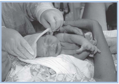
During the immediate postpartum period, the perinatal nurse focuses on maternal and newborn stabilization and recovery from the birth process. Maternal-newborn attachment and breastfeeding (if the woman desires) should be promoted and encouraged (Figs. 17-1 and 17-2). Nursing assessments and interventions should occur concurrently with activities celebrating the joy of childbirth and welcoming the new baby into the family. Family and visitor interactions, including holding the new baby and taking video and still pictures of the first hours of life, should be supported as much as possible based on the condition of the mother and newborn. Every effort should be made to accommodate the wishes of the woman and her family.
FIGURE 17-1. Promoting mother-baby attachment at birth.
Only gold members can continue reading. Log In or Register to continue