Newborn Adaptation to Extrauterine Life


Newborn Adaptation to Extrauterine Life


Debbie Fraser


Transition from fetal to newborn life is a critical period involving diverse physiologic changes. The newborn must move from an organism completely dependent on another for life-sustaining oxygen and nutrients to an independent being, a task that requires intense adjustment carried out over a period of hours to days. In addition to the normal physiologic tasks of transition, some neonates have congenital abnormalities, birth injury, or underlying disease processes. Careful assessment and nursing care is needed during the period of transition to ensure that the neonate who is experiencing problems with transition is recognized and appropriate interventions are initiated.

This chapter focuses on those factors influencing adaptation and physiologic changes during the early newborn period. These factors include the maternal history and medical and obstetric conditions, intrapartum status, delivery issues, and nursing assessment and interventions during transition, such as resuscitative needs and interventions facilitating maternal-newborn attachment.


MATERNAL MEDICAL AND OBSTETRIC CONDITIONS INFLUENCING NEWBORN ADAPTATION

A thorough review of the mother’s prenatal and intrapartum history is essential to identify factors with the potential to compromise successful transition. Table 18-1 lists maternal risk factors and associated fetal and neonatal complications. In addition to identification of current pregnancy complications, it is important to review prior obstetric history. Conditions that predispose the newborn to risk may recur in subsequent pregnancies (Display 18-1). Intrapartum risk factors may also influence adaptation (Table 18-2).

Intrapartum fetal assessment provides important data about the fetal response to labor. Electronic fetal heart rate (FHR) monitoring or intermittent auscultation provides documentation of fetal well-being. Requisite perinatal nursing skills include knowledge of the physiologic basis for monitoring, an understanding of FHR patterns, and the initiation of appropriate nursing interventions based on data from the monitor or from auscultation. The FHR reflects the fetal response to labor. The perinatal nurse focuses on discriminating between reassuring and nonreassuring patterns. If the FHR pattern is nonreassuring, intrauterine resuscitation procedures such as maternal position change, oxygen therapy, and intravenous fluids are initiated. Oxytocin should be decreased or discontinued if infusing, or the next dose of cervical ripening agents should be delayed. Safe passage through the labor and birth process sets the stage for successful transition to extrauterine life.


UNIQUE MECHANISMS OF NEWBORN PHYSIOLOGIC ADAPTATION

The respiratory, cardiovascular, thermoregulatory, and immunologic systems undergo significant physiologic changes and adaptations during the transition from fetal to neonatal life. Successful transition requires a complex interaction among these systems.


RESPIRATORY ADAPTATIONS

Critical to the neonate’s transition to extrauterine life is the ability to clear fetal lung fluid and establish respirations, allowing the lungs to become the organ of gas exchange after separation from maternal uteroplacental circulation. Pulmonary fluid, secreted by the lung epithelium, is essential to the normal growth and development of alveoli (Helve, Pitkänen, Janér, & Andersson, 2009). Toward the end of gestation, production of lung fluid gradually diminishes. The catecholamine surge that occurs just before the onset of labor has been shown to correspond to a more rapid drop in fetal lung fluid levels (Katz, Bentur, & Elias, 2011). Those infants who do not experience labor, those born by elective cesarean section, are more likely to develop transient tachypnea of the newborn (TTN) because of lower levels of serum catecholamine (Jain & Dudell, 2006).


Table 18-1. MATERNAL RISK FACTORS AND POTENTIAL FETAL AND NEONATAL COMPLICATIONS


Risk Factors


Potential Complications


Maternal substance abuse


Drug addiction


SGA, neonatal abstinence syndrome, neonatal HIV, hepatitis B and C


Alcohol use


Fetal alcohol spectrum disorder


Smoking


SGA, polycythemia


Maternal nutritional status


Maternal weight <100 lb


SGA


Maternal weight >200 lb


SGA, LGA, neonatal hypoglycemia


Maternal medical complication


Hereditary CNS disorders


Inherited CNS disorder


Seizure disorders requiring medication


Congenital anomalies (e.g., result of medication [Dilantin] use)


Chronic hypertension


IUGR, asphyxia, SGA


Congenital heart disease with congestive heart failure


Preterm birth, inherited cardiac defects


Hemoglobin <10 g/dL (100g/L)


Preterm birth, low birth weight


Sickle cell disease


IUGR, fetal demise


Hemoglobinopathies


IUGR, inherited hemoglobinopathies


ITP


Transient ITP, intracranial hemorrhage


Chronic glomerulonephritis, renal insufficiency


IUGR, SGA, preterm birth, asphyxia


Recurrent urinary tract infection


Preterm birth


Uterine malformation


Preterm birth, fetal malposition


Cervical incompetence


Preterm birth


Diabetes


LGA, hypoglycemia & hypocalcemia, anomalies, respiratory distress syndrome


Thyroid disease


Hypothyroidism, CNS defects, hyperthyroidism, goiter


Current pregnancy complications


Pregnancy-induced hypertension


IUGR, SGA


TORCH infections


IUGR, SGA, active infection, anomalies


Sexually transmitted diseases


Ophthalmia neonatorum, congenital syphilis, chlamydial pneumonia


Hepatitis


Hepatitis


AIDS or HIV seropositive


Neonatal HIV


Multiple gestation


Preterm birth, asphyxia, IUGR, SGA, twin-to-twin transfusion, birth trauma


Fetal malposition


Prolapsed cord, asphyxia, birth trauma


Maternal blood group antibodies


Anemia, hyperbilirubinemia, immune-mediated hydrops fetalis


Prolonged pregnancy


Postmaturity, meconium aspiration, IUGR, asphyxia


Intraamniotic infection


Newborn sepsis, preterm birth


Group B streptococcal infection


Newborn sepsis, preterm birth


CNS, central nervous system; HIV, human immunodeficiency virus; ITP, idiopathic thrombocytopenic purpura; IUGR, intrauterine growth restriction; LGA, large for gestational age; SGA, small for gestational age; TORCH, Toxoplasma gondii, other agents, rubella virus, cytomegalovirus, herpes virus.


Initiation of breathing is a complex process that involves the interplay of biochemical, neural, and mechanical factors, some of which have yet to be clearly identified (Alvaro & Rigatto, 2005). Pulmonary blood flow, surfactant production, and respiratory musculature also influence respiratory adaptation to extrauterine life. Establishment of independent breathing and oxygen-carbon dioxide exchange depends on these physiologic factors.


Table 18-2. INTRAPARTUM RISK FACTORS AND POTENTIAL FETAL AND NEONATAL COMPLICATIONS


Risk Factors


Potential Complications


Umbilical cord


Prolapsed umbilical cord


Asphyxia


True knot in cord


Asphyxia


Velamentous insertion


Intrauterine blood loss, shock, anemia


Vasa previa


Intrauterine blood loss, shock, anemia


Rupture or tearing of cord


Blood loss, shock, anemia


Membranes


Premature rupture of membranes


Infection, respiratory distress syndrome, prolapsed cord, asphyxia


Prolonged rupture of membranes


Infection


Amnionitis


Infection


Amniotic fluid


Oligohydramnios


Congenital anomalies, pulmonary hypoplasia


Polyhydramnios


Congenital anomalies, prolapsed cord


Meconium-stained fluid


Asphyxia, meconium aspiration syndrome


Placenta


Placenta previa


Preterm birth, asphyxia


Abruptio placenta


Preterm birth, asphyxia


Placental insufficiency


Intrauterine growth restriction, SGA, asphyxia


Abnormal fetal presentations


Breech birth


Asphyxia, birth injuries (CNS, skeletal)


Face or brow presentation


Asphyxia, facial trauma


Transverse lie


Asphyxia, birth injuries, cesarean birth, umbilical cord prolapse


Birth complications


Forceps-assisted birth


CNS trauma, cephalohematoma, asphyxia, facial trauma


Vacuum extraction


Cephalohematoma, subgaleal hemorrhage


Manual version or extraction


Asphyxia, birth trauma, prolapsed cord


Shoulder dystocia


Asphyxia, brachial plexus injury, fractured clavicle


Precipitous birth


Asphyxia, birth trauma (CNS)


Undiagnosed multiple gestation


Asphyxia, birth trauma


Administration of drugs


Oxytocin


Complications of uterine hyperstimulation (asphyxia)


Magnesium sulfate


Hypermagnesemia, CNS and respiratory depression


Analgesics


CNS and respiratory depression


Anesthetics


CNS and respiratory depression, bradycardia


CNS, central nervous system; SGA, small for gestational age.


Chemical Stimuli

A number of factors have been implicated in the initiation of postnatal breathing: decreased oxygen concentration, increased carbon dioxide concentration, and a decrease in pH, all of which may stimulate fetal aortic and carotid chemoreceptors, triggering the respiratory center in the medulla to initiate respiration. Some researchers have questioned the influence of these factors and suggest instead that factors secreted by the placenta may inhibit breathing, and that regular breathing is initiated with the clamping of the umbilical cord (Alvaro & Rigatto, 2005).


Mechanical Stimulation

In utero, the fetal lungs are filled with fluid. Mechanical compression of the chest during vaginal birth forces approximately one third of this fluid out of fetal lungs. As the chest is delivered through the birth canal, it reexpands, creating negative pressure and drawing air into the lungs. This passive inspiration of air replaces fluid that previously filled the alveoli. Further expansion and distribution of air throughout the alveoli occurs when the newborn cries. Crying creates positive intrathoracic pressure that keeps alveoli open and
forces the remaining fetal lung fluid into pulmonary capillaries and the lymphatic circulation.


Sensory Stimuli

The newborn is exposed to numerous tactile, visual, auditory, and olfactory stimuli during and immediately after birth. Tactile stimulation begins in utero as the fetus experiences uterine contractions and descent through the pelvis and birth canal. Stimulation to initiate breathing continues after birth as the neonate is exposed to stimuli such as light, sound, touch, smell, and pain. Vigorously drying the newborn immediately after birth is a significant tactile stimulation.


Contributing Factors


Pulmonary Blood Flow

In utero, the placenta is the organ of gas exchange for the fetus. Oxygenated blood is delivered from the placenta through the umbilical vein, through the ductus venosus into the inferior vena cava, and then to the left and right side of the fetal heart for distribution to the fetal body. Oxygenated blood is diverted away from pulmonary circulation in utero and instead flows through the foramen ovale and ductus arteriosus to the fetal body.

The fluid-filled lungs of the fetus create a state of alveolar hypoxia. Fetal pulmonary arterioles, which are very sensitive to oxygen, have thick musculature because of low oxygen tension in utero (Steinhorn, 2011). This results in constriction of pulmonary arterioles, which causes increased pulmonary vascular resistance (PVR) and decreased pulmonary blood flow. After birth, pulmonary vasodilatation occurs when oxygen enters the lungs as oxygen is a potent pulmonary vasodilator. This significantly decreases PVR. Increased pulmonary blood flow is established as PVR decreases with normal changes in arterial PO2, alveolar PO2, acid-base status, and absence of vasoactive substances such as prostaglandin and bradykinin. Adequate pulmonary blood flow is crucial for newborn gas exchange and successful transition. After the onset of breathing, fluid in the lungs is replaced by air.


Surfactant Production

Pulmonary surfactant is necessary to maintain expanded alveoli. Surfactant lowers surface tension, preventing alveolar collapse during inspiration and expiration. By approximately 34 to 36 weeks’ gestation, there is adequate surfactant production to support respiration and to protect against development of respiratory distress syndrome (Gardner, Enzman-Hines, & Dickey, 2011). Surfactant deficiency results in atelectasis and requires greater than normal breathing efforts. Oxygen and metabolic needs increase as the newborn must use more energy to maintain respirations. Preterm newborns are at high risk for surfactant deficiency, which may significantly jeopardize respiratory adaptation to extrauterine life.


Respiratory Musculature

Intercostal muscles support the rib cage and assist with inspiration by creating negative intrathoracic pressure. Intercostal muscles may not be fully developed at birth, increasing the risk of respiratory compromise by increasing breathing effort.


CARDIOVASCULAR ADAPTATIONS

Transition from fetal to neonatal circulation is a major cardiovascular change and occurs simultaneously with respiratory system adaptation. To appreciate hemodynamic changes, an understanding of structural and blood flow differences between fetal and neonatal circulation is necessary. Figure 18-1 illustrates fetal circulation. Also influencing the cardiovascular system are physiologic changes in the vasculature, which include decreased PVR, resulting in increased pulmonary blood flow, and increased systemic vascular resistance (SVR).


Fetal Circulation

In utero, oxygenated blood flows to the fetus from the placenta through the umbilical vein. Although a small amount of oxygenated blood is delivered to the liver, most blood bypasses the hepatic system through the ductus venosus. The ductus venosus is a vascular structure that forms a connection between the umbilical vein and the inferior vena cava. Oxygenated blood from the inferior vena cava enters the right atrium, and most of it is directed through the foramen ovale to the left atrium, then to the left ventricle, and on to the ascending aorta, where it is primarily directed to the fetal heart and brain. The foramen ovale is a flaplike structure between the right and left atria. Blood flows through the foramen ovale because pressure in the right atrium is greater than that in the left atrium. In addition, the superior vena cava drains deoxygenated blood from the head and upper extremities into the right atrium, where it mixes with oxygenated blood from the placenta. This blood enters the right ventricle and pulmonary artery where again, increased resistance in the pulmonary vessels causes 60% of this blood to be shunted across the ductus arteriosus and into the descending aorta. This mixture of oxygenated and deoxygenated blood continues through the descending aorta, oxygenating the lower half of the fetal body and eventually draining back to the placenta through the two umbilical arteries. The remaining 40% of the blood coming from the right ventricle perfuses lung tissue to meet metabolic needs. The blood that actually reaches the lungs represents about
8% to 10% of fetal cardiac output (Blackburn, 2012; Steinhorn, 2011).


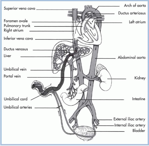
FIGURE 18-1. Fetal circulation.


Neonatal Circulation

During fetal life, the placenta is an organ of low vascular resistance. Clamping the umbilical cord at birth eliminates the placenta as a reservoir for blood, causing increased SVR, an increase in blood pressure, and increased pressures in the left side of the heart. Removal of the placenta also eliminates the need for blood flow through the ductus venosus, causing functional elimination of this fetal shunt. Systemic venous blood flow is then directed through the portal system for hepatic circulation. Umbilical vessels constrict, with functional closure occurring immediately. Fibrous infiltration leads to anatomic closure during the first week of life (Alvaro & Rigatto, 2005).

Several other significant events must take place for successful transition to neonatal circulation. With the infant’s first breath and exposure to increased oxygen levels, the pulmonary blood flow must increase, allowing the lungs to become the organ for exchange of oxygen and carbon dioxide; the foramen ovale must close (this occurs because left atrial pressures exceed right atrial pressures due to the increased pulmonary venous return of blood flow from the lungs) (Goldsmith, 2011); and the ductus arteriosus must close. In utero, shunting of blood from the pulmonary artery through the ductus arteriosus to the aorta occurs as a result of high PVR. After birth, SVR rises and PVR falls, causing a reversal of blood flow through the ductus. The mechanism of closure of the ductus arteriosus is not completely clear; however, it is known that rising arterial oxygen concentrations in the blood plays an important role (Freed, 2006). As the PaO2 level increases after birth, the ductus arteriosus begins to constrict. In utero, elevated prostaglandin levels helped maintain ductal patency. Removal of the placenta decreases prostaglandin levels, further influencing closure (Alvaro & Rigatto, 2005; Bagwell, 2007). Constriction of the ductus arteriosus is a gradual process, permitting bidirectional shunting of blood after birth. PVR may be higher than the SVR, allowing some degree of right-to-left shunting, until the SVR rises above PVR and blood flow is directed left to right.
Smooth muscle constriction in the wall of the ductus arteriosus narrows the diameter of the ductal wall within 18 hours of birth. Permanent anatomic closure of the ductus arteriosus is usually complete within 10 to 21 days (McDaniel, 2010). Any clinical situation that causes hypoxia, with pulmonary vasoconstriction and subsequent increased PVR, potentiates right-to-left shunting (Lott, 2007). Successful transition and closure of fetal shunts creates a neonatal circulation where deoxygenated blood returns to the heart through the inferior and superior vena cava. It enters the right atrium to the right ventricle and travels through the pulmonary artery to the pulmonary vascular bed. Oxygenated blood returns through pulmonary veins to the left atrium, the left ventricle, and through the aorta to systemic circulation.


Relationship between Respiratory and Cardiovascular Adaptation

Successful initiation of respirations and transition from fetal to neonatal circulation are essential to maintain life after birth. Conditions that lead to sustained elevated PVR such as hypoxia, acidosis, sepsis, or congenital heart defects can interrupt the normal sequence of events. Closure of fetal shunts depends on oxygenation and pressure changes within the cardiovascular system as described in the previous section. Foramen ovale and ductus arteriosus closure occurs only if PVR drops with the onset of respiration and subsequent oxygenation. The pulmonary vascular bed is very reactive to low oxygen levels. If the neonate experiences significant hypoxia, PVR will remain elevated with resultant decreased pulmonary blood flow and right-to-left shunting across the foramen ovale and ductus arteriosus. These events may induce a state of hypoxia as deoxygenated blood bypasses the lungs through the patent fetal shunts to be mixed with oxygenated blood entering the systemic circulation. The result is persistent pulmonary hypertension of the newborn (PPHN) requiring aggressive cardiorespiratory support.


THERMOREGULATION

The newborn’s ability to maintain temperature control after birth is determined by external environmental factors and internal physiologic processes. Characteristics of newborns that predispose them to heat loss include a large body surface area in relation to body mass and a limited amount of subcutaneous fat. Newborns attempt to regulate body temperature by non-shivering thermogenesis, increased metabolic rate, and increased muscle activity. Peripheral vasoconstriction also decreases heat loss to the skin surface. Mechanisms of heat loss including evaporation, conduction, convection, and radiation play an integral part in newborn adaptation to extrauterine life. Nursing care is critical in supporting thermoregulation through ongoing assessments and environmental interventions to decrease heat loss.


Mechanisms of Heat Production


Nonshivering Thermogenesis

Newborns have a limited capacity to shiver and, therefore, must generate heat through nonshivering thermogenesis. Heat is produced by metabolism of brown fat, a unique process present only in newborns. This highly vascular adipose tissue is located in the neck, scapula, axilla, and mediastinum and around kidneys and adrenal glands. Production of brown fat begins around 26 to 28 weeks’ gestation and continues for 3 to 5 weeks after birth (Blackburn, 2012). When exposed to cold stress, thermal receptors in skin transmit messages to the central nervous system, activating the sympathetic nervous system and triggering metabolism of brown fat, a process that utilizes glucose and oxygen and produces acids as a byproduct. Once utilized, brown fat stores are not replaced (Brand & Boyd, 2010).


Voluntary Muscle Activity

Heat produced through voluntary muscle activity is minimal in the newborn. Flexion of the extremities and maintaining a fetal position decreases heat loss to the environment. Term newborns have the ability to maintain this flexed posture, whereas preterm and compromised newborns may lack the muscle tone for this posturing, making them more vulnerable to cold stress (Bagwell, 2007).


Mechanisms of Heat Loss


Evaporation

Evaporation and heat loss occur as amniotic fluid on skin is converted to a vapor. Drying the newborn immediately after birth and removing wet blankets decreases evaporative losses and prevents further cooling. The amount of insensible water loss from the skin is inversely related to gestational age. Skin of a preterm newborn is more susceptible to evaporative losses because the keratin layer of the skin has not matured. Absence or greater permeability of this skin layer allows increased heat loss (Brown & Landers, 2011; Bagwell, 2007

Only gold members can continue reading. Log In or Register to continue

Stay updated, free articles. Join our Telegram channel

May 22, 2016 | Posted by in NURSING | Comments Off on Newborn Adaptation to Extrauterine Life

Full access? Get Clinical Tree

Get Clinical Tree app for offline access