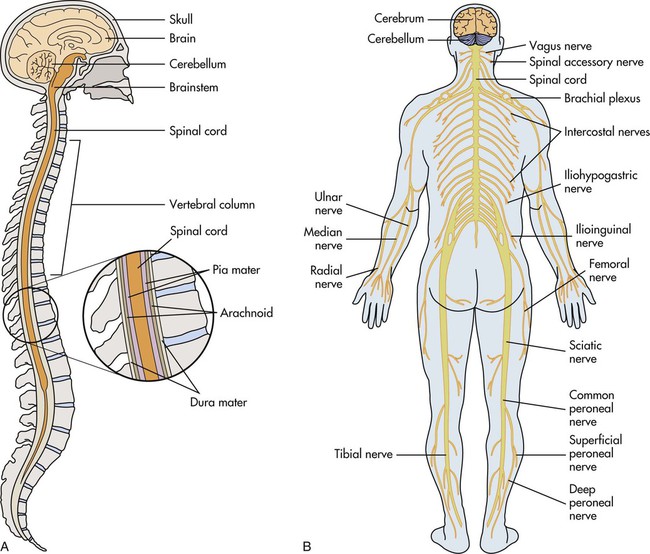
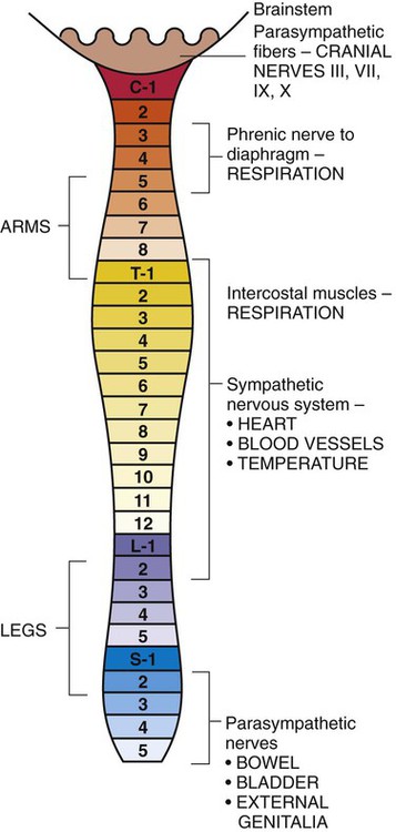
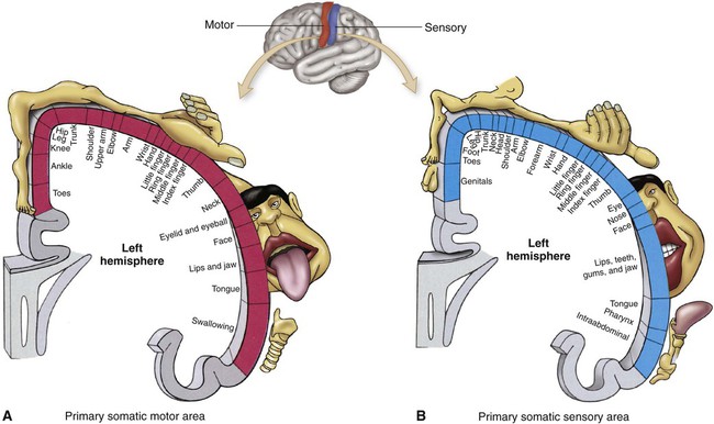
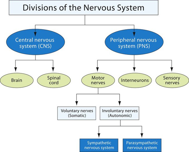
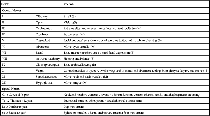
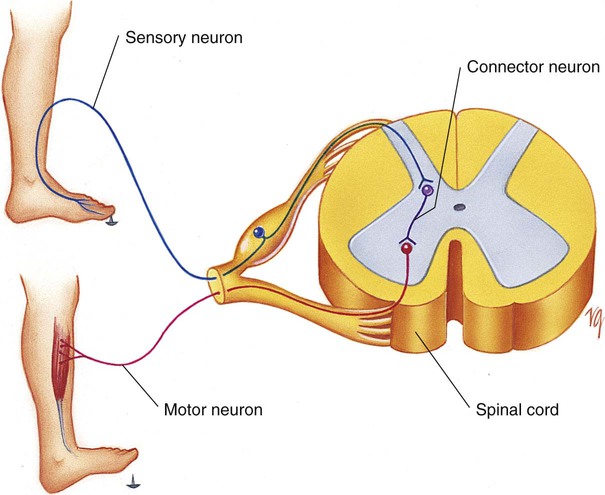
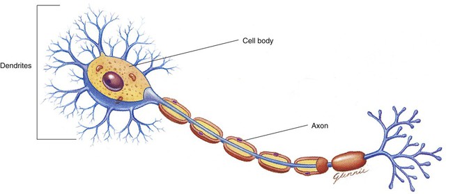
Chapter 19 *A transition syllable or vowel may be added to or deleted from the word parts to make the combining form. The nervous system is one of the most complex and interesting body systems. It is also one of the least understood. New discoveries are made almost daily about the capabilities of the nervous system. The function of the nervous system is to sense, interpret, and respond to internal and external environmental changes to maintain a steady state in the body (homeostasis). The nervous system is divided into two major structures: the central nervous system (CNS) and the peripheral nervous system (PNS) (Fig. 19-1). The CNS is made up of the brain and spinal cord (Fig. 19-2). It functions as the coordinator of the body’s full nervous system and contains the nerves that control connections between impulses coming to and from the brain and the rest of the body. The CNS plays a crucial role in maintaining a healthy, normally functioning body. Because nervous tissues are delicate and easily damaged, tough membranes called meninges surround the tissues. The nervous tissue and meninges are further protected by bones (vertebrae and cranium). The PNS consists of 12 pairs of cranial nerves and 31 pairs of spinal nerves (Table 19-1) that reach all parts of the body. The cranial nerves originate in the brain, and the spinal nerves emerge from the spinal cord. The spinal cord nerves can act independently from the brain in some reflex reactions (Fig. 19-3). Other reflex reactions of the nervous system may lead to the release of glandular secretions. TABLE 19-1 Functions of the Peripheral Nervous System The organs of the PNS contain sensory (afferent) and motor (efferent) neurons (Fig. 19-4). Afferent neurons, or nerves, carry messages from the sensory cells of the body to the brain. Efferent, or motor, nerves carry messages from the brain to the body organs or parts. The connecting nerves (interneurons) of the CNS carry messages from afferent nerves to efferent nerves. Efferent nerves are classified as voluntary (somatic) or involuntary (autonomic). The autonomic (involuntary) nervous system (ANS) is a part of the PNS (Fig. 19-5). It has two parts: the sympathetic system and the parasympathetic system. The sympathetic nerves are stimulated in situations that require action, such as the “fight or flight” reaction. The parasympathetic functions in response to normal, everyday situations. For example, the parasympathetic system would stimulate the digestion of food and slow the heart rate, whereas the sympathetic system would inhibit digestion and increase the heart rate. The neuron has several important parts (Fig. 19-6). The dendrites receive impulses and transmit them to the cell body. The cell body, which contains the nucleus of the neuron, transmits the impulse to the axon. The axon transmits the impulse away from the cell body to the dendrite of the next neuron. These impulse transmissions can travel more than 130 meters per second or 300 miles per hour. Neuroglia may become cancerous when they divide to make new cells.
Nervous System
 Define at least 10 terms relating to the nervous system.
 Describe the function of the nervous system.
 Identify at least 10 structures of the nervous system.
 Identify at least three methods used to assess the function of the nervous system.
Term
Definition
Prefix
Root
Suffix
Anesthesia
Without sensation
an
esthes
ia
Cerebrospinal
Pertaining to the brain and spine
cerebro
spin
al
Craniotomy
Incision into the skull
crani
otomy
Encephalotomy
Incision into the brain
encephal
otomy
Hypnotic
Pertaining to sleep
hypnot
ic
Insomnia
Lack of sleep
in
somn
ia
Meningitis
Inflammation of the meninges
mening
itis
Microencephaly
Small brain
micro
encephal
y
Neuralgia
Nerve pain
neur
algia
Neurology
Study of the nerve
neur
ology

Structure and Function of the Nervous System
Nerve
Function
Cranial Nerves
I
Olfactory
Smell (S)
II
Optic
Vision (S)
III
Oculomotor
Raise eyelids, move eyes, focus lens, control pupil size (M)
IV
Trochlear
Rotate eyes (M)
V
Trigeminal
Facial and head sensation, control muscles in floor of mouth for chewing (B)
VI
Abducens
Move eyes laterally (M)
VII
Facial
Taste in anterior of mouth, control facial expression (B)
VIII
Acoustic (auditory)
Hearing and balance (S)
IX
Glossopharyngeal
Taste and swallowing (B)
X
Vagus
Control muscles of speech, swallowing, and of thorax and abdomen; feeling from pharynx, larynx, and trachea (B)
XI
Spinal accessory
Move neck and back muscles (M)
XII
Hypoglossal
Move tongue (M)
Spinal Nerves
C1-8 Cervical (8 pair)
Neck and head movement; elevation of shoulders; movement of arms, hands, and diaphragmatic breathing
T1-12 Thoracic (12 pair)
Intercostal muscles of respiration and abdominal contractions
L1-5 Lumbar (5 pair)
Leg movement
S1-5 Sacral (5 pair)
Sphincter muscles of anus and urinary meatus; foot movement
Neuron
Neuroglia
 The astrocytes, star-shaped cells, are believed to help transfer substances from the blood to the brain. They make up what is known as the blood-brain barrier.
 The oligodendroglia in the CNS and Schwann cells in the PNS help to develop the myelin sheath.
 The microglia destroy and engulf bacteria and fight infection.
 The ependymal cells line the cavities of the nervous system, producing and circulating fluid in the system.
![]()
Stay updated, free articles. Join our Telegram channel

Full access? Get Clinical Tree


Nurse Key
Fastest Nurse Insight Engine
Get Clinical Tree app for offline access


