12 Muscle
Structure of muscle
The muscular system consists of many muscles through which the movements of the body are carried out. Voluntary muscles are attached to bones, cartilages, ligaments, skin or to other muscles by fibrous structures called tendons and aponeuroses. The individual fibres of voluntary muscle with their sheaths of sarcolemma are bound together into bundles by the endomysium and are covered by the perimysium. The bundles, or fasciculi, are bound together by a denser covering called the epimysium and these groups form the individual voluntary muscles of the body (see Fig. 3.8). All muscles have a good blood supply from nearby arteries. Arterioles in the perimysium give off capillaries which run in the endomysium and across the fibres. Blood vessels and nerves enter the muscle together at the hilum.
Action of muscle
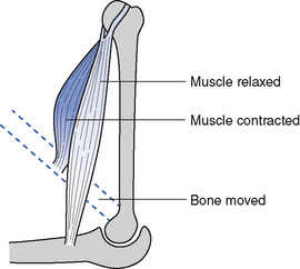
When a muscle contracts, one end normally remains stationary while the other end is drawn towards it (Figs 12.1 and 12.2). The end that remains stationary is called the origin and that which moves is called the insertion. It is not uncommon, however, for a muscle to be used, as it were, the wrong way round so that the insertion remains fixed and the origin moves towards it. The gluteus maximus provides an illustration. Its origin is in the sacrum and it is inserted into the femur. When the insertion moves towards the origin the flexed thigh is extended; when the body is bent forward at the hips the standing position is regained by movement of the origin towards the insertion. This arrangement economizes on the number of muscles required and further economy is achieved by the placing of muscles so that they can carry out more than one action. Muscles must cross the joint they move; some cross two joints producing movement in both, e.g. the biceps crosses both elbow and shoulder, causing flexion of both.
Contraction of muscle
The composition of muscle is as follows:
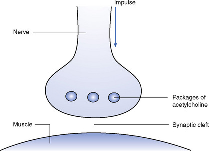
Muscle contraction occurs as a result of nerve impulses. The nerve impulses, which are electrical, are transmitted to the muscle cells by chemical means and this is accomplished by the neuromuscular junction (Fig. 12.3). Nerve impulses arrive at the neuromuscular junction, which contains small packages of acetylcholine. This is released into the space between the nerve and the muscle, the synaptic cleft. When the acetylcholine attaches to the muscle cell it causes depolarization (see Chapter 4), and hence electrical activity spreads over the muscle cell leading to contraction.
Energy is required for muscle fibres to contract; this is obtained from the oxidation of food, particularly carbohydrates. During digestion, carbohydrates are broken down to a simple sugar called glucose. The glucose that is not required immediately by the body is converted to glycogen and is stored in the liver and muscles. Muscle glycogen constitutes the source of heat and energy for muscular activity (Fig. 12.4). During the oxidation of glycogen to carbon dioxide and water, a compound is formed which is rich in energy. This compound is called adenosine triphosphate (ATP). When it is necessary for muscular contraction, the energy from ATP can be released as it changes to adenosine diphosphate (ADP). During the oxidation of glycogen, pyruvic acid is formed. If oxygen is plentiful, as it usually is during ordinary movement, pyruvic acid is broken down to carbon dioxide and water and, during the process, energy is released, which is used to make more ATP. If insufficient oxygen is available, the pyruvic acid is converted to lactic acid, which accumulates and produces muscle fatigue.
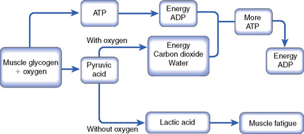
Fig. 12.4 Changes during muscle contraction. ATP, adenosine triphosphate; ADP, adenosine diphosphate.
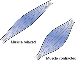
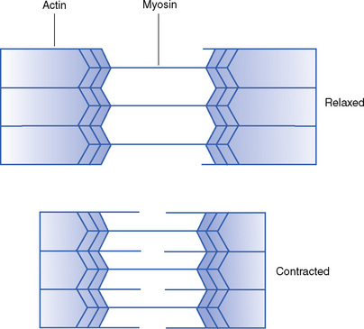
Skeletal muscle is also known as striated (striped) muscle (see Chapter 3) as a result of its microscopic appearance. The striations are due to the structure of the proteins of which the muscle is composed. These proteins are called actin and myosin. When muscle is contracted the striations are narrower – this is thought to result from the movement of one protein relative to the other in what is described as the sliding filament theory (Fig. 12.5). One of the proteins (myosin) has projections which, with the expenditure of energy, allow it to ‘walk’ along the other protein, thus leading to contraction when the muscle is stimulated by an electrical impulse.
Chief muscles of the body
Muscles of the head
The muscles of the head (Fig. 12.6) are divided into two groups according to their function: the muscles of expression and the muscles of mastication.
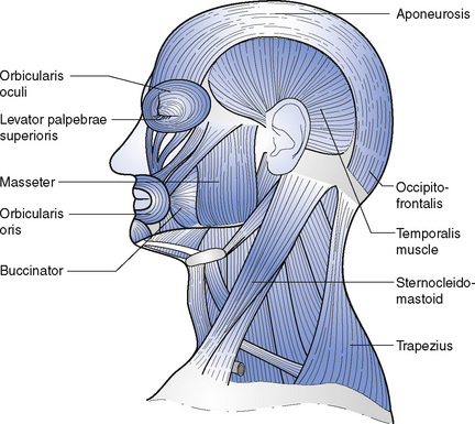
The muscles of expression are attached to the skin rather than the bone so that they move the skin and change the facial appearance. Circular muscles, called orbicularis oculi and orbicularis oris, surround the eyes and mouth, respectively, closing them. Small muscles raise and lower the eyebrows and upper lids, raise and lower the angles of the mouth and dilate the nostrils, causing a look of surprise, worry, happiness or sorrow. Small muscles also move the eyeballs in the orbits (see Chapter 6) both to direct the eyes for sight and also to change the expression.
Muscles of the neck
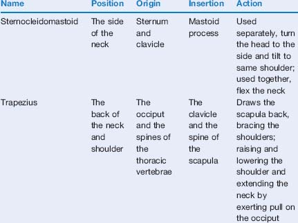
The neck contains two large muscles: the sternocleidomastoid and the trapezius (see Fig. 12.6).
The trapezius lies over the back of the neck and shoulder and is roughly triangular, with the base joining the spine down the back of the neck and chest, from the occiput, to which it is also attached, downwards. The angle is inserted into the scapula and clavicle, over the top and back of the shoulder. It draws the shoulders back when used as a whole, and also draws the scapula up and down, when the upper and lower portions are used separately (Table 12.1).
Muscles of the trunk
The chief muscles of the trunk can be grouped according to their function:
< div class='tao-gold-member'>
Stay updated, free articles. Join our Telegram channel

Full access? Get Clinical Tree


