Maintaining fluid, electrolyte and acid–base balance

CHAPTER 20 Maintaining fluid, electrolyte and acid–base balance


Introduction


Monitoring and manipulating body fluid and electrolytes form a crucial aspect of nursing care. For a lean adult male, about 18% of the body weight is protein, 15% fat and 7% minerals; 60% is water. For health, body water and electrolytes must be maintained within a limited range. Homeostatic mechanisms regulate parameters such as body fluid volume, acid–base balance (pH) and electrolyte concentrations, maintaining a delicate, dynamic balance which can be destabilised during illness. In extreme cases, the fluid or electrolyte deficit or excess can lead to death. Consequently, nurses must have a clear understanding of fluid and electrolyte homeostasis (autoregulatory processes that maintain fluid and electrolyte levels, pH, blood glucose, etc. within set parameters) so that they can assess fluid and electrolyte status, anticipate and recognise deterioration and implement corrective interventions.


Nursing interventions in relation to fluid therapy may range from encouraging the patient to drink an afternoon cup of tea to managing a complicated intravenous fluid regimen. Ill-defined terms, such as ‘encourage fluids’, and instructions to record fluid intake/output or daily weight, are commonly encountered. However, without a knowledgeable appreciation of the physiology and pathophysiology of fluid and electrolyte balance there is a real risk that these activities will be performed in a somewhat mechanistic fashion, without sufficient thought or understanding.


This chapter reviews the normal mechanisms which regulate body fluid and electrolytes and outlines some of the basic adaptive responses to imbalances. The regulation of acid–base balance is also considered, along with basic principles in the management of fluid and electrolyte disorders. Within the chapter, typical clinical situations where fluid and electrolyte control may be compromised are identified. Students who are unfamiliar with the physiology of fluid, electrolyte and acid–base balance or need to refresh their understanding are advised to read this chapter in conjunction with their physiology textbooks.


Nursing goals in the care of patients with existing or potential fluid and electrolyte problems include:


This chapter will provide some of the essential knowledge necessary to achieving these goals.


Fluid in the body


Water is essential to life and has a range of functions within the body including giving form to body structures and acting as a transport medium for nutrients, electrolytes, blood gases, metabolic wastes, heat and electrical currents. Water makes up nearly three quarters of the body’s active tissues and homeostatic regulation of body fluids is essential to normal function and health.


An average young adult man has a total body water (TBW) of about 60% of his body weight, or approximately 40 L in a 70 kg man. The percentage of body weight represented by the TBW varies from one individual to another depending on factors such as age, gender and build. Most of this variation is due to fat (adipose tissue). Adipose tissue contains only about 10% of water and so contributes little towards TBW. A 70 kg woman of average build would have a TBW of about 52% (36 L) due to the greater proportion of adipose (fatty) tissue. In both genders, the percentage of body water decreases with age.


Fluid compartments


Body water is distributed between two major compartments: approximately two thirds as intracellular fluid (ICF) and one third extracellular fluid (ECF) (Figure 20.1). The volume of ICF is around 28 L in a 70 kg male. The ECF has a volume of approximately 12 L. It comprises the intravascular fluid (about 2.5 L within the heart and vessels) and the interstitial fluid (about 9.5 L) surrounding the cells and forming a relatively constant environment. In addition, there are very small quantities of transcellular fluid (e.g. within the eye, cerebrospinal fluid, glandular secretions) and water in bone.
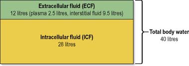


In infants and children, although the actual ECF volume is much smaller than in adults, the ECF:ICF ratio is larger, i.e. the ECF represents a greater percentage of the TBW. Fluid loss in children can rapidly lead to dehydration and is potentially more serious in the infant than in the adult.


The distinction between the different fluid compartments is maintained by the selective permeability of cell membranes. The membrane is freely permeable to water and the movement of other solutes is influenced by amongst other things the size of molecule, concentration gradient and electrical charge. Large proteins which are synthesised by the cell remain inside as they are too big to pass through the membrane. Distribution of electrolytes and water is influenced by selective membrane transport processes.


Electrolytes in the body


Fluid in the body cannot be equated simply with water, as it also contains dissolved substances or solutes as well as larger particles in suspension (colloids). Solutes may be complete molecules (non-electrolytes) or parts of molecules (electrolytes). An electrolyte is a chemical compound that, when in solution, dissociates into electrically charged particles called ions. These ions can be positively charged (cation) or negatively charged (anion). The main electrolytes found within the body are sodium (Na+), chloride (Cl), potassium (K+), magnesium (Mg2+), calcium (Ca2+), phosphorus as phosphates (H2PO4 and HPO42−) and bicarbonate (HCO3). In solution, a molecule of sodium chloride dissociates into a positively charged sodium ion Na+ (cation) and a negatively charged chloride ion Cl (anion). Sodium is the dominant cation in the ECF. Chloride and bicarbonate (HCO3) are the major extracellular anions. Inside the cell, potassium (K+) is the dominant cation. Glucose is a good example of a non-electrolyte in that it dissolves in body water but does not dissociate into ions.


Electrolytes are essential for cellular activity and need to remain within a specific range for optimal function. In clinical practice, serum electrolytes are measured by a simple blood test, urea and electrolytes, often known as ‘U&Es’. Nurses should be aware of normal ranges and factors which may cause electrolyte imbalances in patients for whom they are caring, for example patients with cardiovascular disease or renal disease or those receiving diuretic medication. This knowledge will assist nurses and other health professionals to respond with appropriate care interventions. Box 20.1 provides a more detailed description of the common electrolytes.


Box 20.1 Information


(adapted from Waugh 2003 with permission)


Common electrolytes: functions and features


Water and electrolyte homeostasis


Maintaining fluid and electrolyte balance


Maintaining normal, healthy fluid intake and the constancy of the internal environment is essential for efficient cell function. Fluid balance in the body is maintained by a number of physiological mechanisms. These mechanisms help to ensure that water gain and water loss are balanced (Table 20.1) and prevent the consequences of imbalances such as dehydration and possible death. The main stimulus for increasing intake of fluid is the thirst mechanism.


Table 20.1 Water balance – water gain and water loss (adult)


Input – Water Gain Output – Water Loss
Beverages 1500 mL
Food 750 mL
Water produced during metabolism 250 mL
Urine 1500 mL
Faeces 100 mL
Sweat 200 mL
Skin and respiration 700 mL (insensible loss)
Total 2500 mL Total 2500 mL

Fluid and electrolyte balance is inextricably linked and in health the volume and composition of the different fluid compartments are finely regulated, with daily fluctuations in TBW of less than 0.2%. Water and electrolytes are ingested and absorbed through the gastrointestinal (GI) tract, although a small volume of water is produced through the oxidation of hydrogen in food. Excess water, electrolytes and waste products are excreted via the kidneys and in faeces. Additional water and salt loss occurs via the skin and respiratory tract, known as insensible loss. Since losses from the gastrointestinal and respiratory tract are not subject to fine regulation, the kidney is the main regulator of fluid and electrolyte balance (see Ch. 8). The flow of blood through the kidney is known as the glomerular filtration rate (GFR) and a normal GFR is 125 mL/min. In 24 hours, the total blood flow through the kidneys is approximately 180 L; this means that the plasma contained within the body is filtered and reabsorbed about 60 times a day. The kidney regulates not only fluid volume, but also electrolyte composition, pH and osmolality. Central to the renal regulation of body fluid osmolality and volume is the kidney’s role in the handling of sodium and water.


The osmotic pressure of a solution is expressed as the osmolality (the number of particles present per kilogram of solvent). Osmolality is a standard measure used in medicine and in the very simplest of terms could be considered a measure of the concentration of a solution. Each of the constituents within the ECF and plasma, e.g. the principal ions (sodium, potassium, chloride, etc.) and other non-ionic substances such as glucose and amino acids, exerts a different osmotic pressure. The total sum of these is the measure of the osmolality of the ECF and plasma within 1 kg of the principal solvent within the body, water. The ECF and plasma are said to have an osmolality of around 300 mOsm/kg. The osmotic pressure produced by the proteins, although small, plays an important role in the exchange of fluid between body compartments (Pocock & Richards 2006).


Fluid exchange between the intracellular and extracellular compartments


The movement of body fluids and their constituents between the different compartments is dynamic and involves the processes:


These processes take place through the selectively permeable membrane separating the different fluid compartments, that is, the cell membrane and the capillary wall (single-cell layer of endothelium). The cell membrane separates the ICF from the ECF, and the capillary wall represents the boundary between the intravascular and interstitial compartments of the ECF.


Diffusion is the movement of solutes from an area of higher concentration to an area of lower concentration, which results in an equal concentration in both areas. Diffusion is a form of passive transport because no energy is required and the solutes just move or flow across the membrane.


Osmosis is the passive movement of fluid from an area of lower concentration (of solutes) to an area of higher concentration, in an attempt to dilute the ‘stronger’ solution and achieve an equal balance on both sides of the membrane. The capillary wall is selectively permeable to substances of a molecular weight less than 69 000 Daltons (69.0 kDa) and to lipid-soluble molecules. Lipid-soluble substances diffuse directly through the cell membrane, while water and electrolytes utilise channels formed by membrane proteins. The membrane is freely permeable to water but is only selectively permeable to electrolytes. Permeability is affected by the size and charge of the hydrated ion; for example, the membrane is 50–100 times more permeable to potassium than to sodium.


Some substances move through facilitated diffusion or active transport, for example, glucose moves under the influence of insulin. The most important example of active transport is the sodium–potassium pump. This pump is a membrane protein that couples the active transport of sodium out of the cell with the active transport of potassium inwards. It requires energy derived from the hydrolysis of adenosine triphosphate (ATP) to function.


The exchange of water, electrolytes, metabolites and waste products between the plasma and interstitial fluid occurs in the capillary bed. The capillary endothelium is freely permeable to water and solutes but the larger plasma proteins are retained. The movement of fluid through the capillaries relies on a process called capillary filtration. At the arterial end, fluid is forced out of the capillary by hydrostatic pressure. As water is lost from the capillary, the plasma proteins become more concentrated and the colloid osmotic or oncotic pressure exerted by the plasma proteins increases. At the venous end, the hydrostatic pressure, now lower than the rising osmotic pressure, results in fluid being drawn back into the capillary.


The lymphatic system is central to maintaining interstitial fluid volume. Blind-ended lymphatic capillaries in the interstitium are more permeable than the capillaries and easily take up and remove any plasma proteins that have leaked out and fluid from the interstitial space. The fluid formed in these vessels – lymph – is carried through the lymphatic system, eventually returning to the circulation via the central lymphatic and the thoracic duct.


Disturbances in fluid exchange


As approximately 20% of body fluid is found in the interstitial or tissue spaces, the maintenance of fluid volume in this compartment plays a key role in homeostasis. Normally, a dynamic equilibrium exists which maintains the extracellular fluid content of both the plasma and the tissue spaces. However, this delicate balance can easily be disturbed by the following factors and the resulting clinical picture for many of them will be oedema (Box 20.2). Further information about oedema is available on the website.


image See website for further content


Regulation and adjustments of ECF volume, osmolality and sodium


The circulatory system comprises the arterial system of high pressure and low volume and the venous system which operates with a lower pressure and higher volume. Approximately 55% of the plasma volume is in the venous system, 10% in the arterial system and the remaining 35% distributed in the heart, lungs and capillaries (see Ch. 2). Thus changes in volume are usually accommodated by the venous system. The effect of gravity upon fluid in the circulatory system is marked, causing pooling of blood in the venous system with a consequent reduction in the arterial blood volume. If an individual stands for a prolonged period, particularly in a warm environment, there is a reduction in arterial flow to the cells; inadequate venous return then leads to a fall in end-diastolic volume and hence cardiac output. Inadequate perfusion of the brain can then lead to fainting.


The tissue spaces can accommodate large volumes of fluid, but the process is slow and causes fewer disturbances to the ICF or plasma. Adults can usually tolerate changes of about 2 L in the tissue spaces before there are noticeable signs of a volume shift. This ‘hidden’ accumulation of fluid may, however, be noticed by changes in body weight, 1 L of water (pure) being equivalent to 1 kg.


Volume and osmolality regulation of the ECF involves a series of homeostatic mechanisms. Although the plasma compartment is small, it is dynamic, with shifts in volume and pressure occurring in response to internal and external stimuli. Sodium is also a major factor in the regulation of ECF. The kidney regulates sodium and water ingestion/excretion under the influence of two hormones, aldosterone and antidiuretic hormone (ADH; also known as arginine vasopressin [AVP] or vasopressin).


Influence of antidiuretic hormone


Within the hypothalamus are specialised cells called osmoreceptors which monitor and respond to changes in plasma osmolality (see above). They are very sensitive and respond to changes of as little as ±3 milliosmoles (mOsm). Plasma osmolality is normally in the range of 300 mOsmL/kg water. The osmoreceptors respond to variations in plasma osmolality by stimulating two mechanisms: thirst and ADH release.


The stimulation of the thirst centre will make the active, independent individual seek fluid to drink, thus attempting to correct any dehydration or shortage of water. ADH will act on the epithelial cells of the nephron collecting ducts to increase/decrease tubular permeability to water. If there is fluid loss, or excess ingestion of salt, plasma osmolality is increased and registered by the osmoreceptors and the individual should begin to feel thirsty. The release of ADH (from the posterior pituitary gland) affects the permeability of the nephron collecting ducts, and water will be reabsorbed into the blood to dilute the hypertonic plasma. This will result in a decrease in urine volume and increase in urine osmolality – urine produced will have a darker, more concentrated appearance. Conversely, if a large volume of water is ingested, the plasma sodium is diluted, causing a fall in the plasma osmolality. This fall is registered by the osmoreceptors, with a resultant decrease in ADH release. Lowered plasma ADH then results in decreased tubular permeability, less water is reabsorbed and water is excreted in the form of a more dilute urine.


The presence of a non-absorbable solute in the renal tubular lumen will also increase water loss. For example, when plasma glucose levels are raised, as in poorly controlled diabetes mellitus (see Ch. 5), and filtered glucose exceeds the ability of the nephrons to reabsorb it, urine production is increased. The glucose exerts an osmotic force, keeping water in the tubule. Osmotic diuresis can also be induced therapeutically by the i.v. administration of a non-absorbable molecule such as the sugar mannitol.


Influence of aldosterone


ECF volume is principally determined by sodium, and body sodium is regulated by the kidney, mainly under the influence of aldosterone. Aldosterone is a mineralocorticoid, released from the adrenal cortex, and is essential for sodium (with associated water) reabsorption. Aldosterone release is stimulated by changes in ECF volume, plasma sodium concentration and plasma potassium concentration. Increases in serum potassium levels of as little as 0.2 mmol/L can cause a marked increase in aldosterone release.


The release of aldosterone is part of a physiological mechanism often known as the reninangiotensinaldosterone system. Renin is a proteolytic enzyme released from the kidney; more specifically from the juxtaglomerular apparatus (JGA) situated near the afferent arteriole of the glomerulus. It is released in response to changes in plasma sodium and effective circulating volume. Renin acts on a circulating plasma protein (angiotensinogen) to form angiotensin I. Angiotensin I is in turn altered by a converting enzyme found in the lungs to its active form angiotensin II. Angiotensin II has several actions: it is a potent vasoconstrictor and it stimulates the release of aldosterone. This release of aldosterone will then result in an increase in sodium (and water) reabsorption within the distal tubule of the nephron. The increase in sodium reabsorption is in direct exchange for potassium. A rising serum potassium will directly stimulate the production of aldosterone, which in turn will increase sodium reabsorption from the kidney tubule in exchange for potassium which will be lost in the urine.


Hypovolaemia (reduced volume of blood circulating), which reflects a fall in sodium content (as opposed to concentration), will increase aldosterone secretion via the renin–angiotensin system. The resulting vasoconstriction and reabsorption of sodium and water reflects the body’s attempt to correct the intravascular volume and thereby maintain blood pressure.


In certain types of hypertension, this mechanism malfunctions and is managed by medication such as angiotensin converting enzyme inhibitors (ACE inhibitors) and angiotensin II receptor blockers (ARBs).


Fluid and electrolyte disturbances


Water volume and sodium imbalances frequently occur in combination with other electrolyte problems, although occasionally they occur alone. Principal causes of disturbance can be related to:


Assessment of fluid and electrolyte balance is covered later (pp. 585–589 including Tables 20.4 and 20.5 that outline some of the signs associated with fluid and electrolyte disturbances and measurable parameters and their significance).


Table 20.4 Observable/reportable signs of fluid and electrolyte disturbance and their possible significance


Clinical Signs/Symptoms Clinical Significance Cautions in Interpretation
Mucous membranes will appear dry and the patient will complain of thirst Decreased saliva production will result in a dry mouth and sensation of thirst. Osmoreceptors detecting hypovolaemia will also trigger a feeling of thirst. This acts as a useful backup, as effective oral hygiene often disguises the decreased saliva production Mouth breathing
Oxygen administration
Anticholinergic drugs, e.g. dicycloverine hydrochloride
Tongue furrows A normal tongue has one long longitudinal furrow, but in dehydration additional furrows will be present and the tongue will appear smaller due to fluid loss (Lapides et al 1965)
Sunken eyes The result of decreased intraocular pressure
Increased jugular venous pressure (JVP). With a patient positioned at 45 degrees, venous distension should not exceed 2 cm above the sternal angle Distended veins indicate fluid overload; flat veins indicate decreased plasma volume Assessing the right internal jugular vein gives a more reliable reading than on the left as it is the most anatomically direct route to the right atrium
Reduced capillary refill time Capillary refill taking 2–3 s indicates a mild fluid deficit
Refill times in excess of 3 s signify severe fluid deficits
Peripheral shutdown due to cold will slow capillary refill irrespective of fluid status
Reduced skin turgor Reduction in interstitial and intracellular fluid will reduce skin elasticity In an older person it is difficult to detect changes in skin turgor due to the gradual loss of skin elasticity with age
Cool peripheral temperature and pale skin colour As a result of the renin–angiotensin cycle, hypovolaemia will result in peripheral vasoconstriction and therefore reduced temperature and colour Patients with poor circulation, e.g. peripheral vascular disease or Raynaud’s disease, will normally have cool peripheries
Certain antihypertensives including vasodilators and ACE inhibitors will disguise the body’s normal compensatory mechanism
Dark urine Hypovolaemia will trigger release of ADH, leading to more concentrated urine Patients with liver disease may have bilirubin present in their urine giving it a very dark colour. Diuretic therapy will override the body’s production of ADH
Peripheral oedema Oedema occurs with the movement of fluid into interstitial spaces as a result of fluid excess and/or reduced levels of plasma proteins
A consequent decrease in intravascular fluid may lead to a reduction in blood pressure
Pulmonary oedema (fluid within the lung alveoli), observable through frothy, pink-stained sputum and/or dyspnoea (shortness of breath) Pulmonary oedema will result in decreased gaseous exchange, with a reduction in oxygen saturation and arterial oxygen levels

Table 20.5 Measurable parameters and their significance in fluid and electrolyte imbalance


Clinical Parameters/ Measurements Clinical Significance Cautions in Interpretation
Pulse If there is a reduction in circulating volume and therefore stroke volume, the heart rate will increase to compensate and maintain cardiac output (see Ch. 2)
Initial assessment of rhythm, based on the regularity of the pulse, may be useful and indicate a need for an ECG recording
Cardiac drugs, e.g. beta-blockers, such as atenolol, celiprolol, will inhibit the body’s compensatory mechanism and therefore block an increase in heart rate
Blood pressure Measurement of blood pressure will give an indication of circulating volume. Pulse pressure, the difference between systolic and diastolic pressure, will give an indication of vasoconstriction, i.e. compensation by the body As a result of numerous compensatory mechanisms, blood pressure is maintained by the body for as long as possible. A ‘normal’ blood pressure in the presence of compensation must be acted upon immediately
Central venous pressure (CVP) Will be reduced as a result of hypovolaemia and/or vasodilatation
An increase in CVP does not necessarily indicate fluid overload as CVP is influenced by numerous other factors including cardiac competence, systemic vascular resistance (SVR), intrathoracic and intra-abdominal pressure
For further information, see Chapter 18
Urine volume In health, the body produces 1 mL urine/kg of body weight per h
Acceptable urine output in the critically ill patient is equal to 0.5 mL urine/kg per h
A knowledge of the patient’s weight and calculation of desired urine output based upon that is essential
Administration of diuretics will override normal physiological processes. Their use must be noted when assessing volume of urine produced
Specific gravity (SG) of urine Demonstrates the body’s ability to concentrate urine as an indicator of kidney function and/or response to ADH production Administration of diuretics will override normal physiological processes. Their use must be noted when assessing urinary SG

Stay updated, free articles. Join our Telegram channel

Oct 19, 2016 | Posted by in NURSING | Comments Off on Maintaining fluid, electrolyte and acid–base balance

Full access? Get Clinical Tree

Get Clinical Tree app for offline access