M
Magnesium, serum
Basics the nurse needs to know
Decreased values
Hypomagnesemia often occurs with inadequate food intake because of impaired intestinal absorption or as a result of hemodialysis treatment. It may also occur during long-term hyperalimentation or intravenous fluid replacement. Symptoms of hypomagnesemia do not appear until the serum level is very low. Hypomagnesemia together with hypokalemia is associated with a high rate of ventricular dysrhythmias. Patients with a cardiac disorder or an acute myocardial infarction are particularly vulnerable to a depleted magnesium level, and ventricular dysrhythmia or sudden death can occur. Hypomagnesemia is considered more serious than hypermagnesemia.
REFERENCE VALUES
Newborn: 1.5-2.2 mg/dL or SI: 0.62-0.9 mmol/L
Infant-child 5 months-6 years: 1.7-2.3 mg/dL or SI: 0.70-0.95 mmol/L
Child 6-12 years: 1.7-2.1 mg/dL or SI: 0.0.70-0.86 mmol/L
12-20 years: 1.7-2.2 mg/dL or SI: 0.70-0.91 mmol/L
Adult 21-59 years: 1.6-2.6 mg/dL or SI: 0.66-1.07 mmol/L
Adult 60-90 years: 1.6-2.4 mg/dL or 0.66-0.99 mmol/L

Interfering factors
Nursing Care
Nursing measures are similar to those used in other venipuncture procedures (see Chapter 2), with the following additional measures.
Posttest

Magnetic resonance imaging
Purpose of the test
Magnetic resonance imaging (MRI) is used to assess anatomic structures, organs, and soft tissue, including visualization of any pathologic condition that is present. It can differentiate between benign and malignant growth and may be used to stage cancer or evaluate the response to treatment of a malignancy.
Basics the nurse needs to know
Types of magnetic resonance imaging systems
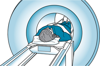
The conventional MRI uses a circumferential whole body scanner. The entire body moves into a narrow chamber that is open at both ends (Figure 66). With an open configuration scanner, the patient is not completely enclosed in the cylindrical chamber, but the images may not be as clear as with the conventional scanner and the procedure takes longer. The third type of MRI is the dedicated extremity MRI (E-MRI). This system uses a special scanner to image the affected extremity only. The imaging is for the middle and more distal joints of the arm or leg.
Safety concerns
Any ferromagnetic metal implant or other metal fragments within the patient’s body are contraindications for this imaging procedure. The magnetic forces of MRI are so great that they will twist, damage, or move the metallic object and cause injury. The metallic items include shrapnel, BBs, bullets, or other metal fragments that are imbedded in the body, particularly those located in or near the eyes or brain. Implants that are contraindications for the test include aneurysmal vascular clips, vascular stents, pacemakers, cochlear implants, joint implants, intrauterine devices, surgical screws, clips, staples, and other implanted therapeutic devices.
Magnetic resonance angiography (MRA)
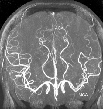
Vascular imaging by MRI is done to evaluate blood flow and the structure and location of the major blood vessels. Some studies require no contrast medium. When contrast is used, there is minimal occurrence of side effects and a lower toxicity than iodinated contrast (Frank, Long & Smith, 2007). In the head and neck, this procedure is used for imaging of the circle of Willis and the carotid arteries (Figure 67). In the evaluation of major arteries of the torso, the aorta and the renal, mesenteric, and iliac arteries can demonstrate an aneurysm, dissection, and coarctation. MRA is also used to visualize impaired blood flow in cases of peripheral vascular disease. In vascular applications, the procedure often is done before medical or surgical intervention. The data help to determine whether the problem can be corrected by endovascular stenting, angioplasty, or surgical repair with bypass or graft replacement of the blood vessel.
Brain and spinal cord imaging
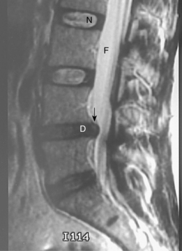
MRI is particularly valuable for assessment of the soft tissue of the brain and spinal cord. Although it cannot image the bones of the skull and vertebrae, it provides clear imaging of the organs and tissue contained within these bones. In the study of the brain, MRI provides images of tumors, cerebral edema, ischemia, multiple sclerosis, and other demyelinating diseases. In the study of the spinal cord, MRI provides images of disk degeneration, spinal cord tumor, epidural fat, and postoperative scar tissue that impinge on the cord or its nerve roots (Figure 68).
Musculoskeletal imaging
The soft tissue structures of the knee, hip, and shoulder joints are most often examined. MRI provides clear images of tears in the menisci, ligaments, tendons, and muscles that provide the structure for the knee or shoulder joint. The procedure can distinguish between an inflammatory problem that will heal with conservative therapy and a tear that will require surgical repair.
Interfering factors
Nursing Care
Pretest
The nurse teaches the patient that as he or she moves into the chamber of the MRI machine, the space will be small, with only 3 to 10 inches of extra space around the patient. The chamber is usually lighted and has a fan to help cool the air. During the procedure, the machine’s magnets cause loud knocking noises. The patient can wear earplugs or headphones to help lower the level of noise. The technologist and the patient can also communicate during the procedure. The nurse should also tell the patient that the procedure is painless and the imaging time lasts 20 to 90 minutes. The patient must remain still throughout that time so that the imaging will be clear.
Stay updated, free articles. Join our Telegram channel

Full access? Get Clinical Tree


