The cell
Cell structure
Cell component
Structure
Function
Cell membrane
The cell membrane is composed of a phospholipid bilayer embedded with various protein structures such as hormone receptors, ion channels and antigen markers
The membrane acts as a differential permeable membrane between the cell and its immediate environment
The nucleus
The nucleus is bound by a membrane, similar to the plasma membrane of the cell; this contains openings referred to as nuclear pores, which allow the movement of substances in and out of the nucleus
The nucleus contains deoxyribonucleic acid (DNA), the genetic instruction for the organism. Most of the time, the DNA is organized as chromatin threads; these condense into chromosomes prior to cell division. The nucleus stores and replicates DNA, which is expressed to synthesize proteins via a second type of nucleic acid, ribonucleic acid (RNA). These proteins determine the structure and function of the cell
Endoplasmic reticulum
This is a system of membranes, enclosing a space, which is continuous with the nuclear membrane. Endoplasmic reticulum (ER) exists as rough (granular) endoplasmic reticulum (RER) and smooth (agranular) endoplasmic reticulum (SER)
RER appears rough because of the attached ribosomes. RER is involved in protein packaging. SER is involved in lipid and steroid synthesis and the regulation of intracellular calcium levels
Mitochondria
Spherical or elongated rod-like structures surrounded by a folded inner membrane and a smooth outer membrane. There are more mitochondria in cells that are metabolically active and have a high energy requirement
Chemical processes involved in the formation of adenosine triphosphate (ATP). The cristae (inner membrane folds) are the site of oxidative phosphorylation and the electron transfer chain of aerobic respiration. Krebs (tricarboxylic acid or TCA) cycle and the oxidation of fatty acids take place within the matrix. Mitochondria contain mitochondrial DNA, which is maternally inherited and contains the genes for mitochondrial proteins
Golgi apparatus (complex)
A series of flattened curved membranous sacs
Modifies proteins from the RER and sorts them into secretory vesicles
Lysosomes
Spherical or oval organelles enclosed by a single membrane
Enclose acidic fluid containing digestive enzymes which act as a ‘cellular stomach’ breaking down cellular debris
Peroxisomes
Similar structure to lysosomes
Destroy reactive oxygen species and protect cell
Cytoskeleton
Filamentous network
Involved in maintaining cell shape and motility
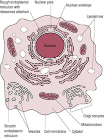
Fig. 1.2
Cells and tissues
Cell group
Epithelial cells
Support cells
Contractile cells
Nerve cells
Germ cells
Blood cells
Immune cells
Hormone-secreting cells
Example
Lining gut and blood vessels Covering skin
Fibrous support tissue, cartilage, bone
Muscle
Brain
Spermatozoa Ova
Circulating
Lymphoid tissues, nodes and spleen
Islets, thyroid adrenal
Function
Barrier; absorption; secretion
Organize and maintain body structure
Movement
Direct cell communication
Reproduction
Defence
Indirect cell communication
Special features
Tightly bound together by cell junctions
Produce and interact with extracellular matrix material
Contractile proteins
Release chemical messengers directly on to other cells
Haploid (i.e. half-normal chromosome number)
Recognize and destroy foreign material
Secrete chemical messengers into blood
Epithelial tissue
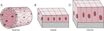
Fig. 1.3
Muscle tissue
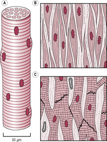
Fig. 1.4
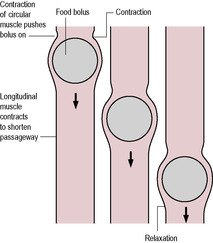
Fig. 1.5
Connective tissue
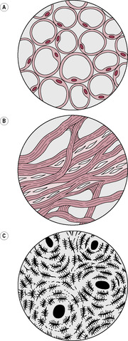
Fig. 1.6
Neural tissue
The structural organization of the body
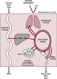
Fig. 1.8
Homeostasis
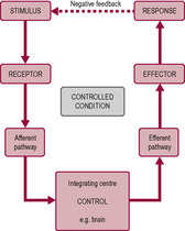
Fig. 1.9
Thermoregulation
The nervous system
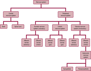
Fig. 1.11
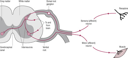
Fig. 1.12
The action potential
![]()
Stay updated, free articles. Join our Telegram channel

Full access? Get Clinical Tree


Introduction to physiology
Zara is a 29-year-old primipara who presents herself at the midwife’s clinic, which is held at her GP’s surgery, giving a history of a positive pregnancy test.
• If Zara had accessed pre-conceptual care what advice do you think she should have been given in her preparation for pregnancy?
• What information would be available in Zara’s medical records that would be useful to the midwife in her initial assessment of Zara’s pregnancy?
• How could this information be used by the midwife to inform Zara of the physiological changes that have started to occur in her body?
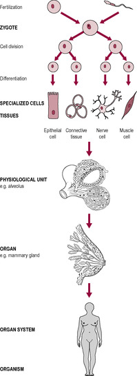
The cell is the fundamental unit of structure and function of all living organisms. The evolution of multicellular organisms has led to the differentiation of cells, which means that different cells have evolved to perform specific functions and processes that contribute to the well-being of the organism as a whole. Differentiated cells form tissues, which combine with other tissues to form organs, which are linked together in physiological systems (Fig. 1.1). However, although cells can be highly specialized, they all share common features of the single cellular organisms from which we evolved. A typical human cell is about 10 μm in diameter. The largest human cell is the oocyte (see Chapter 6); it can just be seen with the naked eye. The follicular cells surrounding the oocyte have a more typical human cell size. The sperm cell is one of the smallest human cells. Smaller cells and organelles can be visualized by light and electron microscopy.
Most cells contain cytoplasm and are bound by a plasma membrane. Within them are various structures, called organelles (see Table 1.1), and a specialized part of the cell, called the nucleus (Fig. 1.2). The fluid surrounding the organelles is called cytosol.
Although about 200 types of cells with different structures can be identified within the body, cells can be grouped together in functional categories (Table 1.2). The study of the physical characteristics of cells is called histology (see Box 1.1). There are four types of tissue: epithelial tissue, muscle, connective tissue and neural tissue.
1. red cells
2. white cells
3. platelets
1. Oxygen transport
2. Defence
1. Proteins bind oxygen
2. Proteins destroy bacteria
3. Blood clotting
Box 1.1
The study of tissue structure is described as histology. The functions of tissues are reflected in the microscopic structure of the cells of which the tissue is composed. For example, cells that are metabolically active contain many mitochondria, whereas cells that produce hormones or enzymes, for instance, will contain a large proportion of ER. Specific tissues and cellular structures are often identified by the application of various chemicals that stain particular tissues. Histology is important in diagnosing cancer, as the cancerous tissue often has histological characteristics different from those of the tissue in which the cancer has developed. Malignant cancerous tumours have highly differentiated cells, which means they often appear different from the normal tissue cells from which they arose; they often have a simpler structure and are usually prolific in their division rate. These cells are less likely to adhere to neighbouring cells as normal cells do, so they are shed into the circulatory system and carried to other parts of the body where they seed more tumours (secondary tumours or metastases). Benign tumours usually have undifferentiated cells, which may closely resemble the cells of the tissue from which they arose and tend only to grow in the one position. Although cancers in pregnancy are rare, many cancerous cells may respond to oestrogen (sometimes referred to as oestrogen dependent); therefore, cancer growth during pregnancy can be quite rapid.
Epithelial cells line the internal and external surfaces of body organs (Fig. 1.3), forming the outer layer of the skin, the mucous membranes, the lining of the lungs, gut, reproductive and urinary tracts, and also the endocrine and exocrine glands. Epithelial cells are often ‘polarized’ and have different characteristics on their apical (top) surface and their basal surface (which is in contact with the basement membrane). Epithelial cells are relatively undifferentiated and tend to undergo frequent mitotic divisions (see Chapter 7). This is because they are often exposed to wear and tear and so replacement epithelial cells are generated from a basal layer where cell division takes place. Epithelial cells form a barrier, which allows secretion and absorption of substances from one compartment to another. The skin is a specialized epithelial layer. The basal layer produces cells that are enriched with the protein keratin. The outer layers of skin cells are dead and so lack cytoplasm; it is these keratinized dead cells that provide the barrier function of the skin. Epithelial cells are classified by shape (cuboidal or columnar) and the number of layers. If there is a single layer of cells, the epithelium is described as simple; if there is more than one layer of cells (such as skin), it is stratified. Pseudostratified cells are a single layer of cells that appear to consist of more than one layer. Glands are derived from epithelial tissue.
(Reproduced with permission from Brooker, 1998.)
Muscle cells contain contractile elements, so the cells can generate the mechanical force required for movement of the body or substances within the body (Fig. 1.4) or change shape and size. Muscle tissue is formed from the mesodermal layer of the embryo (see Chapter 9). There are three types of muscle tissue: skeletal, cardiac and smooth muscle. Skeletal muscle may be attached to bones and controls movement of the skeleton. Skeletal muscle can also be attached to the skin, for instance the muscles of the face involved with expression. Contraction of skeletal muscle is usually under voluntary or conscious control. Skeletal muscle is often described as ‘striated’ because of the striped appearance of the sarcomeres of the muscle observed under the light microscope. Skeletal muscle fibres can be subdivided into slow and fast twitch fibres. Fast twitch fibres contract more strongly but they tire easily, whereas slow twitch fibres can contract for prolonged periods.
(Adapted with permission from Brooker, 1998.)
Cardiac muscle is only found in the heart; it has some structural similarity with skeletal muscle. Smooth muscle and cardiac muscle are usually under involuntary control (meaning there is no conscious awareness of the control). Smooth muscle surrounds many of the ‘tubes’ in the body, maintaining the function of several body systems. Smooth muscle cells are linked by gap junctions, and muscle contraction is relatively slow. Blood pressure is maintained by the contraction of a smooth muscle layer in the walls of the blood vessels. If the smooth muscle constricts, described as ‘vasoconstriction’, the internal lumen of the vessel will decrease and blood pressure will increase. ‘Vasodilatation’ is the opposite condition: the smooth muscle relaxes and the lumen diameter increases, so blood pressure falls. Organized synchronized waves of smooth muscle contraction, for instance in the gut, renal system and uterine tubes, generate peristaltic waves; these produce unidirectional movement of the contents within the lumen of the tube (Fig. 1.5).
(Reproduced with permission from Brooker, 1998.)
Connective tissue functions to connect, anchor and support body structures (Fig. 1.6). Connective tissue cells often produce an extracellular matrix composed of proteins in a ground substance of sugars, proteins and minerals. Bone is a type of connective tissue, whereas collagen is an example of an extracellular matrix. Adipose tissue is composed of specialized cells that store fat for future energy requirements and have an endocrine role. Adipose tissue also acts as an insulating layer to conserve body heat loss and so contributes to the maintenance of the homeothermic status of the organism. Fibrous tissue is an example of dense connective tissue. It is a tough tissue that forms ligaments, tendons and protective membranes.
(Reproduced with permission from Brooker, 1998.)
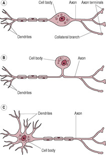
Neurons are cells that are specialized to initiate and conduct electrical signals (Fig. 1.7). Neurons require the presence of glial cells for nourishment and support; glial cells are also involved in the propagation of the electrical impulses in the neurons. As neurons are so highly specialized, they do not usually undergo further mitotic divisions once developed. Therefore, in the fetal and early neonatal period, the number of neurons produced far exceeds the level required for normal neurological function. To survive and function, neurons need regular stimulation. Throughout life, millions of neurons become dysfunctional and die.
The body’s organization can be understood by considering each component organ system separately (see below). However, these systems all work together, as a whole. Together the systems provide nutrients and oxygen for the cells and the excretion of waste products (Fig. 1.8). Movement is controlled and the temperature is maintained. Survival until reproductive function is completed has allowed the species to multiply. Cells are bathed in extracellular fluid, which can be differentiated into the interstitial fluid surrounding the tissue cells and the plasma within the blood vessels.
Homeostasis is the term used to describe the processes of the various physiological systems that maintain the constancy of the internal environment. Multicellular animals are able to maintain an internal stability that is essential for the optimal functioning of all body systems, whereas simple unicellular organisms tend to inhabit stable environments or have adapted to overcome fluctuations in the environment, for instance by forming spores during dry periods. Unicellular organisms rely on basic nutrients being present in the environment to allow cell growth and reproduction.
The evolution of multicellular organisms and the development of motility meant that these animals were able to move within the environment to seek out the conditions that suited them best and so optimize their ability to reproduce. Mammals have developed homeostasis to a high degree. Motility, together with the homeostatic challenge of counteracting fluctuations in the external environment, places a huge energy burden upon these individuals. This increased energy requirement is above the basal metabolic rate, which is the rate of energy required to maintain essential functioning only.
Homeostasis can be considered to have three main components:
• chemostasis: the maintenance of electrolytes and pH balance
• haemostasis: the maintenance of an adequate circulatory system facilitating the passage of nutrients and oxygen into and waste products out of the organism
• thermostasis: the maintenance of a constant internal temperature.
Homeostasis is regulated by the nervous system, the endocrine system and behavioural factors that are dependent on conscious or subconscious action by the organism. A homeostatic control system requires monitoring a variable, detecting changes and generating responses which will restore the composition of the internal environment (Fig. 1.9). (This type of system, involving a process called negative feedback, is dealt with in more detail under Hormonal regulation in Chapter 3, p. 64.)
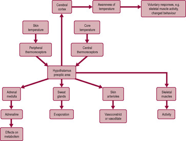
Temperature regulation is one example of homeostasis (Fig. 1.10). Enzymes regulating biochemical changes, and physiological and metabolic functions, have optimal activity within a narrow temperature range. Outside this physiological temperature range, the protein structure of the enzyme begins to denature, so the configuration (shape) of the enzyme distorts, which affects its functional activity. A warm-blooded (homeothermic) animal is well prepared to react quickly and efficiently to changes within the environment, unlike a cold-blooded (poikilothermic) animal, which depends upon the ambient temperature of the environment.
The nervous system coordinates body functions. It monitors physiological processes by processing input from the senses, integrating them and initiating responses or motor output. The nervous system is an organization of millions of neurons, or nerve cells, and glial cells, which support and regulate the composition of the nervous system. It is composed of the brain, the spinal cord (in the centre of the vertebral column) and the neurons throughout the body. The skull and the vertebral column protect the brain and the spinal cord. The brain and spinal cord form the central nervous system (CNS) and the remainder is the peripheral nervous system (Fig. 1.11). Neurons usually consist of a cell body and dendrites (extensions) and an axon or nerve fibre, which carries information from the cell body to or from the CNS. They are of different sizes; some neurons have axon projections over 1 m in length. A nerve is a collection of axons running alongside each other over the same distance. A ganglion is a collection of cell bodies of neurons within the peripheral nervous system. Ganglions are located in dorsal (back) or ventral (front) branches of the spinal cord. The spinal cord and spinal nerves are organized on a segmental basis; this corresponds to the embryonic origin of the dermatomes (see Chapter 9). Cranial nerves carry information between the brain and regions of the head. Neurons that carry information towards the brain, entering the dorsal roots of the spinal cord, are sensory or afferent neurons. Neurons carrying information from the CNS to the skeletal muscles, and leaving the spinal cord at the ventral roots, are motor or efferent neurons (Fig. 1.12). Neurons that carry information between a sensory neuron and the CNS (or between the CNS and a motor neuron) are known as interneurons.
Neurons carry information or nerve impulses by changing the electrical charge along their axon length so the transmembrane polarity changes rapidly from negative to positive and back. This change in electrical charge is termed an ‘action potential’. When the impulse reaches the axon, specific channels known as ‘sodium gates’ open, allowing the movement of extracellular sodium ions across the concentration gradient into the axon. As the sodium ions carry a positive charge, the immediate local area around the sodium gate inside of the axon becomes electrically positive compared with the immediate area outside and so the membrane becomes temporarily depolarized. At the height of the action potential (about 1 ms) the sodium channels close and the membrane becomes leaky to potassium ions; these move out of the axon down the electrochemical gradient. The result is restoration of the membrane potential, described as ‘repolarization’. That segment of the axon then enters a refractory period when no further action potential can be produced. However, depolarization in one small segment of the neuron leads to depolarization in the next segment; the rapid movement of the altered electrical activity is therefore propagated along the length of the neuron.
Get Clinical Tree app for offline access
