CHAPTER 19 Hoarseness
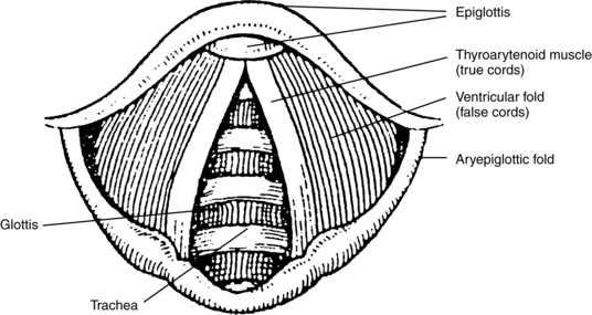
The larynx is a musculocartilaginous structure lined with a mucous membrane connected to the superior part of the trachea and to the pharynx inferior to the tongue and hyoid bone. It is the sphincter that guards the entrance into the trachea and functions secondarily as the organ of voice. Nine cartilages connected by ligaments and eight muscles form the larynx. The lower portion of the thyroarytenoid muscle forms the true vocal fold, or folds, which are highly elastic and account for the extraordinary versatility of the voice and the wide range of pitch, volume, and quality. The glottis is the triangular opening between the true vocal cords. The supraglottic area includes the ventricular folds (false vocal cords), aryepiglottic folds, and the epiglottis (Figure 19-1). The epiglottis is the lidlike cartilaginous structure that overhangs the entrance to the larynx and serves to prevent food from entering the larynx and trachea while swallowing.

FIGURE 19-1 Laryngoscopic view of the interior of the larynx.
(Modified from Greene MCL, Mathieson L: The voice and its disorders, ed 5, London, 1989, Wiley. Copyright John Wiley & Sons Limited. Reproduced with permission.)
Diagnostic reasoning: focused history
Recurrence
Recurrent episodes of hoarseness may indicate allergies or sinusitis with postnasal drip.
Progression
Progressive hoarseness usually indicates a lesion, such as a laryngeal or hypopharyngeal cyst.
Surgical history
Does the presence of risk factors help narrow the diagnosis?
Key questions
 Have you had a recent cold or upper respiratory tract infection?
 Do you have allergies or asthma?
 Do you smoke? How long have you been a smoker?
 How much alcohol do you drink?
 Can you describe your voice habits, such as singing, talking, and shouting?
 Are you frequently exposed to dust, fumes, or loud noise?


