HIV-associated tuberculosis

Chapter 26 HIV-associated tuberculosis


Introduction


Over the last three decades, tuberculosis (TB) and HIV epidemics have become inextricably connected in a bidirectional interaction, often seen as a deadly association or “the cursed duet” [1]. TB remains a major cause of morbidity and mortality in HIV-infected individuals, especially in countries with a high burden of both infections. The HIV pandemic has changed TB epidemiology, natural history, and pathogenesis, affecting its clinical and radiographic presentation, diagnosis, treatment, and prognosis.


In general, people living with HIV are 20 to 37 times more likely to develop TB disease during their lifetimes than HIV-uninfected individuals [2]. Patients with advanced HIV disease who develop TB are more likely to manifest atypical radiographic findings and develop extrapulmonary and disseminated disease. The diagnosis of TB in HIV-infected patients is more difficult, and the treatment is often complicated by drug interactions, cumulative toxicities and decreased efficacy. Furthermore, mortality rates are remarkably higher. Among individuals treated for TB, HIV-infected individuals have higher rates of recurrent TB than those who are HIV-uninfected. Conversely, active TB is associated with increased viral replication in HIV-infected patients, contributing to HIV progression and shortening survival in dually-infected patients.


The goal of this chapter is to provide a general review of TB, focusing on the unique features of HIV-associated TB and addressing specific issues of co-infection management and control strategies.


Epidemiology


TB is a major global health problem, ranking as the leading cause of death from an infectious agent and the seventh cause of death in the world [3]. The combined burden of disease caused by HIV and TB is daunting. More than 33 million people were living with HIV at the end of 2009, and the death toll from HIV/AIDS in the same year was 1.8 million. It is estimated that one-third of the HIV-infected population is co-infected with Mycobacterium tuberculosis, the majority harboring latent TB infection.


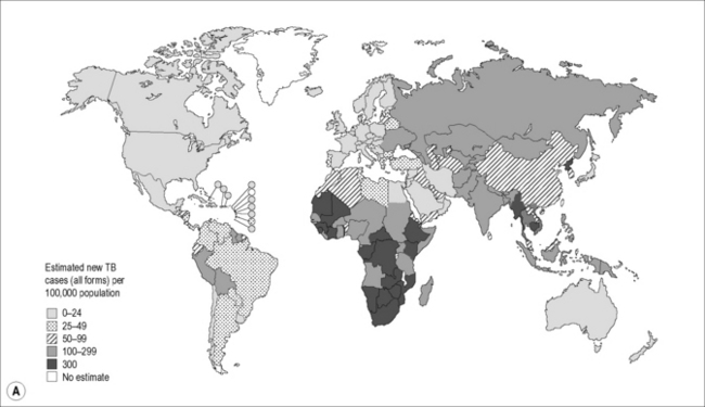
In 2009, there were 9.4 million incident cases of TB globally, equivalent to 137 cases per 100,000 population. TB prevalence was estimated at 14 million cases, equivalent to 200/100,000 [2]. Most of the estimated number of cases in 2009 occurred in Asia (55%) and Africa (30%), and smaller proportions of cases occurred in the Eastern Mediterranean region (7%), the European region (4%), and the Americas (3%). Approximately 80% of all estimated cases worldwide were concentrated in 22 high–burden countries (HBCs), among which India alone accounts for an estimated one-fifth (21%), and China and India combined account for 35% of all TB cases worldwide (Fig. 26.1A).
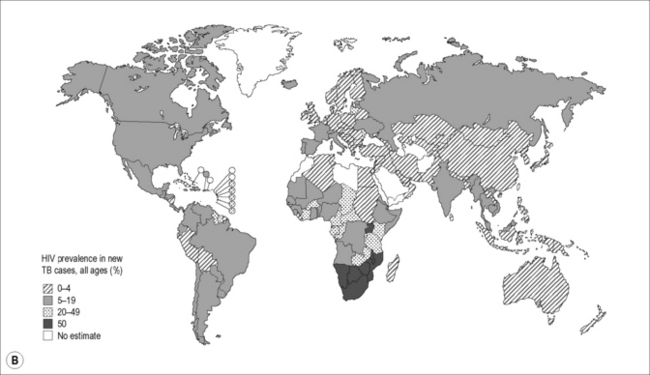

Of the 9.4 million incident cases in 2009, an estimated 1.1 million (12%) were HIV-infected, with approximately 80% of the TB-HIV cases occurring in Africa. In some southern and eastern African countries, more than 50% of TB patients are co-infected with HIV (Fig. 26.1B). Approximately 1.7 million people died of TB in 2009, including an estimated 0.4 million people with HIV, amounting to about one in four of the deaths occurring among HIV-infected individuals.


TB drug resistance, including multi-drug resistant TB (MDR-TB) and extensively-drug resistant TB (XDR-TB) is an increasing global problem. MDR-TB is defined as resistance to at least isoniazid and rifampicin, and XDR-TB is defined as an MDR isolate that is also resistant to any fluoroquinolone and at least one of the injectable second-line drugs (capreomycin, amikacin and kanamycin). There were an estimated 440,000 cases of MDR-TB in 2008, of which 85% occurred in 27 high MDR-TB burden countries; 150,000 deaths from MDR-TB occurred in 2008. The global epidemiology of drug-resistant TB in HIV-infected persons is not known. A systematic review that included 32 studies from 17 countries failed to demonstrate an overall association between MDR-TB and HIV infection [4]. However, HIV is a potent risk factor for institutional outbreaks of MDR-TB. In outbreak settings the case mortality rate has been extremely high.


Since HIV was first recognized 3 decades ago, TB incidence and notification rates have remained tightly associated with HIV prevalence rates. The global incidence of TB per capita appears to have peaked in 2004 and is now in decline, following a similar pattern to the trend in HIV prevalence in the general population, but with a 6-year delay.


Globally, the absolute number of cases is increasing slowly, reflecting population growth, although the number of cases per capita (expressed as the number of cases per 100,000 population) is falling by around 1% per year. In 2009, the per capita TB incidence rate was stable or falling in five of the six World Health Organization (WHO) regions, with the exception of the South-East Asia region, where the incidence rate is stable, largely explained by apparent stability in the TB incidence rate in India. The most recent assessment for the 22 HBCs suggests that incidence rates are falling or stable in all countries except South Africa. Mortality rates (excluding TB deaths among HIV-infected people) are falling in all six WHO regions. Among the 22 HBCs, mortality rates appear to be falling, with the possible exception of Afghanistan and Uganda.


Since monitoring of the scale-up of integrated TB/HIV activities began in 2003, considerable progress has been made. By 2009, 1.6 million TB patients knew their HIV status, equivalent to 22% of notified cases, up from 4% in 2003. In 2009, there were 55 countries in which >75% of TB patients knew their HIV status, including 16 African countries. Of the HIV-infected TB patients, 75% were receiving co-trimoxazole preventive therapy (CPT), and 37% were enrolled on antiretroviral therapy (ART) in 2009. Despite the progress achieved over the past several years, the epidemic of HIV-associated-TB continues to rage in many parts of the world and greatly increased collaboration between programs and services is needed.


Pathogenesis and Natural History


TB is caused by one of the pathogenic mycobacteria belonging to Mycobacterium tuberculosis complex, most commonly Mycobacterium tuberculosis and rarely M. bovis or M. africanum.


HIV infection is the most significant risk factor for TB progression, accelerating the development of TB at all stages that follow the deposition of M. tuberculosis in the pulmonary alveoli.


TB stages are conventionally viewed as “primary TB,” “progressive primary TB,” “latent TB,” and “secondary or post-primary TB”. There is, however, evidence supporting the paradigm that the interaction between M. tuberculosis and the human host represents a continuum of immune responses, pathologic manifestations, mycobacterial metabolic activity and clinical disease [5]. Nonetheless, this chapter will utilize the classical presentation for simplicity.


Transmission of M. tuberculosis


The chain of host–pathogen interactions begins with the inhalation into the pulmonary alveoli of droplet aerosols containing M. tuberculosis. When a patient with active pulmonary or laryngeal tuberculosis coughs, speaks, sings or sneezes, he or she generates respiratory droplets that transform into small droplet nuclei through water evaporation. The infected aerosols can remain dispersed in the air for prolonged periods of time. Particles smaller than 5 μm reach the pulmonary alveoli, a process that is necessary for M. tuberculosis transmission.


Environmental characteristics such as ventilation, humidity or presence of UV light influence the likelihood of transmission. The likelihood of TB transmission also depends on the infectiousness of the source case as measured by the cough strength [6] and the grade of acid-fast bacilli (AFB) sputum smear results. Patients with cavitary lesions and intensely positive sputum smears have a higher risk of TB transmission. Bacterial factors such as virulence and viability also influence M. tuberculosis transmission, as evidenced by the Beijing strain family of M. tuberculosis that has been associated with increased transmission, dissemination and outbreaks. Transmission of MDR-TB is comparable to that of drug-susceptible M. tuberculosis.


In general, TB transmission occurs as a consequence of household exposure, the prolonged and frequent contact with an active TB patient being much more likely to transmit M. tuberculosis than a brief contact. This pattern is observed in both low- and high-prevalence countries, irrespective of HIV status and can be demonstrated by traditional epidemiological studies and confirmed by molecular approaches [7, 8]. Occasionally, M. tuberculosis transmission has been reported after casual contact with an infectious case. Transmission after brief exposure has been linked more often to outbreaks in shelters, nursing homes, hospitals, prisons or air travel. In such cases, transmission may be related to increased virulence of the involved strain, environmental factors or patient characteristics. There is, in fact, evidence that some patients with pulmonary TB are “super-transmitters.”


Remarkably, only about one-half of the household contacts of active TB patients become infected [9], suggesting that in addition to the type of exposure, host-related factors may influence susceptibility to M. tuberculosis. The existence of innate immunity to TB is a certainty, although the immunologic mechanisms that render some populations susceptible and other resistant to TB remain largely uncharacterized. Several studies have demonstrated the association of various human leukocyte antigens (HLA) with disease susceptibility in different ethnic populations [10, 11]. Genetic susceptibility to TB has also been associated with polymorphisms in the human SLC11A1 (formerly NRAMP1) gene, some toll-like receptor (TLR) genes, the genes for the vitamin D receptor and components of the interferon (IFN) gamma-signaling pathways.


The interplay between these multiple factors and possibly others, still unknown, will determine the likelihood that the inhaled mycobacteria reach the alveoli and initiate a host–bacterial interaction that culminates in M. tuberculosis infection.


The effect of HIV on M. tuberculosis transmission has been studied extensively with contradictory results. A meta-analysis concluded that HIV infection does not significantly increase the risk of M. tuberculosis transmission, as patients with HIV-1 infection and TB are no more infectious than HIV–uninfected patients with TB [12].


M. tuberculosis infection


In most cases, the first lesion that develops as a result of M. tuberculosis infection (primary focus) is located in the lung, whereas the initial inoculation of M. tuberculosis at extra-pulmonary sites is uncommon.


Transmitted M. tuberculosis bacilli are usually deposited in the mid-lung, subpleural alveoli and are first ingested by resident alveolar macrophages, dendritic cells and recruited monocytes. Within the alveolar macrophages, M. tuberculosis bacilli multiply, destroy the macrophages (and are taken up by monocytes recruited to the inflammatory focus), giving rise to an initial exudative primary pulmonary focus. The infected macrophages are transported to the regional hilar and mediastinal lymph nodes, where bacilli continue to multiply, events that define the primary TB infection. A discrete lympho-hematogenous dissemination may occur before the development of acquired immunity, giving rise to small, micronodular pulmonary foci (apical Simon foci) or extra-pulmonary foci that can harbor viable bacilli for prolonged periods of time. These are considered to be the origin of the future secondary pulmonary or extra-pulmonary TB (endogenous reactivation).


Adaptive immunity and delayed-type hypersensitivity (DTH) develop after 3–8 weeks. Adaptive immunity is characterized by decreased bacillary multiplication, macrophage apoptosis, and granuloma formation at the site of the primary focus and disseminated areas. The immune response usually controls the infection. Most often, the lesions of primary TB undergo fibrosis and calcification and may be observed radiographically as calcified peripheral lung nodules (Ghon lesion), calcified adenopathy, or both (Ranke complex), hallmarks of primary TB infection. Caseous necrosis at the center of the lesion, tissue destruction, and cavity formation are the result of DTH.


Primary TB in HIV-infected individuals may occasionally progress directly to disease without an intervening period of clinical latency. This is termed progressive primary tuberculosis, a condition characterized by exudative, caseous and ulcerative pulmonary lesions as a result of pneumonic progression of the primary focus. The host adaptive T cell-mediated response, mainly mediated by interleukin (IL)-12 and interferon (IFN)-γ production by Th1 cells and the local expression of tumor necrosis factor (TNF)-α, is important in the control of M. tuberculosis infection, granuloma formation and prevention of mycobacterial dissemination. HIV infection causes CD4 T cell depletion and impairment of cytokine expression, which accounts for poor granuloma formation and TB dissemination (extrapulmonary/miliary forms) in co-infected patients.


TB disease


The reactivation of pulmonary TB in the host with pre-existing immunity leads to a vigorous inflammatory response with the development of caseating granulomas and ultimately cavitary lesions. Pathologically, the lungs of HIV-infected patients dying with TB are frequently characterized by caseous lesions containing tubercle bacilli; however, the type of lesion is correlated with the degree of immunosuppression. In patients with high CD4 counts, typical caseating granulomas can be seen, while in patients with low CD4 counts, the lesions tend to be diffuse, with more extensive caseous necrosis, poor granuloma formation, reduced fibrosis and less frequent cavity formation, reflecting the impaired immune activation.


In HIV-infected individuals, post-primary TB most commonly develops through the endogenous reactivation of the small foci of hematogenous dissemination occurring in the course of primary infection, as in HIV-uninfected populations. In both HIV-infected and uninfected individuals, the most common site of TB reactivation is the lung. Extrapulmonary TB, which may occur in any organ, is more common in HIV-infected patients. In contrast to HIV-uninfected individuals, active TB occurring as a result of TB re-infection, proved through restriction fragment length polymorphism (RFLP) analysis of strains, has been observed frequently in HIV-infected patients, especially in countries where the prevalence of TB is high [13, 14].


In HIV-uninfected individuals, the lifetime risk of developing TB disease by reactivating latent TB infection is approximately 5–10%. In HIV-infected individuals, this risk increases to 5–15% annually, rising as immune deficiency worsens. The increased risk is manifest even in the absence of immunodeficiency. In South African gold miners, the risk of active TB was increased two- to threefold within the first 2 years of HIV infection despite the absence of significant CD4 cell depletion [15]. In general, the risk of TB progression correlates with the CD4 count. In an African cohort, the TB incidence rate was 17.5 cases/100 person-years in HIV-infected patients with CD4 counts <200/mm3 versus 3.6 cases/100 person-years for CD4 counts >350/mm3 [16].


Immunologic aspects


Multiple distinct receptors, such as complement receptors CR1, CR3, CR4, the mannose receptor, CD14, surfactant protein A (Sp-A) receptors and scavenger receptors, have the potential to recognize and bind M. tuberculosis in vitro. M. tuberculosis can activate the alternative pathway of complement and become opsonized by complement products that facilitate uptake by complement receptors. M. tuberculosis also expresses surface polysaccharides that can directly interact with complement receptors.


Besides expressing traditional phagocytic receptors for antibody and complement, macrophages and dendritic cells also express Toll-like receptors (TLRs) that recognize conserved antigens expressed on pathogens. Binding of TLRs to these pathogen-specific ligands initiates a signal transduction pathway in the host cell that culminates in the activation of NFκB and the induction of cytokines and chemokines that are crucial to eliciting the adaptive immune response against M. tuberculosis. The sequence of the initial immune events following interaction of M. tuberculosis with TLRs and other receptors is not completely understood. Nevertheless, it is clear that in the vast majority of individuals, the interaction culminates in the development of a protective Th1 dominant immune response [17]. Th1 lymphocytes are characterized by expression of IL-2 and IFN-γ.


Several mechanisms, including induction of Th2 and T-regulatory cells, contribute to host susceptibility to TB. Th2 cells are characterized by secretion of IL-4 and IL-10. T-regulatory cells are a distinct subset of T cells that suppress Th1 responses and are characterized by the cell surface expression of CD25, cytoplasmic expression of FOXP3, and secretion of IL-10 and transforming growth factor-β. TB disease and particularly disease with a poor prognosis have been associated with increased production of IL-4 and IL-10. IL-10-secreting CD8 T cells have also been described in anergic TB patients, and increased levels of CD4 CD25 regulatory T cells have been reported in bronchoalveolar lavage of untreated TB patients. These studies imply that an imbalance in effector cell populations and modulation by regulatory T cells may determine progression from latent infection to disease.


Although the precise immune mechanisms engendering protection in the acute or latent phase of TB remain incompletely defined, current evidence supports the notion that effective acquired cellular immunity to M. tuberculosis is critically dependent on the activation of the Th1 subset of CD4 T cells.


Clinical Manifestations


TB is a complex, dynamic and multifaceted disease, with protean manifestations in both HIV-uninfected and HIV-infected individuals. Depending of the involved organ and the underlying immunological state (reflected by the CD4 count), TB may present as a myriad of clinical syndromes ranging from asymptomatic disease to fever of unknown origin to a fulminant presentation mimicking septic shock. In general, HIV-infected patients with high CD4 counts tend to present with clinical and radiographic manifestations of secondary TB disease similar to those seen in HIV-uninfected individuals, whereas HIV-infected patients with advanced degrees of immunosuppression are more likely to present with findings consistent with progressive primary TB. Patients with low CD4 counts often present with atypical radiographic findings or extrapulmonary and disseminated TB, reflecting the inability of their immune system to contain the infection.


Primary tuberculosis


Primary tuberculosis with complications


The primary TB complex may be associated with relatively benign local complications, such as sero-fibrinous pleural effusion, epituberculosis, bronchial compression or bronchial perforation by lymph nodes, complications that may regress spontaneously in the absence of advanced immunosuppression.


A transient small sero-fibrinous pleural effusion associated with DTH may complicate primary TB, resolving without specific treatment in patients able to develop an adaptive immune response. Patients may experience pleuritic chest pain, and a pleural rub may be noted on the physical exam.


Epituberculosis is characterized radiographically by an extensive pneumonic opacity, related to the inflammatory changes associated with DTH and/or compression atelectasis from enlarged lymphadenopathy and with additional components related to bronchogenic spread. In contrast to the extensive radiologic changes, the clinical symptoms may be mild and include fever, cough, and dyspnea. The physical findings may reveal signs consistent with lung consolidation.


Upper or middle lobe collapse due to bronchial compression by enlarged lymph nodes may also be seen. Bronchial stenosis may be manifested clinically by non-productive cough, dyspnea, and localized wheezing. The enlarged lymph nodes may perforate into the adjacent bronchus, causing a fistula, through which the infectious caseous material can be expectorated. Clinical symptoms include low-grade fever, productive cough with muco-purulent sputum and rarely hemoptysis. Radiologic examination is not revealing. Fistulas may be diagnosed through bronchoscopic examination, and AFB smear may be positive, allowing bacteriologic confirmation.


The spectrum of progressive primary TB includes extensive lobar consolidation or bronchopneumonia, miliary TB and TB meningitis, and is a syndrome clinically indistinguishable from secondary (active) TB. It represents a severe complication of primary TB, more likely to manifest in patients with advanced immunosuppression. The extensive parenchymal involvement may lead to tissue destruction with cavity formation, fibrosis, and bronchiectasis, similar to secondary TB. Caseous bronchopneumonia evolves rapidly with altered general status, fever, weight loss, productive cough, often with hemoptysis, and the physical exam may reveal findings of consolidation or show other HIV stigmata such as oral thrush or wasting. Sputum may be positive for AFB. Radiographically, there are extensive bilateral dense, bronchopneumonic infiltrates. Miliary TB and TB meningitis are discussed below.


Secondary tuberculosis


Pulmonary tuberculosis


Classically, the onset of post-primary pulmonary TB is insidious, with non-specific constitutional symptoms such as fever, malaise, anorexia, weight loss, and nocturnal sweats that are often the earliest indicator of disease. Respiratory symptoms such as cough, dyspnea, or pleuritic chest pain develop subsequently. A pattern of onset with flu-like symptoms with fever, and upper respiratory tract congestion, may be more common in HIV-infected patients. An acute onset with hemoptysis or pleuritic chest pain is sometimes seen.


Cough, fever, fatigue, weight loss, and night sweats are the most frequent symptoms of pulmonary TB used in clinical algorithms for TB screening in both HIV-infected and HIV-uninfected individuals. Several studies were conducted to determine the performance of these symptoms in predicting the diagnosis of TB in HIV-infected patients. Screening algorithms that combine multiple symptoms have demonstrated higher sensitivity, but lower specificity. A Southeast Asian study of HIV-infected individuals has shown that the presence of cough, fatigue, fever or weight loss in the previous 4 weeks had a sensitivity greater than 70% for each of the individual symptoms, with relatively lower specificities. However, the performance of clinical indicators was greatly increased if a combination of these symptoms was used for TB screening [19]. Although the presence of cough for 2–3 weeks has traditionally served as a criterion for identifying the TB suspects, this duration was found to have a relatively low (22–33%) sensitivity in this study, whereas a cough of any duration in the previous 4 weeks had a sensitivity greater than 70%. The combination of cough of any duration, fever of any duration and night sweats lasting 3 or more weeks in the preceding 4 weeks was 93% sensitive for TB and had a negative predictive value of 97%. A subsequent WHO meta-analysis of 12 studies that included more than 8,000 HIV-infected patients concluded that the absence of current cough, fever, night sweats, and weight loss (all inclusive) had a negative predictive value of ~98% in a setting with TB prevalence >5% [20].


If present, cough is usually characterized by mucopurulent sputum production, reflecting the expectoration of caseous material. If present, hemoptysis is usually mild, manifested as bloody-streaked sputum. Moderate or severe hemoptysis may occur secondary to arterial rupture in the wall of a cavity (Rasmussen’s aneurysm) or in the setting of bronchiectasis associated with fibrotic changes, as well as secondary to the development of a mycetoma or fungus ball within an old cavity (aspergilloma). Occasionally, patients may have dysphonia, suggesting laryngeal involvement, often associated with active pulmonary TB, as a result of laryngeal contamination with highly contagious caseous sputum. Pulmonary TB may often remain asymptomatic, particularly in the setting of HIV co-infection.


The contribution of physical examination to the diagnosis of TB is inferior to the radiologic examination. Fever, tachypnea, tachycardia, clubbing, or cyanosis may be seen. The chest examination may be normal or reveal rhonchi, rales, wheezing, or altered breath sounds. Occasionally, amphoric (hollow, resonant) breath sounds may be heard in large cavitary disease. Dullness to percussion or decreased breath sounds may be present over pleural effusions. The chest examination is often completely normal, contrasting with the advanced anatomic and radiographic abnormalities.


Laboratory evaluation may reveal mild leukocytosis with monocytosis. HIV-infected patients may show lymphopenia. Normochromic, normocytic anemia is often associated with TB. Elevation of ESR and CRP is non-specific. Hyponatremia secondary to SIADH is associated with extensive lung involvement. ABG usually shows normal PaO2, except for extensive parenchymal disease or miliary TB, which can be associated with hypoxia. Findings of hypercapnia and respiratory acidosis can be encountered in the post-tuberculous syndrome or chronic pulmonary TB, in relation to chronic structural lung disease. Pulmonary function testing is non-specific in active TB, although a restrictive pattern with decreased DLCO may be seen in patients with chronic disease.


Radiographically, the typical lesions are located in the apico-posterior lung segments or in the apical segments of the lower lobes, most commonly manifesting as heterogeneous opacities. Early stages are usually characterized by ill-defined areas of increased opacity often associated with nodular and linear opacities. If the disease is minimal it may be best seen on apical lordotic chest radiographs or CT scan. HRCT scan may reveal early bronchogenic spread as 2- to 4-mm centrilobular nodules and linear branching opacities that represent caseous necrosis containing bacilli around terminal and respiratory bronchioles (“tree-in-bud”). This pattern is not specific to mycobacterial infection and may be seen in other infectious or inflammatory conditions. As the disease progresses, additional opacities develop that may coalesce and include small areas of increased lucency. These areas may establish communication with the tracheobronchial tree to form cavities. Aspergilloma may form within old cavities. Coughing may result in bronchogenic spread in the ipsi- or contralateral lower lung zones (apico-caudal dissemination). There may be an associated pleural effusion or pyopneumothorax if the cavities rupture into the pleural space. A marked fibrotic response may be associated with atelectasis of the upper lobe, retraction of the hilum toward the apex, compensatory lower lobe hyperinflation, and mediastinal shift towards the fibrotic lung. Complete destruction of a whole lung can be seen in advanced cases.


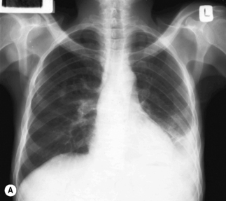
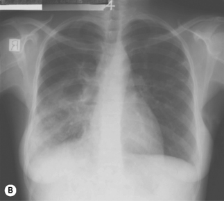
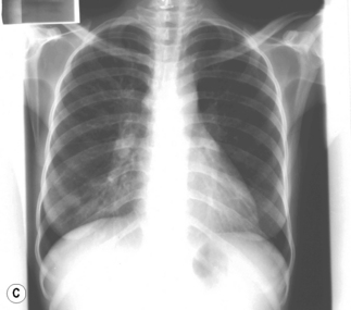
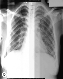
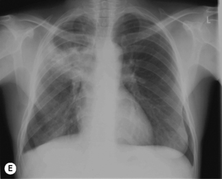
In TB associated with HIV infection, radiographic manifestations correlate with the level of immunosuppression. In patients with CD4 counts >350 cells/mm3, TB presents with classical radiographic findings of reactivation disease. In patients with fewer CD4 cells, the likelihood of atypical radiographic findings increases, and up to 20% of patients may have a normal or near-normal chest radiograph. In a study of radiographic patterns of TB in HIV-infected patients, diffused or localized infiltration were more frequent, as well as hilar or mediastinal lymphadenopathy. Pleural disease, cavitation, and normal radiography were the least common findings [21]. However, studies conducted in Africa demonstrated a greater frequency of cavitation in HIV-infected patients with TB, possibly related to the high incidence of TB in this region. Several radiographical forms of pulmonary TB are illustrated in Fig. 26.2.


Extrapulmonary tuberculosis


HIV infection is associated with a higher frequency of extra-pulmonary disease, including the more serious forms, disseminated (miliary) TB and TB meningitis.


Pleural TB


Pleural TB occurs by direct extension from an adjacent sub-pleural pulmonary focus or through hematogenous seeding. Typical presentation is the abrupt onset of fever, pleuritic chest pain, and cough. Occasionally there is an insidious presentation with fever, weight loss, and malaise. If the pleural effusion is large enough, there may be shortness of breath. Physical examination shows dullness to percussion and decreased breath sounds. Egophony is a helpful sign if present. Chest radiograph typically shows unilateral pleural effusion more frequently in the right hemithorax. Bilateral disease is seen in 10% of cases. Pleural fluid analysis demonstrates high protein concentration, low glucose concentration, and lymphocytosis. The presence of >5% mesothelial cells in the pleural fluid usually argues against a diagnosis of TB pleural effusion, as the chronic pleural inflammation is thought to prevent the exfoliation of mesothelial cells in the pleural cavity. The AFB smear is positive in less than 10% of cases, and the yield of culture has been less than 30% in most series, with a range between 12% and 70%. In patients co-infected with HIV, the yield of pleural fluid and pleural biopsy culture may be higher than in HIV-uninfected individuals with TB pleurisy [22]. Pleural biopsy may show granulomas in approximately half of the cases, and the yield is higher on pleural biopsy culture. An elevated concentration (>70 U/L) of pleural fluid adenosine deaminase (ADA) may suggest the diagnosis of TB.

Only gold members can continue reading. Log In or Register to continue

Stay updated, free articles. Join our Telegram channel

Apr 16, 2017 | Posted by in NURSING | Comments Off on HIV-associated tuberculosis

Full access? Get Clinical Tree

Get Clinical Tree app for offline access