CHAPTER 1 Growth and Development Characteristics
The nurse often encounters concerns regarding the development of an infant, child, adolescent, or young adult. Increasingly, school nurses are working with this wide age range in a variety of school settings. These concerns center on cognition, communication, psychomotor domains, emotional/social skills, psychosexual status, and moral, spiritual, and physical traits. To assess, evaluate, treat, seek consultation, or make a referral, the nurse needs knowledge of normal growth and development in all domains. This chapter is a guide for facilitating this process, and it covers the life cycle from the time of conception through young adulthood. Developmental characteristics for individual age groups are presented in a systematic and sequential form for easy reference.
PRENATAL AND POSTNATAL DEVELOPMENT
Brain Findings
Neurodevelopment of the human brain is defined as the production of nerve cells (both neurons and glia), their migration, differentiation, and establishment of connections (the “wiring diagram” of the nervous system). Neurodevelopment occurs in a predetermined genetic pattern. Production, position assignment, and migration of brain neurons occur mostly during embryonic and early postnatal life.
Brain development occurs in a sequential and hierarchical pattern. The brain organizes from the lower (brainstem and areas of the midbrain) to the higher, most complex areas (limbic and cortical areas). The brainstem and midbrain are the first to develop and are necessary for survival by regulating body functions, such as respiratory and cardiovascular function, appetite, and sleep cycles. Some structures such as the cerebellum (Figure 1-1) continue to produce new neurons well into the postnatal period.
The limbic areas (emotion regulation and experience processing) and cortical areas (executive and cognitive function) develop during the first 3 years of life, but the prefrontal lobes and neocortex continue to develop throughout childhood and adolescence. Development, organization, and functionality of the various areas take place at different times throughout the life span (Perry, 2002; Davies, 2004).
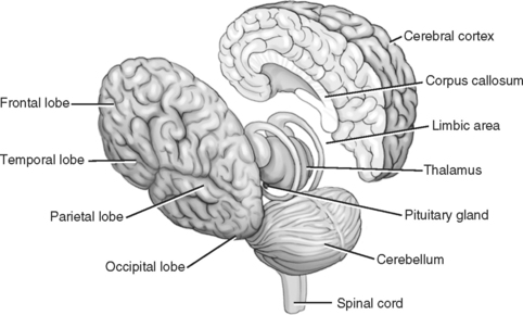
STRUCTURES OF THE BRAIN
The cerebral cortex, also called the neocortex, is the outer covering of the brain. The brain has two hemispheres, and each hemisphere has four major lobes that can be further subdivided into many specialty areas. Further division also occurs, including cortical and subcortical structures, such as the hypothalamus. Figure 1-1 and Figure 1-2 present the basic structural areas of the brain.
The four lobes are frontal, occipital, parietal, and temporal.
• The frontal lobes are responsible for thinking, conceptualizing, planning, emotional regulation, social interaction, abstract thinking, decision making, and memory.
• The occipital lobes are the primary centers for visual processing.
• The parietal lobes are concerned with functions of calculation, orientation, movement, and particular types of recognition, somatic interpretation, and integration.
• The temporal lobes are responsible primarily for sound, hearing, speech comprehension (left side generally), and the formation of long-term memory.
• The fibrous corpus callosum connects the two hemispheres and allows them to pass information back and forth.
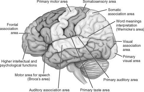
The premotor cortex is dedicated to initiation and sequencing of movements and is part of the frontal lobe.
During the first 7 months of gestation, neurons generate at an awesome speed and migrate to designated places; approximately 50% are pruned away before birth. This is a normal process called apoptosis, or programmed cell death (Diamond and Hopson, 1998; Bear, Connors, and Paradiso, 2007). This usually is thought to contribute to the efficiency of the brain. Unfinished apoptosis may contribute to the dramatic abilities of autistic savants and may also be one factor affecting their other deficits (Carter, 1999). Conversely, overpruning may lead to the cognitive impairment in a child with Down syndrome. This pruning of cells and connections occurs from 7 months gestation and continues throughout childhood and adolescence (Diamond and Hopson, 1998; Huttenlocher and Dabholkar, 1997).
• Wernicke’s area is located in the frontal lobe near the junction of the temporal lobe and is associated with comprehension of speech. This area is also near the auditory cortex, which receives sound signals from the ears.
• Broca’s area is located at the posterior of the frontal lobe in the left hemisphere and is associated with generating speech. It also is near the motor cortex area that controls muscles of the lips and mouth.
OTHER AREAS OF THE BRAIN
The hippocampus, a structure shaped like a ram’s horn, is located deep within the temporal lobe near the amygdala and hypothalamus. It is associated with conscious long-term memory, spatial ability, learning, and motivation. Damage to the hippocampus causes severe memory problems.
EARLY CHILDHOOD: BIRTH TO 5 YEARS
Newborn: Birth to 1 Month
Brain Findings
Basic connectivity of the newborn brain has been established, although it does not function the same as an adult brain. After birth, the infant demonstrates utilization of networks of neurons by moving arms and legs, opening and closing eyes, and by crying and breathing. The brain’s early developmental task is to continue forming and reinforcing some connections and pruning others. This process is a function of genetic predisposition and experience with the outside environment. If experience does not occur during a critical period, synaptic connections do not develop, and the potential ability is gone. For example, cataracts impair visual acuity, and if they are not detected and surgically removed by 2 years of age, visual acuity does not develop, and the synapses in the visual cortex are pruned away and the child is forever blind.
A. Brain weight at birth is approximately 400 g.
B. At birth, the cortex is approximately 1 mm thick—about one half the thickness of an adult brain.
C. Synapses form in the newborn’s brain at the rate of approximately 3 billion per second at given times.
D. Each neuron can be connected to as many as 15,000 other neurons and, with some, many more thousands of synapses are made.
E. The brain uses about 20% oxygen; and in the cerebral cortex, from birth to about 4 years of age, glucose usage is twice that of an adult’s brain.
F. The brain’s capacity is not fixed at birth and is most plastic during infancy. The more complex areas of the brain, such as the cortex, are more plastic; the life sustaining areas of the brain, such as the brainstem, are less plastic.
G. The brain does not grow uniformly but has peaks that correspond to the learning of various skills.
H. The brain constitutes approximately 12% of body weight at birth and approximately 2% to 3% of body weight in adulthood.
I. Brain growth is reflected in the head circumference; growth charts (see Appendix B) indicate that brain growth occurs more rapidly in the first year of life than at any subsequent age. The brain reaches one half its eventual size by 12 months.
J. Neurons are still being produced postnatally, and the neurons are increasing in size and complexity (M. Martone, personal communication, July 15, 2006). A significant part of the increase in brain weight observed at this time can be attributed to the development of glial cells, primarily those that produce myelin.
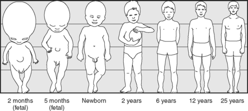
K. The newborn’s head is approximately one fourth of the total body length (see Head and Body Proportions in Figure 1-3 on page 8).
I. Physical and Anatomical Traits
NOTE: A child continues to follow own unique growth curve, which is more important than conformity to averages.
II. Gross and Fine Psychomotor Traits
III. Cognitive and Language Traits
IV. Emotional and Social Traits
VI. Moral and Spiritual Traits
INFANCY: 2 TO 12 MONTHS
3 Months (12 Weeks)
6 Months (24 Weeks)
I. Physical and Anatomical Traits
II. Gross and Fine Psychomotor Traits
III. Cognitive and Language Traits
IV. Emotional and Social Traits
VI. Moral and Spiritual Traits
TODDLER: 1 TO 3 YEARS
Brain Findings
The number of neurons remains relatively stable while neurons are undergoing vigorous growth and remodeling during the first 3 years. The brain’s neuroplasticity is reflected by change in response to experience. Prime times for growth occur in specific brain areas at different times. For example, optimal development of the brain systems responsible for attachment is dependent upon the positive interaction between caregiver and infant during the first year of life. Unhealthy early bonding of caregiver and infant can lead to a fragile, neurological, and emotional foundation for later development of relationships (Perry, 2002).
Animal research supports the fact that more synaptic growth occurs when living in enriched environments versus impoverished environments, which can lead to thinning of neuronal networks (Diamond and Hopson, 1998). Brain functions remain dynamic through practice and use, although current knowledge does not prove that more stimulation will necessarily create smarter children with denser brain systems (Nelson, 2000).
Stay updated, free articles. Join our Telegram channel

Full access? Get Clinical Tree


