CHAPTER 2
Genetics
1 Discuss the potential importance of the National Human Genome Project on patient care.
2 Define the terms commonly used in genetic conditions.
3 Describe the implications of an increased amount of chromosomal material, the deletion of genetic material, and the translocation process in chromosomal disorders.
4 Explain the basic mendelian modes of inheritance.
5 Identify client situations that indicate the need for a chromosomal analysis.
6 Assess the emotional effect on couples of the birth of an infant with a genetic disorder.
7 Discuss the responsibility and needed competencies of the nurse in initial counseling and referral of patients for further genetic testing and counseling.
INTRODUCTION
A The National Human Genome Project
1. First discussed in the 1980s; officially completed in 2003
2. Goals included sequencing the entire human genome (achieved in April 2003), identifying human deoxyribonucleic acid (DNA) sequence variations, identifying and determining the function of individual genes, and studying the ethical, legal, and social implications of information and technologic outcomes of the Human Genome Project on human beings and society.
3. Increased understanding of the genotype has allowed:
a. Development of targeted health promotion strategies
b. Potential disease prevention
c. Genotype-tailored drugs to optimize therapeutic effects while minimizing negative drug interactions and side effects
d. Eventual correction of disease states by development of gene transfer technology (gene therapy)
B Definition: Genetics is a medical science concerned with the transmission of characteristics from parent to child (Nussbaum, McInnes, & Willard, 2007)
1. Gene: the basic hereditary unit; a DNA sequence required for production of a functional product, usually a protein
2. Genotype: an individual’s genetic makeup
3. Phenotype: the outward appearance or expression of the genes
4. Allele: an alternative form of a gene; the wild type, or major sequencing allele is the most common form of a gene found within a population
5. Mutation: a rare alternative form of an allele; occurs in less than 1% of the population
6. Polymorphism: a common alternative form of an allele; occurs in more than 1% of the population
7. Genome: complete DNA sequence containing all of the genetic information for an individual
1. Cell division: all beings begin life as a single cell (zygote). The single cell continues to reproduce itself by the process of either mitosis or meiosis.
a. Mitosis is the process of cell division in which new cells are made. The new cells have the same number and pattern of chromosomes as the parent cell (46 chromosomes comprising 44 autosomes and 2 sex chromosomes); mitosis occurs in five stages (Figure 2-1).
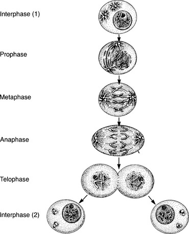
(1) Interphase: before cell division, the DNA replicates itself
(2) Prophase: the strands of chromatin shorten and thicken; the chromosomes reproduce; spindles appear, and the centrioles migrate to the opposite poles of the cell; the membrane separating the nucleus from the cytoplasm disappears
(3) Metaphase: the chromosomes line up along the poles of the spindle
(4) Anaphase: the two chromatids separate and move to the opposite ends of the spindle
(5) Telophase: a nuclear membrane forms, the spindles disappear, and the centrioles relocate to the outside of the new nucleus; toward the end of this phase, the cells divide into two new cells, each with its own nucleus and each having the same number of chromosomes as the parent cell
b. Meiosis is a process of cell division that occurs involving the sperm and ova and is known as gametogenesis; this process decreases the number of chromosomes by 50% (from 46 to 23 per cell) and occurs in two successive cell divisions (Figure 2-2)
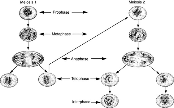
(1) The first division consists of four phases.
(a) Prophase: the chromosomes move close together
[i] Crossover of genetic material from each parent takes place at this time.
[ii] Crossover accounts for the wide individual variation of features seen within same-parent siblings.
(b) Metaphase: spindle fibers attach to separate chromosomes
(c) Anaphase: intact chromosome pairs migrate to opposite ends of the cell (the distribution of maternal and paternal chromosomes is random)
(d) Telophase: the cell divides into two cells, each with 50% (23) of the usual number of chromosomes (22 autosomes and 1 sex chromosome)
(a) The chromatids of each chromosome separate and move to the opposite poles of each of the daughter cells.
(b) This is followed by each of the cells dividing into two cells, which results in four cells (spermatogenesis and oogenesis).
(c) During the meiotic division, two of the chromatids might not move apart when the cell divides; this lack of separation is called autosomal nondisjunction; this is also the stage at which breakage can occur, resulting in abnormalities of chromosomal structure such as that producing cri du chat syndrome and Down syndrome
(d) Thus meiosis mixes up the chromosomes and crossing over mixes up the genes within a chromosome.
2. Genetic information is present on the chromosomes (Jarvi & Chitzyat, 2008; Lashley, 2007; Nussbaum et al., 2007).
a. Chromosomes are composed of DNA, a complex protein that carries the genetic information.
b. DNA occurs as a double-stranded helix found in the cell nucleus.
(1) Two long strands of DNA molecules are wound around each other.
(2) The strands are linked by chemical bonds.
(3) The strands are complementary.
(4) The chains comprise sequences of four nitrogen base subunits (adenine, guanine, thymine, and cytosine).
c. Genes are the smallest known unit of heredity.
(1) Genes are present on the chromosomes and are made up of coding exons and noncoding introns.
(2) Each gene codes for a particular cellular function, such as specific protein structure.
(3) Genes occur in pairs (alleles) derived from the mother and father during reproduction.
(4) Each gene has a specific location on the chromosomes.
(5) Genetic errors often occur when there are changes, such as deletions, substitutions, or duplications in the location of the gene.
3. Chromosomes form a genetic blueprint that is composed of tightly coiled structures of DNA.
a. Chromosomes are threadlike structures within the nucleus of the cell that carry the genes.
b. Humans have 46 chromosomes in each body cell (22 pairs of autosomes and 1 pair of sex chromosomes [diploid]).
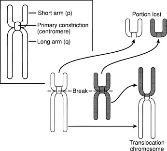
c. Chromosomes have a primary central constriction called the centromere (Figure 2-3).
d. The sex cells contain 23 chromosomes (haploid).
e. Abnormalities of chromosome number are as follows (Lashley, 2005, 2007; Nussbaum et al., 2007):
(1) Paired chromosomes fail to separate during cell division (nondisjunction).
(2) If nondisjunction occurs during meiosis (before fertilization), the fetus usually will have abnormal numbers of chromosomes in every cell (trisomy or monosomy).
(a) Trisomy is a product of the union between a normal gamete (egg or sperm) and a gamete that contains an extra chromosome.
[i] The individual will have 47 chromosomes; one “pair” will have three chromosomes instead of two.
[ii] Examples of trisomies are Down syndrome (47, XY, +21 [the extra chromosome is in the 21st pair]); trisomy 18 (47, XX, +18); and trisomy 13 (47, XY, +13).
(b) Monosomy is the product of a union between a normal gamete and a gamete with a missing chromosome.
(3) If nondisjunction occurs after fertilization, the fetus might have two or more chromosomes that evolve into more than one cell line (mosaicism), each with a different number of chromosomes (Lashley, 2007).
f. Abnormalities in chromosome structure are as follows:
(1) Abnormalities of the chromosomes involving only a part of the chromosome
(2) Abnormalities that can occur by translocation, by deletions, or by additions (Lashley, 2007)
(a) Translocation occurs when the individual has 45 chromosomes, with one of the chromosomes fused to another chromosome (usually number 21 fused to number 14) (see Figure 2-3).
[i] The person has the correct amount of chromosomal material (45, t[14q21q]), but it has been rearranged; this individual is known as a balanced translocation carrier.
[ii] When such an individual and a structurally normal mate have a child, there is a possibility that the offspring:
• Will receive the carrier parent’s abnormal 21/14 chromosome and a normal 14 and 21 from the other parent; thus the child will be a carrier.
• Will receive the abnormal chromosome, plus a normal 21 from the carrier parent; this will result in an extra amount of chromosomal material for the 21 pair (unbalanced translocation), and the child will have Down syndrome (46,14,t[14q21q]).
(b) Additions and/or deletions: a portion of a chromosome can be added or lost, which will result in adverse effects on the infant
D Modes of inheritance (mendelian)
1. Many diseases are caused by an abnormality of a single gene or a pair of genes.
2. Autosomal dominant inheritance: autosomal dominant disorders occur when an individual has a gene that produces an effect whenever it is present (homozygous or heterozygous); this gene overshadows the other gene of the pair
a. Mode of transmission (Figure 2-4)
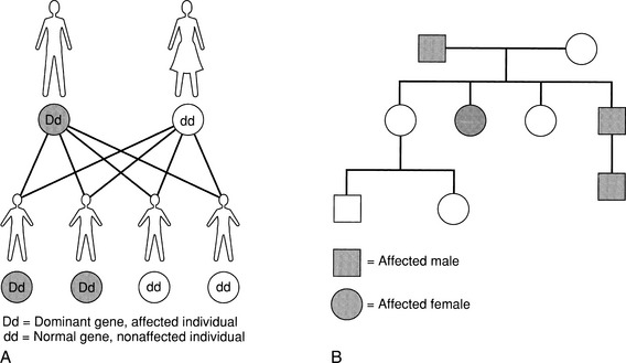
(1) Affected individuals generally have an affected parent; the family tree (genogram) might show several generations of individuals with the condition
(2) The affected individual has a one in two (50%) chance with each pregnancy of passing the abnormal gene on to his or her child.
(3) Males and females are equally affected.
(4) An unaffected individual cannot transmit the disorder to his or her children.
(5) A mutation (a gene that has been spontaneously altered) can result in a new case.
(6) Autosomal dominant disorders vary greatly in the degree of characteristics that are seen within a family (e.g., in Marfan syndrome, the parent might have only elongated extremities but the child might have a more involved condition, including dislocation of the lens of the eye and severe cardiovascular abnormalities).
3. Autosomal recessive inheritance: an individual has an autosomal recessive disorder if he or she has a gene that produces its effects only when there are two genes on the same chromosome pair (homozygous trait).
a. A carrier state can occur (heterozygote).
(1) An individual with the abnormal gene does not manifest obvious symptoms.
(2) When two carriers pass on the same abnormal gene, the condition might appear.
b. Mode of transmission (Figure 2-5)
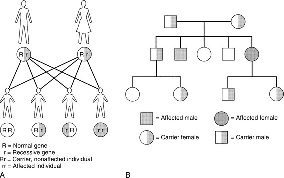
(1) An affected individual has clinically normal parents, but the parents are both carriers for the abnormal gene.
(2) The carrier parents have a one in four (25%) chance with each pregnancy of passing the abnormal gene on to their offspring; in this case, when a recessive gene is received from each parent, the child will have the disorder.
(3) If the offspring of two carrier parents is clinically normal, there is a one in two (50%) chance that he or she will be a carrier, like the parents.
(4) Males and females are affected equally.
(5) The family genogram usually shows siblings affected in a horizontal pattern.
(6) There is an increased risk if intermarriage occurs (consanguineous matings); individuals who are closely related are more likely to have the same genes in common.
(7) Recessive disorders tend to be more severe in their clinical manifestations.
(8) The presence of certain autosomal recessive genes can be detected in the normal carrier parent; examples of diseases for which carrier screening is available are sickle cell anemia, Tay-Sachs disease, and cystic fibrosis.
Stay updated, free articles. Join our Telegram channel

Full access? Get Clinical Tree


