Fluid balance and the urinary system

22 Fluid balance and the urinary system


Maintaining adequate hydration and the correct distribution of fluids between the compartments of the body is vital. This chapter will look at what the normal distribution of fluid between the intracellular and extracellular compartments of the body should be. The urinary system plays a key role in ensuring that excess fluid is removed from the body. It also has an excretory function, which requires it to be constantly removing soluble toxic material from the body in the form of urine. This can be achieved even when fluid has to be conserved. This chapter looks at the anatomical structure of the urinary system and considers its ability to maintain fluid balance and respond to different fluid requirements of the body.


Fluid and electrolyte balance


Water is one of the essential foodstuffs but is a simple substance which can be absorbed and used in the body without chemical change. It enters into many of the metabolic changes that occur in the body, combining with proteins, carbohydrates and fats in digestion and being split off from them when they are used as fuel to produce energy. It is general today to consider the balance rather than the metabolism of water and salts, and the electrolytes that the salts form in the body.


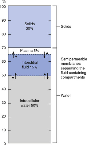
Water forms the greater part of the body cells and the body fluids. Approximately two-thirds (more accurately 60%) of the body weight consists of water. This proportion must be maintained. Of this water, 70% is inside the body cells (intracellular) and the remaining 30% is in the body fluids (extracellular); 15–20% is in the interstitial spaces in the tissues, bathing all the body cells, even the bone cells, and the remaining 10–15% forms the fluid of the blood, i.e. the plasma and the lymph (Fig. 22.1). These three volumes of fluid are separated only by thin semipermeable membranes, the cell walls and the capillary walls; water constantly passes through these walls from one of these areas to the others, though the volume of each remains remarkably constant in normal health.


The water in our bodies is, however, not static. Fresh water is taken in by the body each day and passes out of it by a number of channels. The total quantities taken in and passed out must balance one another. Water is taken in as water and other fluids drunk, and also in the foods eaten, which, like the body, consist largely of water. On average, the healthy person takes in 1.5 L of fluid as water and other drinks daily, and slightly over 1 L in food, an average of 2600–2800 mL. A similar quantity is passed out of the body by the lungs as water vapour (400–500 mL), by the skin as sweat (500–600 mL), by the kidneys as urine (1000–1500 mL) and in the faeces, a small quantity (100–150 mL).


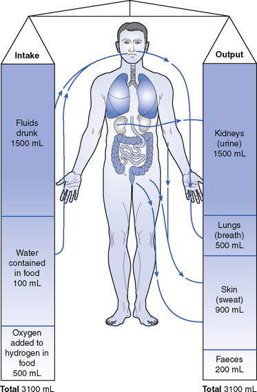
The quantities lost in urine, sweat and water vapour from the lungs vary with conditions. In hot weather and heavy work, more sweat is produced to cool the body, and less urine is passed; at the same time the loss of fluid causes thirst, so that more fluid is drunk. In fever the same changes take place; there is increased loss of fluid and there must be increased intake to balance it (Fig. 22.2).


The urinary system


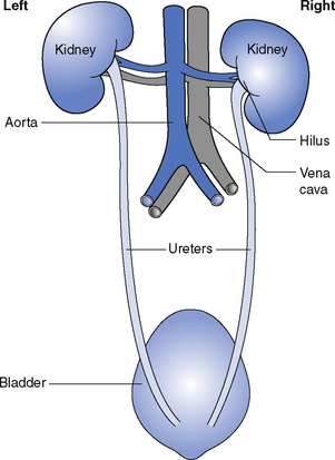
The urinary system consists of the following components (Fig. 22.3):


The kidneys


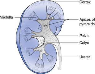
The kidneys are two bean-shaped organs (Fig. 22.4) situated in the posterior part of the abdomen, one on each side of the vertebral column, behind the peritoneum. They lie at the level of the twelfth thoracic to the third lumbar vertebrae, though the right kidney is usually slightly lower than the left because of its relationship to the liver.


Each kidney is approximately 11 cm long, 6 cm wide and 3 cm thick and is embedded in the perirenal fat.


The medial border of each kidney is concave in the centre. This area is called the hilus and it is the point at which the blood vessels, nerves and ureters enter or leave the kidney.


The kidney is enclosed in a capsule of fibrous tissue, which can be easily stripped off. In vertical section the kidney has two distinct parts. The dark outer part is called the cortex and the paler inner portion the medulla. This leads into the collecting space, which is called the renal pelvis.


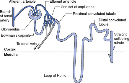
The kidney substance consists of minute twisted tubules called nephrons (Fig. 22.5); there are over a million in each kidney. Each of the nephrons begins in a cup-shaped expansion called the glomerular capsule, from which the tubule leads (Fig. 22.6). Into the cup of each capsule comes a fine branch of the renal artery, forming a tuft of capillaries in close contact with its inner wall; the capillary tuft is called the glomerulus. The arteriole bringing blood to the tuft is called the afferent vessel, and the arteriole that carries the blood away is the efferent vessel; it is slightly smaller than the afferent vessel. The blood in the tuft is under high pressure because of this and because of its nearness to the abdominal aorta.


< div class='tao-gold-member'>

Stay updated, free articles. Join our Telegram channel

Jul 18, 2016 | Posted by in NURSING | Comments Off on Fluid balance and the urinary system

Full access? Get Clinical Tree

Get Clinical Tree app for offline access