Nervous System
Endocrine System
Source of signal
Brain
Endocrine gland
Signal
Neurotransmitter and action potential
Hormone
Usual route
Efferent nerve
Blood
Response rate
Fast
Slow
Specificity
Specific
Diffuse
Target
Single
Multiple
Type of effect
Immediate effect
Long-term control and integration
What is endocrinology?
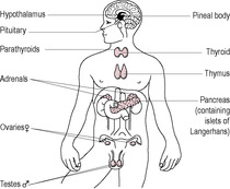
Fig. 3.1
The evolution of endocrinology
Classification of hormones
Hormone structure
Adapted from Johnson and Everitt, 1995.
Lipid hormones
Steroid hormones
Sex steroids, e.g. androgens, oestrogens and progestagens
Glucocorticoids, e.g. cortisol
Mineralocorticoids, e.g. aldosterone
Thyroid hormones; 1,25-Dihydrovitamin D3
Eicosanoids
Prostaglandins
Leukotrienes
Protein hormones
Gonadotrophic glycoproteins
Follicle-stimulating hormone (FSH)
Luteinizing hormone (LH)
Human chorionic gonadotrophin (hCG)
Thyroid-stimulating hormone (TSH)
Somatotrophic polypeptides
Prolactin (PRL)
Human placental lactogen (hPL)
Growth hormone (GH)
Cytokines
Insulin
Activins and inhibins
Anti-Müllerian hormone (AMH)
Interferons
Growth factors
Small peptides
Gonadotrophin-releasing hormone (GnRH)
Oxytocin (OXY)
Antidiuretic hormone (ADH or vasopressin)
β-Endorphin
Vasoactive intestinal peptide (VIP)
Monoamines
Catecholamines
Adrenaline, noradrenaline and dopamine
Melatonin
Dopamine
Steroid hormones
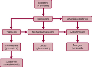
Fig. 3.2
Adapted from Johnson and Everitt, 1995.
Sex Steroid
Family Members (and Approximate Biological Activity)
Main Effects
Androgens
5α-dihydrotestosterone (100%)
Testosterone (50%)
Androstenedione (8%)
Dehydroepiandrosterone (4%)
Differentiation of male embryo
Secondary sex characteristics
Spermatogenesis
Male secondary sex characteristics
Sexual and aggressive behaviour
Growth promoting, protein anabolism, ossification and erythropoiesis
Oestrogens
Oestradiol-17β (E2) (100%)
Female secondary sex characteristics
Oestriol (E3) (10%)
Prepares uterus for ovulation and fertilization
Oestrone (E1) (1%)
Vascular effects – increased blood flow, neovascularization
Growth-promoting effects on endometrium and breasts
Primes endometrium for progesterone action
Mildly anabolic
Increases calcification of bones
May be associated with sexual behaviour
Progestagens
Progesterone (100%)
Prepares uterus for pregnancy
17α-hydroxyprogesterone
Maintains pregnancy (17α-OHP) (40–70%)
Stimulates glandular growth of breasts (but suppresses milk secretion)
20α-hydroxyprogesterone (5%)
Affects sodium and water excretion
Mildly catabolic
Relaxes smooth muscle tone
Affects appetite and thirst, metabolic rate, sensitivity to carbon dioxide
![]()
Stay updated, free articles. Join our Telegram channel

Full access? Get Clinical Tree


Endocrinology
Zara is now just 6 weeks pregnant. She feels concerned about feeling increasingly nauseated, especially late in the evening; she actually vomited last night. Zara rings you, as her midwife, the next day expressing her concern, especially as her sister has told her that this is not the typical morning sickness of pregnancy.
• What explanations could you give Zara to the likely cause of her nausea and what advice should you give her to help her cope with her nausea?
• How is nausea and vomiting of pregnancy differentiated from hyperemesis gravidarum?
• What are the possible complications of hyperemesis and what specific treatment is required to minimize complications?
Later on in her pregnancy, Zara has a routine appointment at about 26 weeks gestation with her midwife who undertakes routine urinalysis and discovers that Zara has glucosuria.
• What are the possible causes for this, what further investigations need to be undertaken and what advice and treatment may be required?
The endocrine system originally appeared to be a relatively simple system of discrete glands (Fig. 3.1) that secreted chemical messengers, or hormones, into the blood where they would be carried to specific target cells at a distant site, inducing a reaction. However, it is now clear that the endocrine system is more complex. Some hormones are secreted into ducts and not into blood; for instance, androgens are secreted into the seminiferous tubules. Some organs that have other functions also produce hormones. For instance, the atrium of the heart produces atrial natriuretic peptide (ANP), which inhibits reabsorption of sodium chloride in the kidneys and hence affects blood pressure. Some hormones are produced by several different glands, for instance somatostatin, which is produced by the hypothalamus, pancreas, stomach and intestine. Although the trophoblast is the prime site of human chorionic gonadotrophin (hCG) production, it can also be produced by other tissues, albeit in very low concentrations (Iles and Chard, 1991). The placenta appears to be capable of synthesizing a very broad range of hormones and releasing factors that interact with both maternal and fetal physiology. Some substances such as noradrenaline can act as both hormone and neurotransmitter depending on their mode of delivery and whether they are released from a gland or from a nerve. The hypothalamus produces neurohormones that are important in the interaction between the endocrine and nervous systems.
(Reproduced with permission from Brooker, 1998.)
Overall, the endocrine system (in partnership with the neural system) has the following functions:
• coordinates the homeostatic balance
• regulates various physiological systems such as the digestive system and reproductive system
• facilitates differentiation of the sexes in the embryonic stage and the manifestation of the secondary sexual characteristics at puberty
• modifies and induces behavioural changes within the individual.
The evolution of the endocrine system has its rudiments within the activity of single-cell (unicellular) organisms. The unicellular organisms developed the ability to be attracted to chemicals, described as a chemotactic response, or to chemicals that were vital for the functioning of the organism, described as a chemotrophic response. Equally, these organisms developed the ability to recognize noxious chemicals (toxins) and were thus able to avoid them. The cell reacting to chemical signals interacting with receptor sites upon the cell membrane and within the cytoplasm led to the development of active mobility.
As multicellular organisms developed, the group of unicellular organisms that were the prototypes of multicellular organisms evolved chemical communication as an extension of the chemotrophic response. As multicellular evolution progressed further, the cells became more differentiated and specialized. Regulation therefore became the function of more specialized types of cells. This is reflected in the developmental sequence of a fetus, beginning with the division of a single cell (see Chapter 7). With each successive division, the resulting cells are slightly different from the original zygote cell (although differentiation during the initial divisions may be induced by the presence of maternally derived factors within the cytoplasm of the zygote). Although this differentiation is primarily under genetic control, it is achieved through a process of induction from chemical signals produced by one cell type that influence the division of other neighbouring cells. The altered gene expression of the dividing cells results in a changed morphology and developmental pathway.
As organisms became larger and more complex, cell-to-cell communication became more complicated. It evolved in two ways: the endocrine system (of chemical transmission via the circulating blood system) and the neural system (via transmission of an action potential; see Chapter 1). Under the traditional approach to biological science, the endocrine and neural systems were always considered in isolation; however, they are now considered to be extensions of the same system that are highly interactive. Many endocrine responses are initiated by a neuronal influence. Many neurotransmitters and neuromodulators have also been found to be endocrine hormones.
Hormones regulate metabolism, activate or inhibit the immune system, stimulate or inhibit growth, induce or suppress apoptosis (see below) and prepare the body to respond (such as fleeing or fighting) or undergo transition to a new stage of life such as puberty, pregnancy or the menopause. Hormones are produced by almost every organ and type of tissue; they function as cellular messengers. The action of hormones depends on the responses of the target cells and the pattern of hormonal secretion. Endocrine means ‘secreted inwards’ and is applied to hormones that fit the classical description of being secreted into the bloodstream and having an effect at a distant target. There are also exocrine hormones, which are ‘secreted outwards’ into ducts. These include hormones that are secreted into the vas deferens and uterine tubes.
A number of hormones have a local or paracrine effect, diffusing short distances to act on neighbouring cells or cells separated only by an intracellular space. Examples of a paracrine response are the effects of testosterone and anti-Müllerian hormone (AMH; also known as Müllerian-inhibiting hormone or substance, MIH or MIS) on sexual differentiation (see Chapter 5). If the hormone produced acts upon the same cell that produced it, it is described as autocrine. For example, an autocrine hormone may induce cellular division or signal the programmed death of the cell (apoptosis). If it affects adjacent cells and has a very localized action, it is described as a juxtacrine hormone. Therefore, the effect of a hormone depends on how and where it is secreted, the mode of transport (e.g. whether it is soluble or carried by a binding protein) and how quickly it is metabolized or inactivated.
Neuroendocrine hormones are synthesized in specialized neurons, and their effects can also be paracrine in nature (these are usually described as neurotransmitters and neuromodulators). Oxytocin is an example of a neuroendocrine hormone. It is released from the posterior lobe of the pituitary gland and influences the contractility of the myometrium (see Chapter 13) and myoepithelial cells in the breast (see Chapter 16). In these respects, oxytocin has an endocrine effect, but in many mammals it also modifies female behaviour by inducing parental behaviour in the presence of the sex steroids (Insel, 1992). Oxytocin is thought to influence the successful transition to parenthood in different ways; maternal behavioural changes tending to influence affectionate behaviour and emotional bonding and paternal parenting behaviour tending to affect play and social interaction with their infants (Gordon et al., 2010).
A pheromone is a hormone produced by an individual that induces a response, usually social, within another member of the same species. Releaser pheromones stimulate rapid behavioural responses such as attracting potential mates. Primer pheromones act via the olfactory and neuroendocrine system to produce delayed responses which are usually developmental. Receptors for pheromones are found on the vomeronasal organ close to the nasal cavity of mammals, which use pheromones to indicate identity of kin or family territory. It is controversial as to whether the human vomeronasal organ retains a function; some scientists think it is involved in social behaviour such as pair bonding, parental attachment, sexual attraction and synchrony of menstrual cycles (Halpern and Martinez-Marcos, 2003). The synchronization of menstrual cycles within a group of women, responses between lactating women and their offspring and female responses to non-odorants in male perspiration are suggested to be examples of pheromone effects in humans (Bhutta, 2007).
Secretion of hormones is influenced by a number of factors, including the nervous system, hormone-binding proteins, plasma concentrations of nutrients and ions, environmental changes and other hormones, such as stimulating and releasing hormones.
Hormones can be classified according to their structure (see Table 3.2). Steroid hormones and eicosanoids (the prostaglandin family of hormones) are lipids. The other classes are protein and peptide hormones and monoamines.
The steroid group of hormones consists of the sex steroids (progestagens, androgens and oestrogens), the glucocorticoids, mineralocorticoids, thyroid hormones and 1,25-dihydroxyvitamin D3. Steroid hormones are derived from cholesterol which is synthesized from acetate (Fig. 3.2). As well as being the precursor for the steroid hormones, cholesterol is also an important structural component of cell membranes, providing rigidity.
The first and common step in the biosynthesis of sex steroids is the formation of pregnenolone, which is rate-limiting, and therefore important in controlling production of sex steroids. Pregnenolone is produced on the inner mitochondrial membrane whereas the next stages take place in the smooth endoplasmic reticulum (SER).
The three classes of sex steroids are structurally related, which offers the opportunity for interconversion. This means that a genetic defect in one of the steps can result not only in a deficiency of the normal amount of the product but also in an excess of another sex steroid. For instance, a genetic deficiency of the enzyme that converts 17α-hydroxyprogesterone to the precursor of cortisol results in increased levels of 17α-hydroxyprogesterone, which is converted into androstenedione and then into androgens. The unusually high level of androgens can cause masculinization of the female fetus. These structural similarities mean that the steroid hormones can affect the activity of other steroid hormones by exerting agonistic and antagonistic properties at the receptor level (see below). However, the effects of the hormones vary depending on their structure (see Table 3.3).
The main role of androgens is in the development and maintenance of masculine characteristics and fertility. Similarly, the dominant role of oestrogens is in development and maintenance of feminine characteristics and fertility. The key role for progesterone is the preparation for pregnancy and its maintenance. However, all the steroid hormones are produced in men and women but with varying profiles; therefore, for instance, men produce more androgens than women but also produce some oestrogen. Although androgens are primarily associated with the development and maintenance of male sex characteristics, they also affect sexual behaviour in women (Johnson, 2007).
As steroid hormones are lipid-soluble, they are able to diffuse freely across the cell membrane and have their effect within the target cell. In the cytoplasm, the steroid hormones may be altered. The receptor sites for thyroid hormone and the sex steroids are within the nucleus. Specific receptors for the other steroid hormones are within the cytoplasm; binding usually results in cleavage of smaller ‘heat-shock’ proteins from the receptors. Steroid hormones exert their effect by altering ribonucleic acid (RNA) synthesis and subsequent protein synthesis (Box 3.1). The steroid-receptor ligand binds to specific segments of DNA, steroid response elements (SRE) in promoter regions of the gene in that section of DNA, affecting the rate of transcription and gene expression. Protein synthesis can be increased (or decreased) within 30 min, and the effects of steroid hormones are therefore relatively slow in action compared with those of protein hormones. The term ‘anabolic steroids’ describes the effect of steroid hormones in influencing new tissue growth.
Box 3.1
• Transported in plasma bound to binding protein
• Hormone released and diffuses into target cell
• Hormone diffuses into nucleus
• Binds to specific receptor
• Affects DNA transcription
• Affects mRNA synthesis
• Affects protein synthesis
• Altered functional response of cell
The other class of lipid hormones is the eicosanoids (prostaglandins and leukotrienes) which have an important role in reproduction. Eicosanoids are formed from an arachidonic acid precursor, generated by the activity of either phospholipase C or phospholipase A2 (Fig. 3.3). Arachidonic acid production appears to be the rate-limiting step. Phospholipase A2 is present in an inactive form in lysosomes in cells that are released if the cell membranes become unstable. Most tissues of the body including the myometrium, cervix, ovary, placenta and fetal membranes synthesize prostaglandins. They have a short half-life and are metabolized quickly. They have an important role in amplifying signals at the onset of labour (see Chapter 13). Leukotrienes are also synthesized from arachidonic acid by the enzyme 5-lipoxygenase in leukocytes and macrophages. They are involved in inflammatory reactions particularly in asthma and allergy and also seem to be important in pregnancy.
Get Clinical Tree app for offline access
