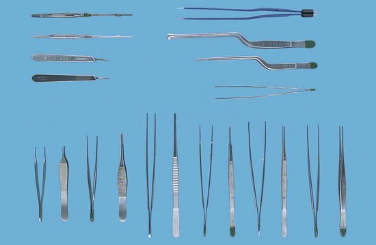
CHAPTER 113 Craniotomy is an incision made in the head through the skull that allows the performance of surgery on the brain. Possible equipment needed for the procedure includes: 1. A Midas Rex drill with craniotome blades, used to open the skull. 2. A Cavitron ultrasonic surgical aspirator (CUSA), used for tumor removal. 3. An operating microscope, used for visualization. 4. An electrosurgical unit, used for hemostasis. 5. A neuroplating set of screws and plates, used to repair fractures and to replace the bone flap. A brief description of the procedure follows: 1. Raney clip appliers and clips are placed on the skin flaps for hemostasis. 2. Strully scissors are used to incise the scalp muscle. 3. An Adson periosteal elevator is used to strip periosteum from the bone and enlarge the burr holes. 4. A craniotome is used to turn a bone flap. 5. A Penfield dissector is used to remove dura mater from the bone. 6. A Ruskin double-action rongeur is used to remove jagged bone edges. 7. Cushing tissue forceps with teeth are used to grasp and elevate the dura. 8. An Adson dura hook is used to elevate the dura. 9. Metzenbaum dissecting scissors are used to incise the dura. 10. A Leyla retractor is used to expose the brain. 11. Cushing bipolar cautery forceps are used for hemostasis. 113-1 Top to bottom, left to right: 2 Bard-Parker knife handles #7; 2 Bard-Parker knife handles #3; 1 Cushing bipolar cautery forceps, microtip, insulated bayonet shaft; 1 Adson hypophyseal forceps, bayonet shaft, round-cup tip; 1 Gerald dressing forceps, bayonet shaft, narrow tips, serrated; 1 Adson dressing forceps, bayonet shaft. Bottom, left to right: 2 Adson tissue forceps with teeth (1 × 2), front view and side view; 2 Gillies tissue forceps with teeth (1 × 2), front view and side view; 2 DeBakey vascular Autraugrip tissue forceps, medium, front view and side view; 2 Gerald tissue forceps with teeth (1 × 2), front view and side view; 2 Cushing tissue forceps with teeth (1 × 2), front view and side view; 2 Cushing tissue forceps with teeth (1 × 2), Gutsch handle, front view and side view.
Craniotomy

Stay updated, free articles. Join our Telegram channel

Full access? Get Clinical Tree


