Chapter 34 Congenital Abnormalities
Definition and causes
By definition, a congenital abnormality is any defect in form, structure or function that is present at birth. Identifiable defects can be categorised in five ways:
Chromosome and gene abnormalities
Each human cell carries 44 chromosomes (autosomes) and two sex chromosomes. Each chromosome comprises a number of genes. The zygote should have 22 autosomes and one sex chromosome from each parent.
Genetic disorders (Mendelian inheritance)
Genes are composed of DNA and each is concerned with the transmission of one specific hereditary factor. Genetically inherited factors may be dominant or recessive.
A dominant gene will produce its effect even if present in only one chromosome of a pair. An autosomal dominant condition can usually be traced through several generations: for example, achondroplasia, osteogenesis imperfecta, adult polycystic kidney disease or Huntington’s chorea.
A recessive gene needs to be present in both chromosomes before producing its effect: for example, cystic fibrosis or phenylketonuria.
In an X-linked recessive inheritance the condition affects males almost exclusively, although females can be carriers. X-linked recessive inheritance is responsible for conditions such as haemophilia A and B and Duchenne muscular dystrophy.
Mitochondrial disorders
These are inherited from the mother. Signs and symptoms are diverse but tend to occur in brain and muscles.
Teratogenic causes
A teratogen is any agent that raises the incidence of congenital abnormality. The list of known and suspected teratogens is continually growing (Box 34.1).
Box 34.1 Known and suspected teratogens
Multifactorial causes
These stem from a genetic defect in addition to one or more teratogenic influences.
Gastrointestinal malformations
Most of the abnormalities affecting this system call for prompt surgical intervention.
Gastroschisis and exomphalos
Gastroschisis (Fig. 34.1) is a paramedian defect of the abdominal wall with extrusion of bowel that is not covered by peritoneum.

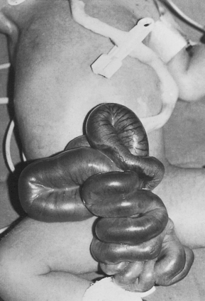
Fig. 34.1 Gastroschisis showing prolapsed intestine to the right of the umbilical cord.
(From Rennie & Roberton 1999, with permission of Churchill Livingstone.)
Exomphalos or omphalocele is when the bowel or other viscera protrude through the umbilicus. These babies may have other abnormalities, for example heart defects.
Immediate management of both is as follows:
Atresias
Oesophageal atresia
Oesophageal atresia occurs when there is incomplete canalisation of the oesophagus in early intrauterine development. It is commonly associated with tracheo-oesophageal fistula. The most common type of abnormality is where the upper oesophagus terminates in a blind upper pouch and the lower oesophagus connects to the trachea. This abnormality should be suspected in the presence of maternal polyhydramnios. At birth the baby has copious amounts of mucus coming from the mouth.
The midwife should attempt to pass a wide orogastric tube but it may travel less than 10–12 cm.
Radiography will confirm the diagnosis.
The baby must be given no oral fluid but a wide-bore oesophageal tube should be passed into the upper pouch and connected to gentle continuous suction. Usually a double-lumen 10FG (Replogle) tube is used and the baby nursed head up.
He or she should be transferred immediately to a paediatric surgical unit and continuous suction must be available throughout the transfer.
Duodenal atresia
This is persistent vomiting within 24–36 hours of birth.
A characteristic double bubble of gas may be seen on radiological examination. Prognosis is good if the baby is otherwise healthy, but this abnormality is often associated with other problems, such as Down syndrome.
Pyloric stenosis
Pyloric stenosis arises from a genetic defect which causes hypertrophy of the muscles of the pyloric sphincter. The characteristic clinical presentation is projectile vomiting, usually around 6 weeks of age but possibly earlier.
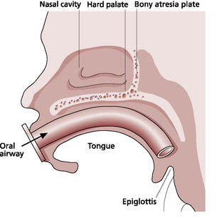
Cleft lip and cleft palate
Cleft lip may be unilateral or bilateral and is very often accompanied by cleft palate.
Clefts may affect the hard palate, soft palate, or both. Some defects will include alveolar margins and sometimes the uvula. The palate is examined by means of a good light source rather than by digital palpation.
If the defect is limited to unilateral cleft lip, breastfeeding is encouraged.
Where there is the additional problem of cleft palate, arranging for the baby to be fitted with an orthodontic plate may facilitate breastfeeding but will not afford the same stimulus as nipple to palate contact.
Cup or spoon feeding is an alternative method, and if bottle feeding there is a wide variety of specially shaped teats available to accommodate the different sizes and positions of palate defects.
Pierre robin syndrome
Pierre Robin syndrome is characterised by micrognathia (hypoplasia of the lower jaw), abnormal attachment of muscles controlling the tongue, which allows it to fall backward and occlude the airway, and a central cleft palate. To maintain a clear airway the baby is nursed prone and may require the insertion of an oral airway. Nasal and nasopharyngeal constant positive airways pressure may be necessary for some time after birth. This is one of the few exceptions to the rules given in the ‘Back to sleep’ campaign aimed at reducing cot deaths. There is a high risk of aspiration occurring when feeding. Suction catheter and oxygen equipment should be ready to hand. An orthodontic plate may be fitted to facilitate feeding.
Abnormalities relating to respiration
Diaphragmatic hernia
There is a defect in the diaphragm, which allows herniation of abdominal contents into the thoracic cavity. The extent to which lung development is compromised as a result depends on the size of the defect and the gestational age at which herniation first occurred.
Chest X-ray confirms the diagnosis. Continuous gastric suction should be commenced. Prognosis relates to the degree of pulmonary hypoplasia. There is also the possibility of coexistent abnormalities, such as cardiac defects or skeletal anomalies.

Full access? Get Clinical Tree

