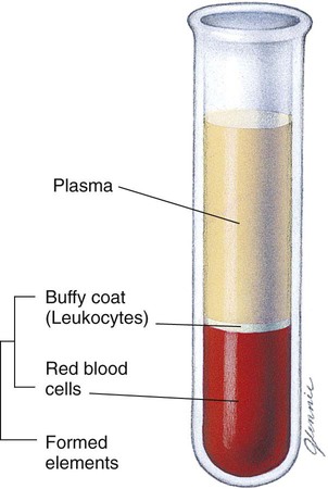
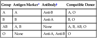
Chapter12 Circulatory System Terminology* *A transition syllable or vowel may be added to or deleted from the word parts to make the combining form. Blood divides into solid and liquid portions when spun in a centrifuge (Fig. 12-1). The solid parts, called formed elements, are red blood cells (RBCs), white blood cells (WBCs), and platelets (thrombocytes). Table 12-1 shows the formed elements of the blood. The buffy coat is a mixture of the WBCs and the platelets. The remaining liquid portion of the blood is the plasma. TABLE 12-1 More than 25 trillion RBCs, also called erythrocytes, circulate in the body’s 4 to 5 L of blood (Fig. 12-2). Erythrocytes contain a protein called hemoglobin that carries oxygen to all cells and removes carbon dioxide. Each RBC lives only 90 to 120 days. New cells are manufactured by the red marrow or myeloid tissue in bones (see Chapter 14). A few million new RBCs are made each second in a process called hemopoiesis. The liver and spleen remove dead RBCs and reuse the material. The five types of WBCs are listed as follows (Table 12-1): The four major blood types are A, B, AB, and O (Table 12-2). Type AB blood is called the universal recipient because it has no antibodies in the plasma to react with other blood cells and can receive any type of blood safely. Type O blood is the universal donor because the blood cells have no antigens to react with the antibodies in the plasma of the other blood types. Therefore type O blood can be given safely to a person of any blood type. TABLE 12-2 Blood Types and Blood Group Components are available on the Evolve website: http://evolve.elsevier.com/Gerdin Another important aspect of blood typing is the identification of the antigen known as the Rh factor. The Rh factor is found in the RBCs. About 85% of North Americans have this factor and are said to be Rh-positive (Table 12-3). If Rh-positive blood is given to someone with Rh-negative blood, that person’s blood considers the Rh-positive blood a foreign particle and tries to combat it by forming antibodies. A second transfusion of Rh-positive blood can be fatal to an Rh-negative person. The Rh factor also becomes important in the Rh-negative mother having a second Rh-positive baby (Fig. 12-3). TABLE 12-3 Approximate Distribution of Blood Types in the U.S. Population*
Circulatory System
 Define at least 10 terms relating to the circulatory system.
 Describe two functions of the circulatory system.
 Describe the function of lymph.
 Identify at least three methods of assessment of the circulatory system.
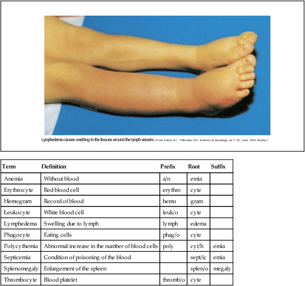
Term
Definition
Prefix
Root
Suffix
Anemia
Without blood
a/n
emia
Erythrocyte
Red blood cell
erythro
cyte
Hemogram
Record of blood
hemo
gram
Leukocyte
White blood cell
leuk/o
cyte
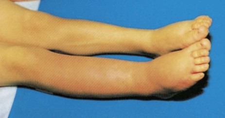
Lymphedema
Swelling due to lymph
lymph
edema
Phagocyte
Eating cells
phag/o
cyte
Polycythemia
Abnormal increase in the number of blood cells
poly
cyt/h
emia
Septicemia
Condition of poisoning of the blood
sept/ic
emia
Splenomegaly
Enlargement of the spleen
splen/o
megaly
Thrombocyte
Blood platelet
thromb/o
cyte
Structure and Function of the Circulatory System
Blood
 Transporting nutrients, oxygen, and hormones
 Removing metabolic wastes and carbon dioxide
 Providing immunity (resistance to disease) through antibodies
 Maintaining body temperature and electrolyte balance
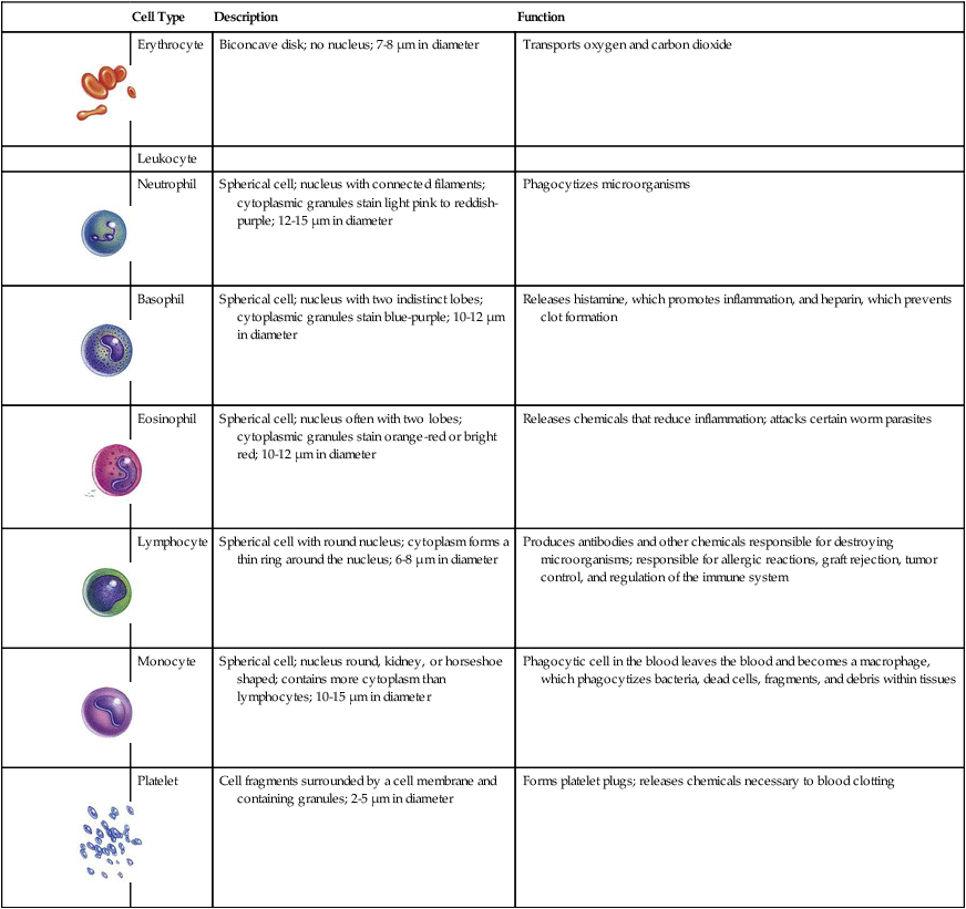
Cell Type
Description
Function

Erythrocyte
Biconcave disk; no nucleus; 7-8 µm in diameter
Transports oxygen and carbon dioxide
Leukocyte

Neutrophil
Spherical cell; nucleus with connected filaments; cytoplasmic granules stain light pink to reddish-purple; 12-15 µm in diameter
Phagocytizes microorganisms

Basophil
Spherical cell; nucleus with two indistinct lobes; cytoplasmic granules stain blue-purple; 10-12 µm in diameter
Releases histamine, which promotes inflammation, and heparin, which prevents clot formation

Eosinophil
Spherical cell; nucleus often with two lobes; cytoplasmic granules stain orange-red or bright red; 10-12 µm in diameter
Releases chemicals that reduce inflammation; attacks certain worm parasites

Lymphocyte
Spherical cell with round nucleus; cytoplasm forms a thin ring around the nucleus; 6-8 µm in diameter
Produces antibodies and other chemicals responsible for destroying microorganisms; responsible for allergic reactions, graft rejection, tumor control, and regulation of the immune system

Monocyte
Spherical cell; nucleus round, kidney, or horseshoe shaped; contains more cytoplasm than lymphocytes; 10-15 µm in diameter
Phagocytic cell in the blood leaves the blood and becomes a macrophage, which phagocytizes bacteria, dead cells, fragments, and debris within tissues

Platelet
Cell fragments surrounded by a cell membrane and containing granules; 2-5 µm in diameter
Forms platelet plugs; releases chemicals necessary to blood clotting
Red Blood Cells
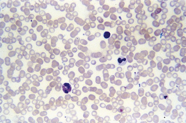
White Blood Cells
 Neutrophils (NOO-truh-filz) engulf and digest bacteria in a process called phagocytosis.
 Basophils (BAY-suh-filz) contain the anticoagulant substance heparin and participate in the inflammatory response of the body.
 Eosinophils (ee-uh-sin-uh-filz) defend the body from allergic reactions and parasitic infections and may help remove toxins from the blood.
 Lymphocytes (LIM-fuh-sites) participate in the production of antibody and plasma cells and help destroy foreign particles.
 Monocytes (MON-uh-sites) help remove foreign materials and bacteria in the process of phagocytosis.
Blood Typing
Group
Antigen Marker*
Antibody†
Compatible Donor
A
A
Anti-B
A, O
B
B
Anti-A
B, O
AB
A, B
None
A, B, AB, O
O
None
Anti-A, Anti-B
O
![]() Case Study 12-1
Case Study 12-1
Answers to Case Studies
Blood Type
Prevalence (%)
O Rh-positive
38
O Rh-negative
7
A Rh-positive
34
A Rh-negative
6
B Rh-positive
9
B Rh-negative
2
AB Rh-positive
3
AB Rh-negative
1 ![]()
Stay updated, free articles. Join our Telegram channel

Full access? Get Clinical Tree


Circulatory System
Get Clinical Tree app for offline access


