3 Caring for the patient with a haematological disorder
ANATOMY AT A GLANCE
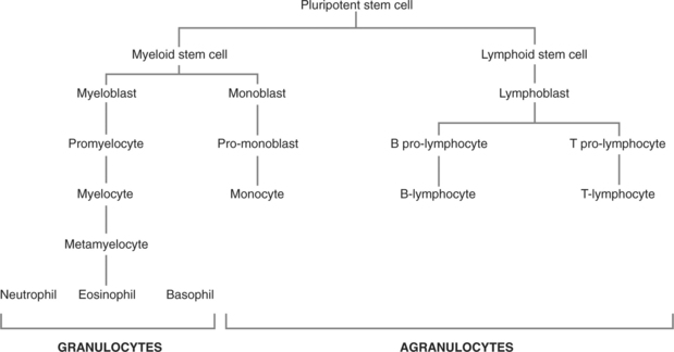
The haematological system consists of the blood and the sites of blood production (especially red bone marrow and lymphatic tissue). The main components of blood are shown in Figure 3.1. Different types of blood cell all develop from their common origin, the pluripotent stem cell. Red blood cells (RBCs), for example, follow a pathway from the pluripotent stem cell via the myeloid stem cell stage, develop into a proerythroblast, then a reticulocyte when they expel their nucleus to finally become a mature RBC or erythrocyte. The pathways followed by other types of blood cell are shown in Figure 3.2.
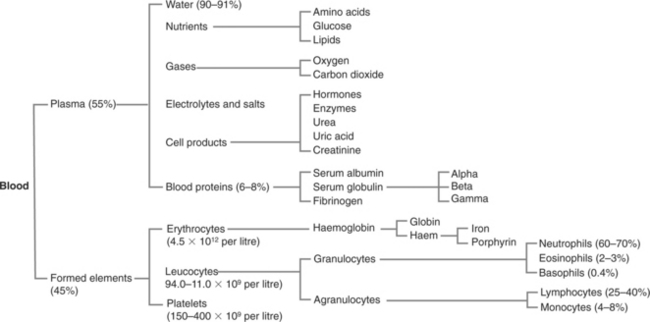
SOME KEY NORMAL VALUES FOR BLOOD
| Blood volume: | |
| Haemoglobin: | |
| Red cell count: | |
| Mean lifespan of RBC | 120 days |
| Platelets | 150–400 g/L |
| Leucocytes | 4.0–11.0 3 109 g/L |
| Erythrocyte | Male 0–5 mm/hour |
| Sedimentation Rate: | Female 0–7 mm/hour |
| Prothrombin Time | 12–14 seconds. |
PHYSIOLOGY YOU NEED TO KNOW
MAIN FUNCTIONS OF BLOOD
 Transport medium for metabolic requirements (e.g. oxygen, nutrients, hormones) and waste products of metabolism (e.g. carbon dioxide).

CONSTITUENTS OF BLOOD
 Erythrocytes (RBCs) are biconcave, circular discs with no nucleus. They are packed with haemoglobin whose main function is to carry oxygen and some carbon dioxide. A haemoglobin molecule consists of a protein called globin and a pigment called haem, which contains iron. The more oxygen is attached to the molecule, the redder the colour appears. Four oxygen molecules combine with each haemoglobin molecule to form oxyhaemoglobin which can readily disassociate back into oxygen and haemoglobin when required.
 Leucocytes (white blood cells). There are three main types:

 Blood Groups. Erythrocytes contain on their surface complex molecules known as isoantigens or agglutinogens which act as antigens. These isoantigens occur in characteristic combinations which give rise to a complex series of different blood groups. The original discovery of this system led to the following simple model based upon four possibilities:
| Type A antigens | Group A |
| Type B antigens | Group B |
| No antigens | Group O |
| A and B antigens | Group AB |
Plasma also contains antibodies or agglutinens which will react with certain antigens:
A person does not possess the antibody that will react with the antigen they display on their own erythrocytes. Grouping and cross matching for blood transfusion ensures that a person only receives blood that is compatible with their own antibodies so that a potentially fatal antigen–antibody reaction does not occur. Blood group O is known as the universal donor because it does not display any antigens which can provoke an antigen–antibody reaction. Research has shown that there are many different types of blood beyond the basic four types originally described in the ABO system, making grouping and cross matching an essential but time-consuming laboratory procedure.
ANAEMIA (P374)
PATHOLOGY: Key facts
Stay updated, free articles. Join our Telegram channel

Full access? Get Clinical Tree


