9 Caring for the patient with a disorder of the nervous system
ANATOMY AT A GLANCE (P653)
If the nervous system is considered in terms of its function, it can again be split into two parts:
 The somatic (voluntary) nervous system which transmits messages to and from the non-visceral part of the body (skeletal, skin, etc.), and is generally under conscious control.
The nervous system consists of two main groups of cells, neurones and neuroglia.
Neurones
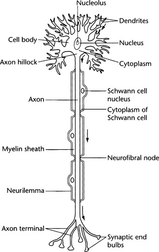
These are the functional cells responsible for initiating or transmitting messages (see Figure 9.1). Each neurone has a nucleus, one or more projections known as dendrites, which conduct the nerve impulse into the cell body, and a single long projection known as an axon, which conducts the nerve impulse away from the cell body to the next cell. Cell bodies are generally known as grey matter, while axons make up the white matter. Where the axon of one neurone meets the dendrite of the next, a synapse is said to occur. Although the axon and dendrite of the two neurones are in close proximity, they are not connected. The nerve impulse is transmitted via neurotransmitter molecules which cross the synapse. Neurones can have a sensory, motor or connecting function (interneurones).
The Central Nervous System
 Basal ganglia are groups of nerve cells deeply imbedded within the white matter whose function is not clearly understood.

 Brainstem consists of three portions


Cranial Nerves
The 12 pairs of cranial nerves are conventionally numbered in Roman numerals from I to XII. Cranial nerves III through XII emerge from the brainstem, whilst I and II are purely sensory and are directly connected to the appropriate sensory centre in the brain (I is olfactory and II is the optic nerve). The Xth cranial nerve is the vagus nerve, and its motor branch is part of the autonomic nervous system which is responsible for regulating smooth muscle function in a wide range of visceral organs.
The Autonomic Nervous System
 The Sympathetic Nervous System: this originates in the lumbar and thoracic regions of the spinal cord and produces generalized responses that help the body cope with threat and stress known as the ‘fight or flight response’. These include, for example, bronchodilation and increasing heart rate.
PHYSIOLOGY YOU NEED TO KNOW
Nerve impulse transmission depends upon the following series of events:
CEREBROVASCULAR ACCIDENT (CVA) [P672]
PATHOLOGY: Key facts


Most CVAs occur in people aged over 60. However, a significant number occur in young adults when the cause is a congenital weakness in a cerebral artery known as a cerebral aneurysm. The person is born with a weak area lacking muscle and elastic tissue in one of their cerebral arteries. Gradually over the years, this protrudes as a swelling from the arterial wall (hence the common name berry aneurysm), which may then start to leak or in worst cases, rupture completely, which is usually fatal. The bleeding may be into the subarachnoid space (subarachnoid haemorrhage) or into the brain substance itself.
WHAT TO LOOK OUT FOR
 Level of consciousness is the key observation for all neurological patients whether they have had a CVA, head injury or any other neurological disorder. Any deterioration in their condition will usually show itself by a decrease in the level of consciousness. This is assessed objectively by the Glasgow Coma Scale (GCS) (see Table 9.1), and these observations may be carried out as frequently as every 15 minutes in an unstable patient. The GCS is based upon the best responses to objective measures of how awake the patient is (eye opening), motor ability and how oriented they are (verbal responses).
Stay updated, free articles. Join our Telegram channel

Full access? Get Clinical Tree


